Abstract
Purpose
To investigate the clinical features and treatment outcomes between methicillin-sensitive Staphylococcus epidermidis (MSSE) and methicillin-resistant Staphylococcus epidermidis (MRSE) keratitis groups.
Methods
A retrospective analysis of case series was conducted of all patients with keratitis caused only by Staphylococcus epidermidis from January 1997 through December 2008. Sex, age, history of trauma, systemic disease, previous ocular history, antibiotic sensitivity test results, and treatment outcomes were evaluated. Patients were categorized into two groups as MSSE and MRSE according to methicillin-sensitivity result, and a comparative analysis was performed.
Results
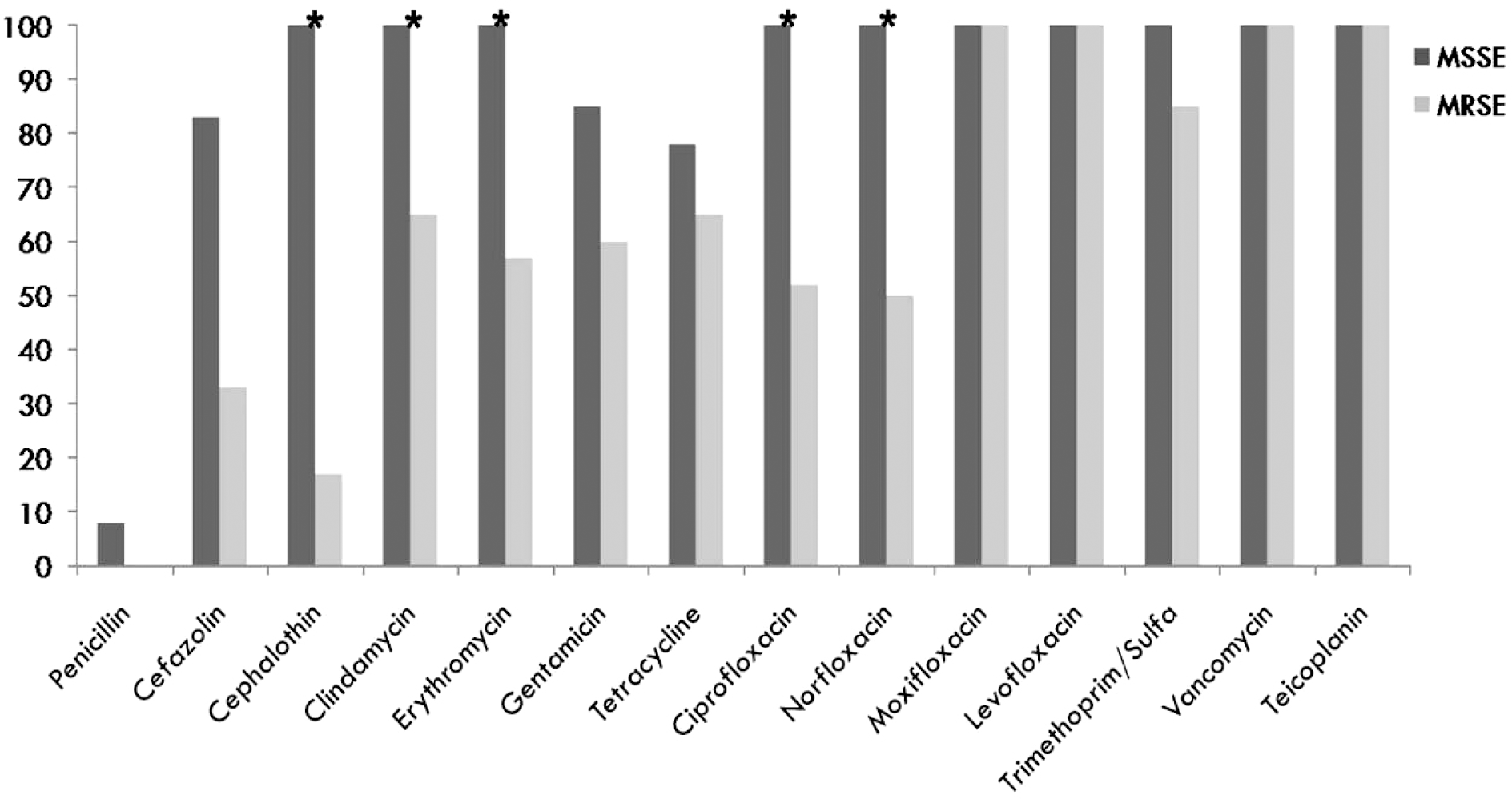
There were no significant differences in clinical features, such as risk factors or size or location of keratitis between the two groups. All MSSE and MRSE isolates were sensitive to vancomycin, moxifloxacin, and levofloxacin. All MSSE and 17%, 50%, 52%, and 57% of MRSE isolates were sensitive to cephalothin, norfloxacin, ciprofloxacin, and erythromycin, respectively (p < 0.05). There was no significant difference in visual acuity between the two groups.
Conclusions
All MSSE and MRSE isolates were sensitive to vancomycin and to third- or fourth-generation fluoroquinolones In addition, approximately 50% of MRSE isolates were sensitive to norfloxacin and ciprofloxacin. There were no significant differences in clinical features of keratitis caused by MSSE versus those of MRSE isolates. Both keratitis groups had relatively good visual prognoses.
Go to : 
References
1. Jones DB. Decision-making in the management of microbial keratitis. Ophthalmology. 1981; 88:814–20.

2. Erie JC, Nevitt MP, Hodge DO, Ballard DJ. Incidence of ulcerative keratitis in a defined population from 1950 through 1988. Arch Ophthalmol. 1993; 111:1665–71.

3. Franklin D, Scott M. Staphylococcus epidermidis infection. Ann Intern Med. 1983; 99:834–9.
4. Cokingtin CD, Hyndiuk RA. Bacterial kratitis. Tabbara KF, Hyndiuk RA, editors. Infection of the Eye. 2nd ed.Boston: Little Brown and Company;1996. p. 338–9.
5. Schaefer F, Bruttin O, Zografos L, Guex-Crosier Y. Bacterial keratitis: a prospective clinical and microbiological study. Br J Ophthalmol. 2001; 85:842–7.

6. Toshida H, Kogure N, Inoue N, Murakami A. Trends in microbial keratitis in Japan. Eye Contact Lens. 2007; 33:70–3.

7. Hahn YH, Hahn TW, Tchah H, et al. Epidemiology of infectious keratitis(II): A multicenter study. J Korean Ophthalmol Soc. 2001; 42:247–65.
8. Jang YS, Hahn YH. Epidemiology of staphylococcus epidermidis keratitis. J Korean Ophthalmol Soc. 2002; 43:665–71.
9. Park JH, Lee SB. Analysis on inpatients with infectious keratitis: causative organisms, clinical aspects and risk factors. J Korean Ophthalmol Soc. 2009; 50:1152–66.

10. Davis JL, Koidou-Tsiligianni A, Pflugfelder SC, et al. aberrations staphylococcal endophthalmitis. Increase in antimicrobial resistance. Ophthalmology. 1988; 95:1404–10.
11. Goldstein MH, Kowalski RP, Gordon YJ. Emerging fluoroquinolone resistance in bacterial keratitis: a 5-year review. Ophthalmology. 1999; 106:1313–8.
12. Miño De Kaspar H, Hoepfner AS, Engelbert M, et al. Antibiotic resistance pattern and visual outcome in experimentally-induced Staphylococcus epidermidis endophthalmitis in a rabbit model. Ophthalmology. 2001; 108:470–8.
13. Mukerji N, Vajpayee RB, Sharma N. Technique of area measurement of epithelial defects. Cornea. 2003; 22:549–51.

14. Hahn YH, Lee SJ, Hahn TW, et al. Antibiotic susceptibilities of ocular isolates from patients with bacterial keratitis: A multicenter study. J Korean Ophthalmol Soc. 1999; 40:2401–10.
15. Sotozono C, Inagaki K, Fujita A, et al. Methicillin-resistant Staphylococcus aureus and methicillin-resistant Staphylococcus epidermidis infections in the cornea. Cornea. 2002; 21:S94–101.

16. Tabbara KF, El-Sheikh HF, Aabed B. Extended wear contact lens related bacterial keratitis. Br J Ophthalmol. 2000; 84:327–8.

17. Miller DM, Vedula AS, Flynn HW Jr, et al. Endophthalmitis caused by staphylococcus epidermidis: in vitro antibiotic susceptibilities and clinical outcomes. Ophthalmic Surg Lasers Imaging. 2007; 38:446–51.

18. Recchia FM, Busbee BG, Pearlman RB, et al. Changing trends in the microbiologic aspects of postcataract endophthalmitis. Arch Ophthalmol. 2005; 123:341–6.

19. Mather R, Karenchak LM, Romanowski EG, Kowalski RP. Fourth generation fluoroquinolones: new weapons in the arsenal of ophthalmic antibiotics. Am J Ophthalmol. 2002; 133:463–6.
20. Betanzos-Cabrera G, Juárez-Verdayes MA, González-González G, et al. Gatifloxacin, moxifloxacin, and balofloxacin resistance due to mutations in the gyrA and parC genes of Staphylococcus epidermidis strains isolated from patients with endophthalmitis, corneal ulcers and conjunctivitis. Ophthalmic Res. 2009; 42:43–8.
Go to : 
 | Figure 1.Antibiotic sensitivity in patients with Staphylococcus epidermidis keratitis (%). MSSE = methicillin-sensitive Staphylococcus epidermidis; MRSE = methicillin-resistant Staphylococcus epidermidis. * p-value < 0.05. |
Table 1.
Demographics and patient characteristics in patients with Staphylococcus epidermidis keratitis
Table 2.
Predisposing factors in patients with Staphylococcus epidermidis keratitis
Table 3.
Clinical manifestations in patients with Staphylococcus epidermidis keratitis
Table 4.
Results of treatment for keratitis caused by Staphylococcus epidermidis
| Result | MSSE group | MRSE group | p-value |
|---|---|---|---|
| Recovery | 12 (60%) | 16 (70%) | 0.512 |
| Treatment failure | 4 (20%) | 5 (22%) | 0.889 |
| No follow-up | 4 (20%) | 2 (8%) | 0.286 |
| Total | 20 | 23 | |


 PDF
PDF ePub
ePub Citation
 Print
Print


 XML Download