

 PDF
PDF ePub
ePub Citation
 Print
Print


Abstract
Purpose
To report a case of proptosis occurring during a Valsalva maneuver in a neurofibromatosis patient with an arachnoid cyst.
Case summary
A 42-year-old man diagnosed with type I neurofibromatosis visited our hospital with a 20-year history of enophthalmos in the left eye. The patient also complained of exophthalmos during the abdominal straining. On exoph-thalmometry, a 4 mm enophthalmos was found. The patient also presented with a limited motion to the superior and lateral direction. There was an ocular pulsation corresponding to the heart rate. During the Valsalva maneuver, a marked exophthalmos of the left eye occurred. The patient had light brown spots on the skin of the face and body trunk. Following an orbital computed tomography (CT), defects of the left sphenoid bone were present. Posteriorly, an arachnoid cyst was found; however, there was a lack of varix. The archnoid cyst was also observed to expand into the orbit during the Valsalva maneuver and forward the globe.
References
1. Bullock JD, Bartley GB. Dynamic proptosis. Am J Ophthalmol. 1986; 102:104–10.

2. Riccardi VM. Neurofibromatosis: clinical heterogeneity. Curr Probl Cancer. 1982; 7:1–34.
3. Hamedani M, Pournaras JA, Goldblum D. Diagnosis and management of enophthalmos. Surv Ophthalmol. 2007; 52:457–73.
4. Huson S, Jones D, Beck L. Ophthalmic manifestations of aberrations. Br J Ophthalmol. 1987; 71:235–8.
5. Bosch MM, Boltshauser E, Harpes P, Landau K. Ophthalmologic findings and long-term course in patients with neurofibromatosis type 2. Am J Ophthalmol. 2006; 141:1068–77.
6. Klatte EC, Franken EA, Smith JA. The radiographic spectrum in neurofibromatosis. Semin Roentgenol. 1976; 11:17–33.
7. Wiesenfeld D, James PL. Pulsating exophthalmos associated with neurofibromatosis. J Maxillofac Surg. 1984; 12:11–3.
8. Fukuta K, Jackson IT. Orbital neurofibromatosis with enophthalmos. Br J Plast Surg. 1993; 46:36–9.
9. Rufa A, Zicari E, Cerase A, et al. Pulsating enophthalmos in an adult patient with type 1 neurofibromatosis. Neurology. 2006; 67:2169.
10. Menon V, Vashisht S, Gupta KK, et al. Pulsating enophthalmos in aplasia of sphenoid wing. Indian J Ophthalmol. 1993; 41:88–90.
11. Morris SR, Desousa JL, Francis I, et al. Radiological pitfalls in patients with inducible dynamic proptosis. Open Ophthalmol J. 2008; 2:91–3.
12. Shehu BB, Hassan I. Cervicothoracic arachnoid cyst in a patient with neurofibromatosis: case report. East Afr Med J. 2006; 83:515–7.
13. Wegener M, Prause JU, Thygesen J, et al. Arachnoid cyst causing an optic neuropathy in neurofibromatosis I. Acta Ophthalmol. 2010; 88:497–9.
Figure 1.
Proptosis during Valsalva maneuver. Figure A shows left enophthalmos by 4 mm and figure B shows 3 mm protrusion of the left eye ball during Valsalva maneuver.
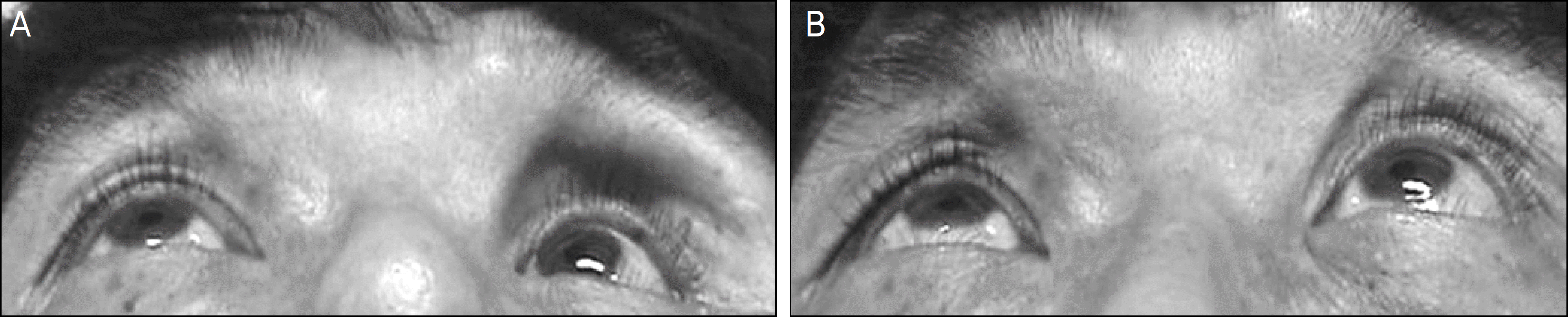
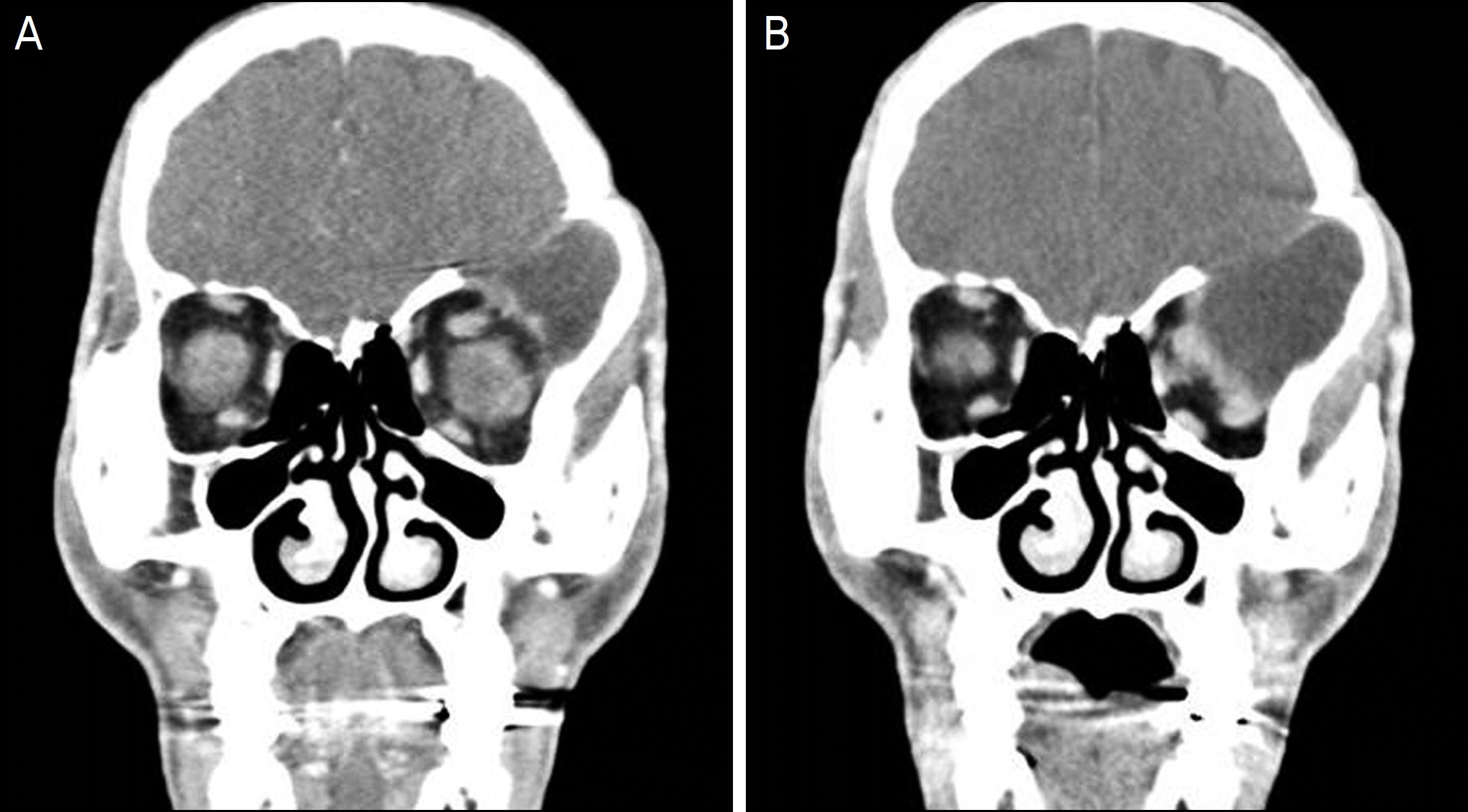
Figure 2.
Orbital computed tomography of the paitent. (A) The coronal section shows the defect of the left sphenoid wing and arachnoid cyst in the middle cranial fossa. (B) Same section during Valsava maneuver shows the arachnoid cyst expands into the orbit through the sphenoid wing defect and displacement of orbital contents.
 XML Download