Abstract
Purpose
To evaluate the differences in corneal endothelial cell density in patients undergoing intraocular lens insertion at 2 different sequences during the triple procedure.
Methods
The present retrospective study divided 37 eyes of 37 patients and into 2 groups: trephination, phacoemulsification and intraocular insertion, followed by graft suturing (20 eyes, Group I), and trephination, phacoemulsification, graft suturing leaving 3 mm gap, followed by intraocular lens insertion and suturing of the gap (17 eyes, Group II). Intraocular pressure, corneal thickness, and endothelial cell density were measured for 3 months postoperatively.
Results
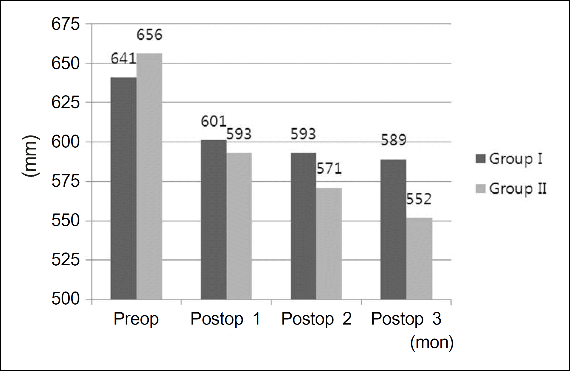
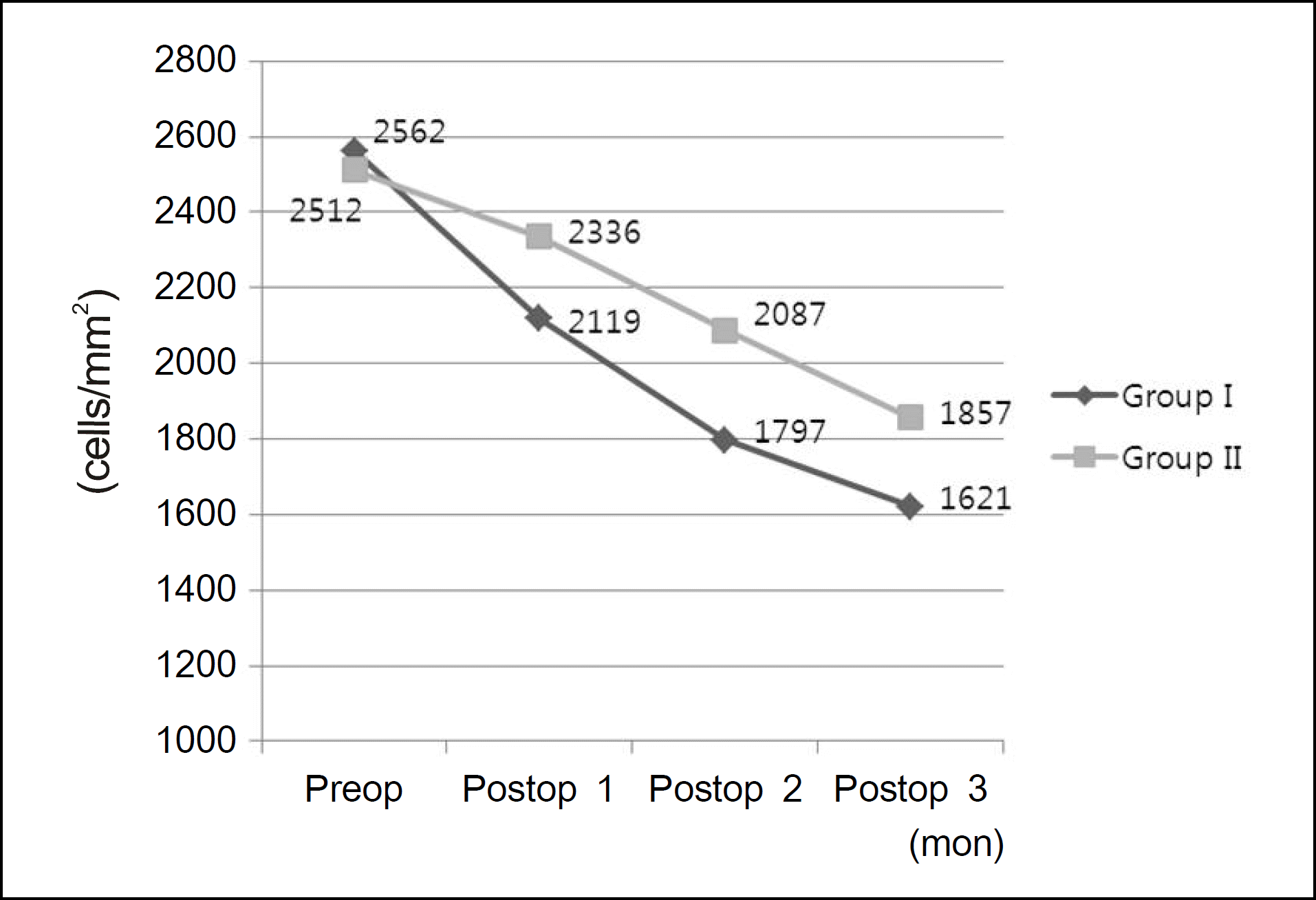
No significant difference in intraocular pressure between the 2 groups (p = 0.14) was observed. However in Group II, the mean corneal thickness showed a greater decrease (p = 0.02) during the 3 months following surgery, and the mean corneal endothelial cell density in this group was higher at 1, 2, and 3 months postoperatively than that of Group I (p = 0.01, 0.02 and 0.04 respectively). There were no significant difference in the rate of endothelial cell loss during the post-operative period between the 2 groups.
Go to : 
References
1. Katzin HM, Meltzer JF. Combined surgery for corneal transplantation and cataract extraction. Am J Ophthalmol. 1966; 62:556–60.

3. Taylor DM, Khaliq A. Keratoplasty and intraocular lenses: follow-up study. Ophthalmic Surg. 1977; 8:49–57.

4. Taylor DM, Khaliq A, Maxwell R. Keratoplasty and intraocular lenses: current status. Ophthalmology. 1979; 86:242–55.

5. Hunkeler JD, Hyde LL. The triple procedure: combined penetrating keratoplasty, extracapsular cataract extraction and lens implantation. An expanded experience. J Am Intraocul Implant Soc. 1983; 9:20–4.

6. Skorpik C, Menapace R, Gnad HD, Grasl M. The triple procedure– results in cataract patients with corneal opacity. Ophthalmologica. 1988; 196:1–6.
7. Binder PS. The triple procedure. Refractive results. 1985 update. Ophthalmology. 1986; 93:1482–8.
8. Meyer RF, Musch DC. Assessment of success and complications of triple procedure surgery. Am J Ophthalmol. 1987; 104:233–40.

9. McCartney DL, Gottsch JD, Stark WJ. Managing posterior pressure during pseudophakic keratoplasty. Arch Ophthalmol. 1989; 107:1384–6.

11. Kim MK, Lee JH. Long-term outcome of graft rejection after penetrating keratoplasty. J Korean Ophthalmol Soc. 1997; 38:1553–60.
12. Patel SV, Hodge DO, Bourne WM. Corneal endothelium and postoperative outcomes 15 years after penetrating keratoplasty. Am J Ophthalmol. 2005; 139:311–9.

13. Bourne WM, O'Fallon WM. Endothelial cell loss during penetrating keratoplasty. Am J Ophthalmol. 1978; 85:760–6.

14. Kramer SG, Stewart HL. Maintenance of the anterior chamber during penetrating keratoplasty. Trans Sect Ophthalmol Am Acad Ophthalmol Otolaryngol. 1976; 81:794–805.
15. Kirkness CM, Ling Y, Moshegov C. Penetrating keratoplasty and raised intraocular pressure. A brief review of the problems and its management. Ann Acad Med Singapore. 1989; 18:168–70.
16. Simmons RB, Stern RA, Teekhasaenee C, Kenyon KR. Elevated intraocular pressure following penetrating keratoplasty. Trans Am Ophthalmol Soc. 1989; 87:79–91. discussion. 91–3.
17. Seitz B, Langenbucher A, Nguyen NX, et al. Long-term follow-up of intraocular pressure after penetrating keratoplasty for keratoconus and Fuchs' dystrophy: comparison of mechanical and Excimer laser trephination. Cornea. 2002; 21:368–73.
18. Nguyen NX, Langenbucher A, Seitz B, et al. Impact of increased intraocular pressure on long-term corneal endothelial cell density after penetrating keratoplasty. Ophthalmologica. 2002; 216:40–4.

19. Burke S, Sugar J, Farber MD. Comparison of the effects of two viscoelastic agents, Healon and Viscoat, on postoperative intraocular pressure after penetrating keratoplasty. Ophthalmic Surg. 1990; 21:821–6.

20. Langenbucher A, Seitz B, Nguyen NX, Naumann GO. Corneal endothelial cell loss after nonmechanical penetrating keratoplasty depends on diagnosis: a regression analysis. Graefes Arch Clin Exp Ophthalmol. 2002; 240:387–92.

21. Gil SY, Park CK, Hahn TW. Evaluation of donor corneal endothelium after keratoplasty. J Korean Ophthalmol Soc. 2006; 47:519–24.
22. Chung SH, Kim HK, Kim MS. Corneal endothelial cell loss after penetrating keratoplasty in relation to preoperative recipient endothelial cell density. Ophthalmologica. 2010; 224:194–8.

23. Sud RN, Loomba R. Achievement of surgically soft and safe eyes–a comparative study. Indian J Ophthalmol. 1991; 39:12–4.
24. Guindon B, Harvey J, Peacocke A, et al. Factors modifying vitreous pressure in cataract surgery. Can J Ophthalmol. 1981; 16:73–5.
Go to : 
 | Figure 1.Comparison of preoperative and postoperative corneal central thickness in Group I and II. |
 | Figure 2.Comparison of preoperative and postoperative corneal endothelial cell density in Group I and II. |
Table 1.
Clinical characteristics in Group I and II
| | Group I | Group II | ‡ p-value |
|---|---|---|---|
| Recipient | | | |
| Age (mean ± SD, yr) | 55.3 ± 10.9 | 58.2 ± 12.1 | 0.23 |
| Sex (M/F) | 11/9 | 10/7 | |
| Laterality (OD/OS) | 7/13 | 9/6 | |
| Follow-up period (mean ± SD, mon) | 7.4 ± 3.1 | 7.9 ± 3.9 | 0.10 |
| Donor | | | |
| Age (mean ± SD, yr) | 63 ± 7.8 | 64 ± 7.2 | 0.17 |
| Trephine size (mean ± SD, mm) | 7.68 ± 0.45 | 7.72 ± 0.42 | 0.08 |
| * CD (mean ± SD, cells/mm2) | 2562 ± 299 | 2512 ± 315 | 0.19 |
| † CT (mean ± SD, um) | 641 ± 33 | 656 ± 40 | 0.14 |


 PDF
PDF ePub
ePub Citation
 Print
Print


 XML Download