

 PDF
PDF ePub
ePub Citation
 Print
Print


Abstract
Purpose
To analyze the morphologic differences in blowout fracture seen on preoperative CT images compared to intraoperative images.
Methods
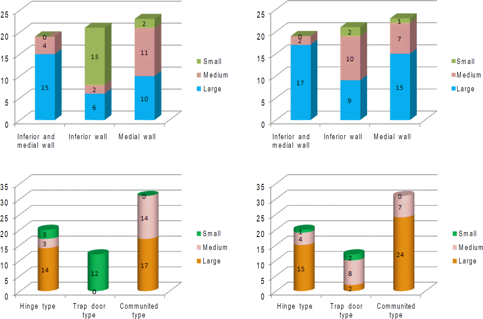
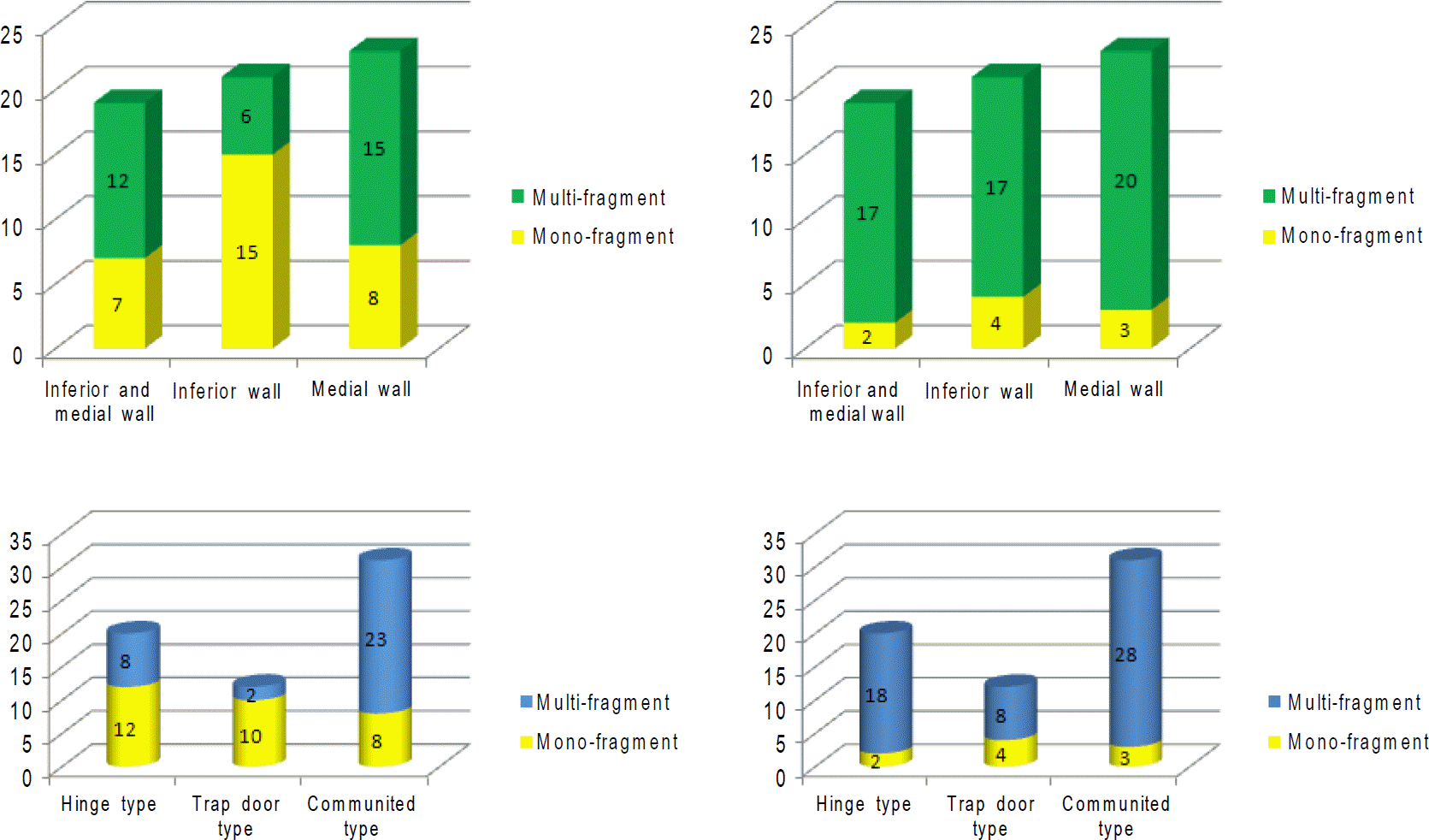
The present study included 63 patients (63 eyes) with orbital fractures that were repaired by orbital reconstruction between January 2009 and April 2010. We assessed the medial, inferior, and inferomedial orbital fractures and excluded superior and lateral wall fractures. We captured intraoperative blowout fracture images using a camera or endoscope and compared the fracture type (fracture size, fracture surface) seen on intraoperative images with that observed on the pre-operative CT images.
Results
The study consisted of patients between 20 and 50 years of age with a mean age of 27.76 years (men: 49 eyes, women: 14 eyes). The proportions of small fractures and medium fractures were similar on the preoperative CT images; however, large fractures were observed more frequently on the intraoperative images. The proportions of mono-fragment fractures and multi-fragment fractures were similar on the preoperative CT images, but multi-fragment fractures, especially inferior orbital fractures, were more frequent on the intraoperative images. Features of the trapdoor fracture differed most between images in terms of fracture size and surface.
Go to : 
References
1. Paek SH, Kim YS, Lee TS. A clinical study of blowout fracture. J Korean Ophthalmol Soc. 1993; 34:1194–8.
2. Lee SY, Kim SY, Kim HB. Orbital fractures evaluated by computed tomography. J Korean Ophthalmol Soc. 1990; 31:249–53.
3. Ahn SK, Jung SW. The clinical aspects of orbital fractures proven by computed tomography. J Korean Ophthalmol Soc. 1997; 38:2077–83.
4. Lee MS, Ahn JH, Kim HY, Lee SY. Clinical study of orbital wall fracture. J Korean Ophthalmol Soc. 1997; 38:1687–93.
5. Kwon YH, Park DW, Chung J-Y, Ahn HB. A clinical study of pediatric orbital wall fracture. J Korean Ophthalmol Soc. 2006; 47:7–12.
6. Park SH, Rah SH, Kim YH. Clinical evaluation of the associated ocular injuries of orbital wall fracture patients. J Korean Ophthalmol Soc. 2002; 43:1474–81.
7. Kim HE, Lew H, Yun YS. The size of extraocular muscles estimated by computed tomography in patients undergoing orbital wall fracture repair. J Korean Ophthalmol Soc. 2009; 50:1447–54.

8. Bansagi ZC, Meyer DR. Internal orbital fractures in the pediatric age group: characterization and management. Ophthalmology. 2000; 107:829–36.
9. Putterman AM. Management of blow out fractures of the orbital floor. III. The conservative approach. Surv Ophthalmol. 1991; 35:292–8.
10. Hawes MJ, Dortzbach RK. Surgery on orbital floor fractures. Influence of time of repair and fracture size. Ophthalmology. 1983; 90:1066–70.
11. Yang HW, Bae JH, Lee HC. The postoperative recovery of ocular motility in pediatric blowout fracture. J Korean Ophthalmol Soc. 2003; 44:259–64.
12. Paek SH, Kim YS, Lee TS. A Clinical study of blowout fracture. J Korean Ophthalmol Soc. 1993; 34:1194–8.
13. Lee SJ, Park KS. Relationship between preoperative clinical features and postoperative recovery of ocular motility restriction in blowout fractures. J Korean Ophthalmol Soc. 2001; 42:1202–9.
14. de Man K, Wijngaarde R, Hes J, de Jong PT. Influence of age on the management of blowout fractures of the orbital floor. Int J Oral Maxillofac Surg. 1991; 20:330–6.
15. Koltai PJ, Amjad I, Meyer D, Feustel PJ. Orbital fractures in children. Arch Otolaryngol Head Neck Surg. 1995; 121:1375–9.
16. Jordan DR, Allen LH, White J, et al. Intervention within days for some orbital floor fractures: the white-eyed blowout. Ophthal Plast Reconstr Surg. 1998; 14:379–90.
17. Anderson PJ, Poole MD. Orbital floor fractures in young children. J Craniomaxillofac Surg. 1995; 23:151–4.
18. Sires BS, Stanley RB Jr, Levine LM. Oculocardiac reflex caused by orbital floor trapdoor fracture: an indication for urgent repair. Arch Ophthalmol. 1998; 116:955–6.
19. Kwon YH, Park DW, Chung J-Y, Ahn HB. A clinical study of pediatric orbital wall fracture. J Korean Ophthalmol Soc. 2006; 47:7–12.
Go to :
 | Figure 1.The size of the orbital wall fracture (arrow) classified into small (A, D), medium (B, E) and large (C, F, H) at inferior (A, B, C), medial (D, E, F) and inferomedial (G, H) orbital wall fracture. |
 | Figure 2.The surface of the orbital wall fracture (arrow) classified into mono-fragment fracture (A, C, E), multi-fragment fracture (B, D, F) at inferior (A, B), medial (C, D) and inferomedial (E, F) orbital wall fracture. |
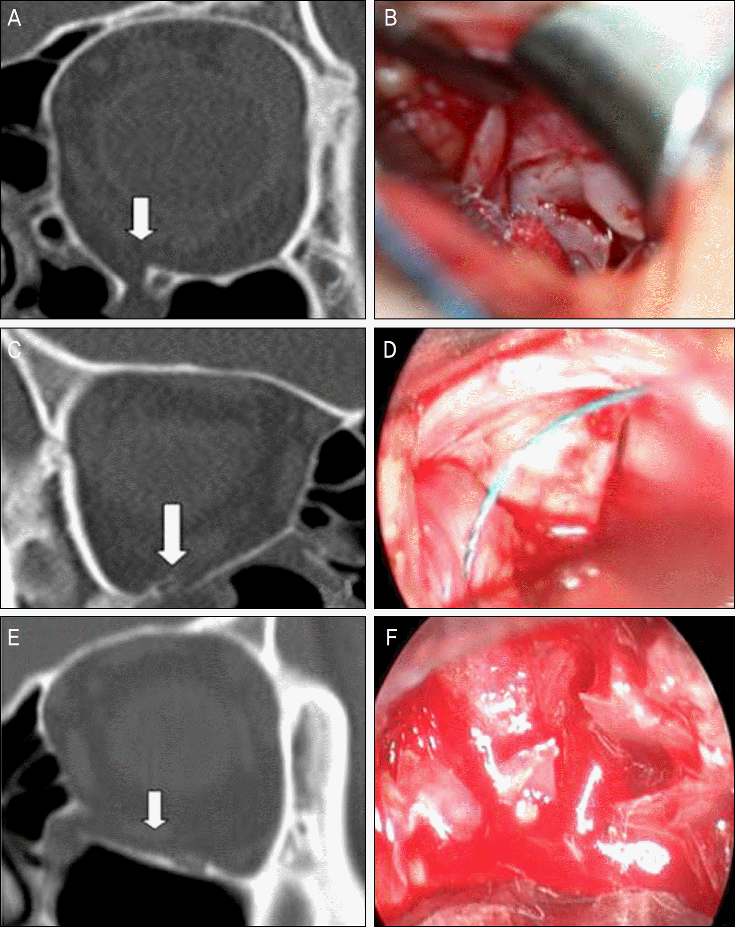 | Figure 3.Different fracture pattern in preoperative CT image and intraoperative real image. (A) Preop CT image: small, mono-fragment, inferior, trapdoor fracture. (B) Intraop real image: medium, multi-fragment, inferior fracture. (C) Preop CT image: small, inferior, trapdoor fracture. (D) Intraop real image: large, inferior fracture. (E) Preop CT image: large, mono-fragment, inferior fracture. (F) Intraop real image: large, multi-fragment inferior fracture. |
 XML Download