

 PDF
PDF ePub
ePub Citation
 Print
Print


Abstract
Purpose
To present a case of a patient with decreased visual acuity and anterior ischemic optic neuropathy diagnosed with advanced Moyamoya disease.
Case summary
A 46-year-old woman presented sudden episodes of headache and decreased visual acuity. On her first visit, her best corrected visual acuity was 20/25 in the right eye and 20/70 in the left eye. The left eye pupil was dilated with a relative afferent papillary defect. Fundus examinations revealed disc swelling in the left eye. After being admitted, steroid pulse therapy was started and Magnetic Resonance Imaging (MRI) studies revealed Moyamoya disease. The diagnosis was confirmed via Magnetic Resonance Angiography (MRA). After steroid pulse therapy, the headaches and visual acuity improved and the patient is continuing follow-up visits at neurosurgery and ophthalmology clinics.
Go to : 
References
1. David CA, Nottmeier E. Intracranial occlusion disease: Moyamoya. In: Winn HR ed. Youmans Neurological Surgery. 5th ed.Philadelphia: Saunders;2004. 2:chap. 103.
2. Noda S, Hayasaka S, Setogawa T, Matsumoto S. Ocular symptoms of moyamoya disease. Am J Ophthalmol. 1987; 103:812–6.

3. Takeuchi K. Occlusive diseases of the carotid artery. Shinkei Shimpo. 1961; 5:511–43.
4. Suzuki J, Takaku A. Cerebrovascular “moyamoya” disease. Disease showing abnormal net-like vessels in base of brain. Arch Neurol. 1969; 20:288–99.
5. Kuriyama S, Kusaka Y, Fujimura M, et al. Prevalence and clin-icoepidemiological features of moyamoya disease in Japan: findings from a nationwide epidemiological survey. Stroke. 2008; 39:42–7.
6. Phi JH, Wang KC, Cho BK, Kim SK. Pediatric cerebrovascular disease. Korean J Pediatr. 2008; 51:1282–9.
7. Robertson RL, Burrows PE, Barnes PD, et al. Angiographic changes after pial synangiosis in childhood moyamoya disease. AJNR Am J Neuroradiol. 1997; 18:837–45.
8. Miyamoto S, Kikuchi H, Karasawa J, et al. Study of the posterior circulation in moyamoya disease. Part 2: Visual disturbances and surgical treatment. J Neurosurg. 1986; 65:454–60.
9. Chen CS, Lee AW, Kelman S, Wityk R. Anterior ischemic optic neuropathy in moyamoya disease: a first case report. Eur J Neurol. 2007; 14:823–5.
10. Barrall JL, Summers CG. Ocular ischemic syndrome in a child with moyamoya disease and neurofibromatosis. Surv Ophthalmol. 1996; 40:500–4.
11. Ushimura S, Mochizuki K, Ohashi M, et al. Sudden blindness in the fourth month of pregnancy led to diagnosis of moyamoya disease. Ophthalmologica. 1993; 207:169–73.
12. Harissi-Dagher M, Sebag M, Dagher JH, Moumdjian R. Chorioretinal atrophy in a patient with moyamoya disease. Case report. J Neurosurg. 2004; 101:843–5.
13. Chace R, Hedges TR 3rd. Retinal artery occlusion due to moyamoya disease. J Clin Neuroophthalmol. 1984; 4:31–4.
14. Lee YW, Lee SN. A case of bilateral upgaze palsy associated with unilateral midbrain hemorrhage in moyamoya disease. J Korean Ophthalmol Soc. 2004; 45:1772–6.
15. Khan N, Schinzel A, Shuknecht B, et al. Moyamoya angiopathy with dolichoectatic internal carotid arteries, patent ductus arterio-sus and pupillary dysfunction: a new genetic syndrome? Eur Neurol. 2004; 51:72–7.
16. Krishnan C, Roy A, Traboulsi E. Morning glory disk anomaly, choroidal coloboma, and congenital constrictive malformations of the internal carotid arteries (moyamoya disease). Ophthalmic Genet. 2000; 21:21–4.
Go to :
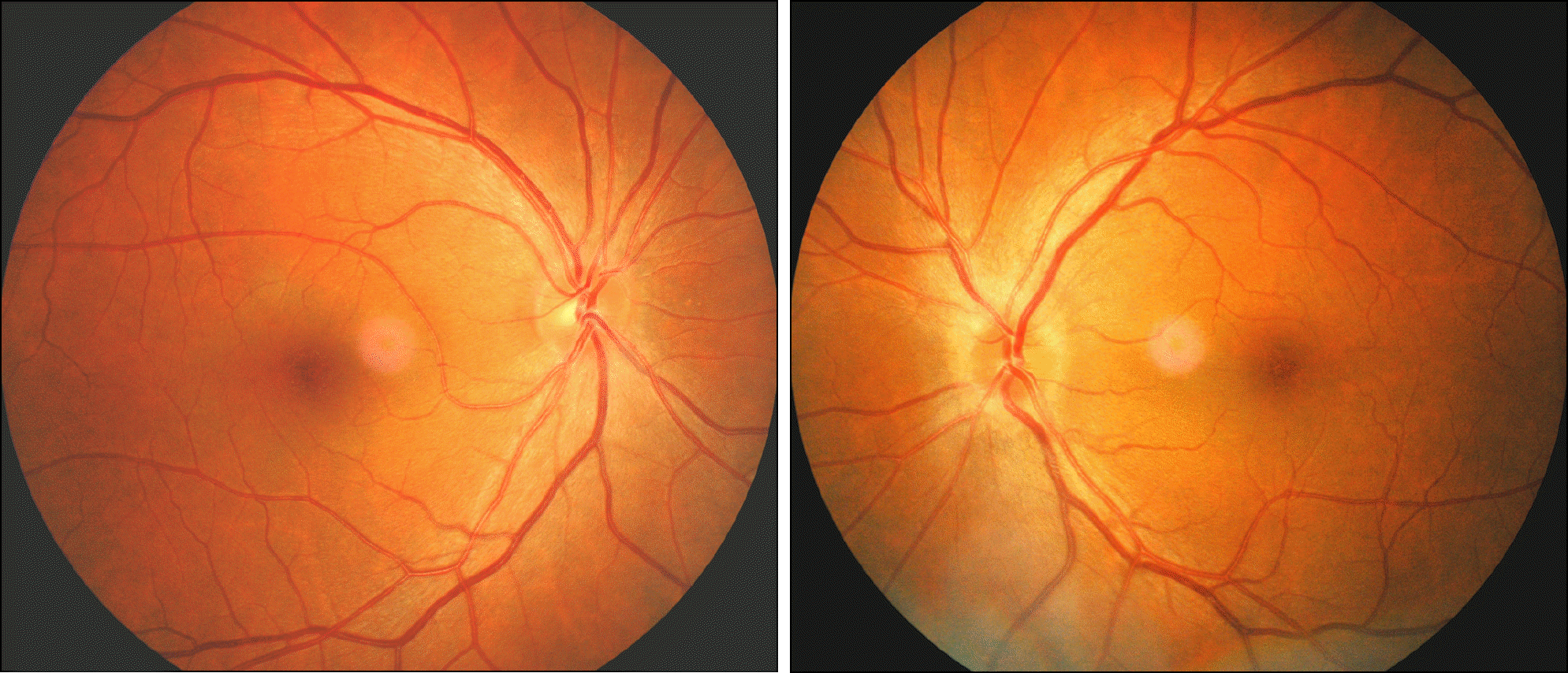 | Figure 1.Fundus photographs show marked disc swelling in the left eye and mild disc swelling in the right eye. |
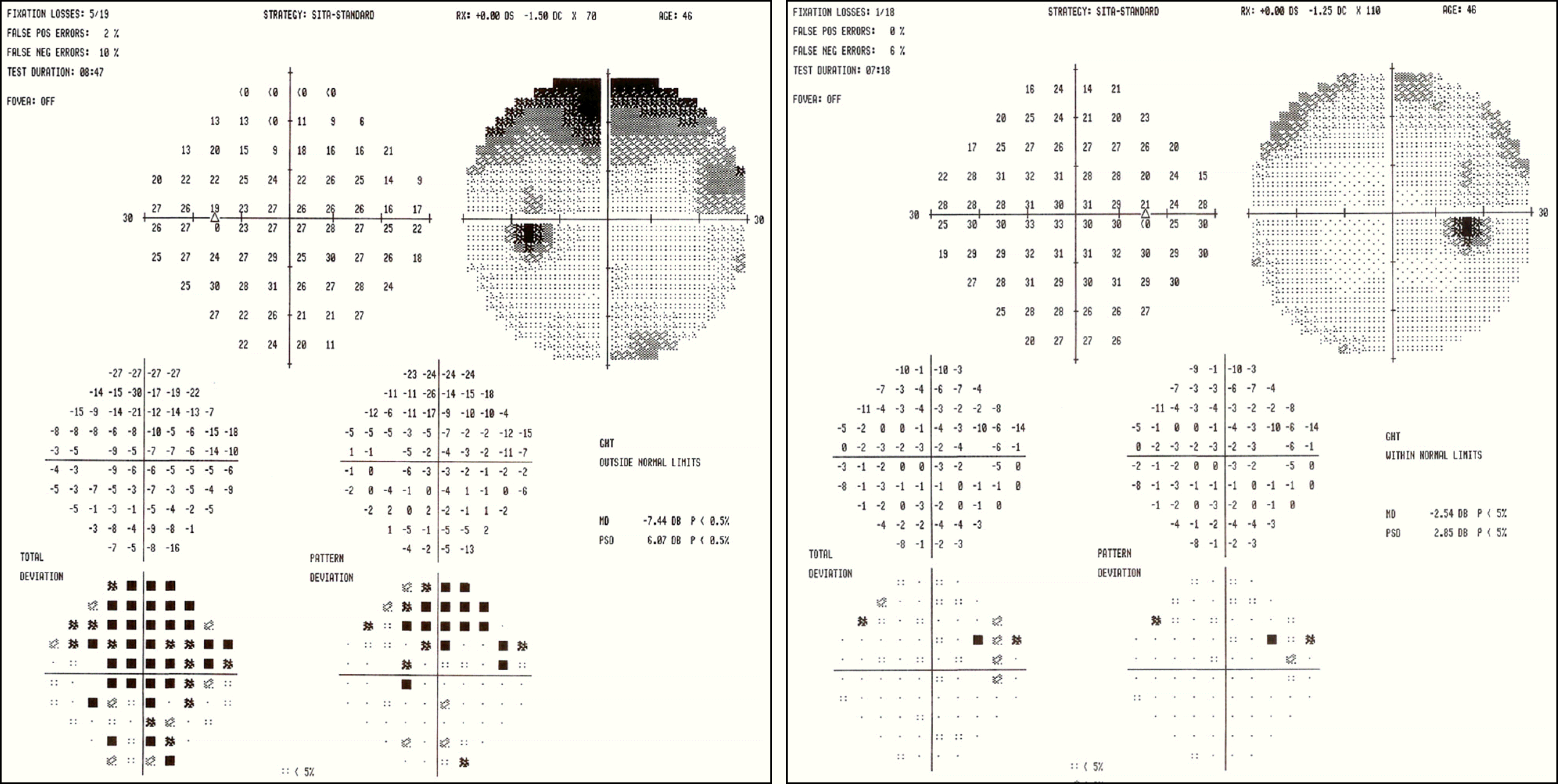
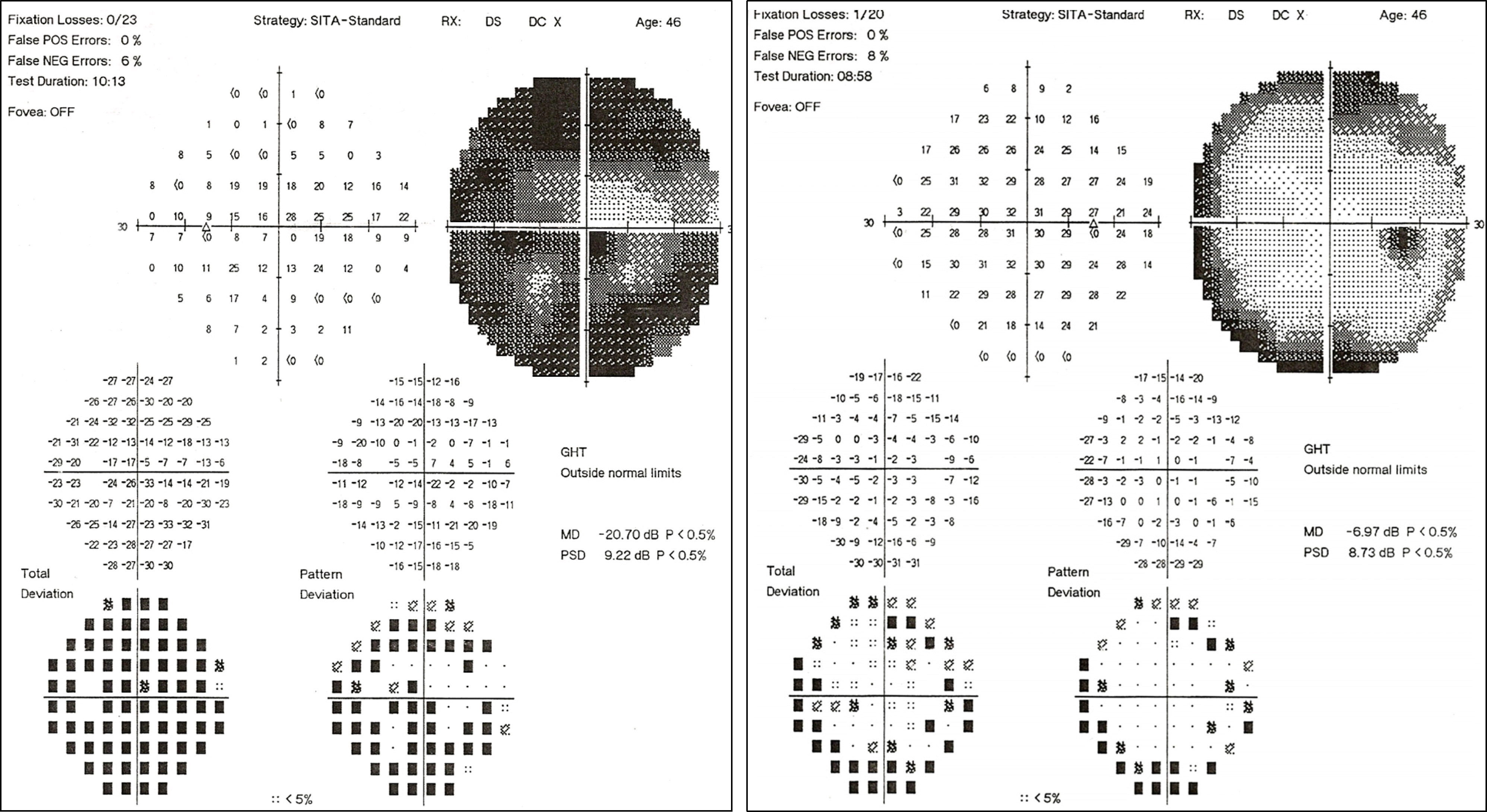 | Figure 2.The visual field exams on first visit show pericentral scotomas in the right eye and diffuse scotoma in the left eye. |
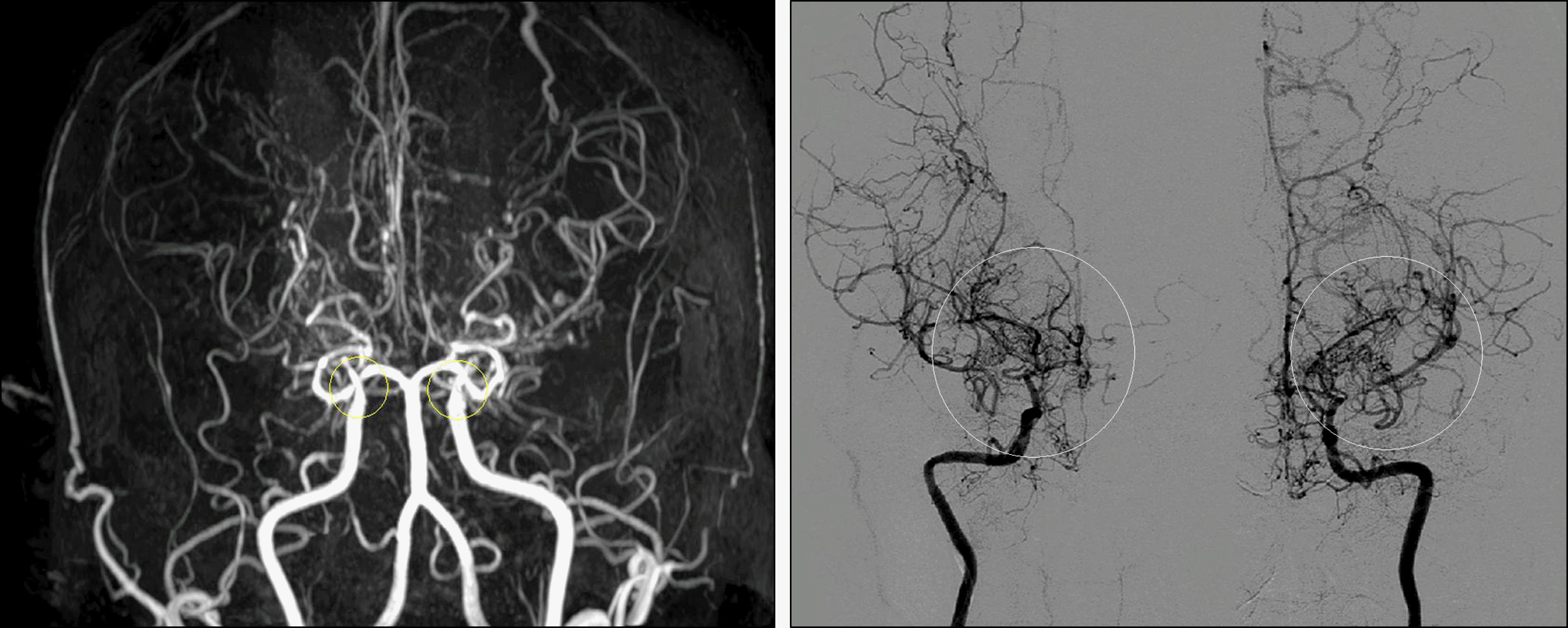
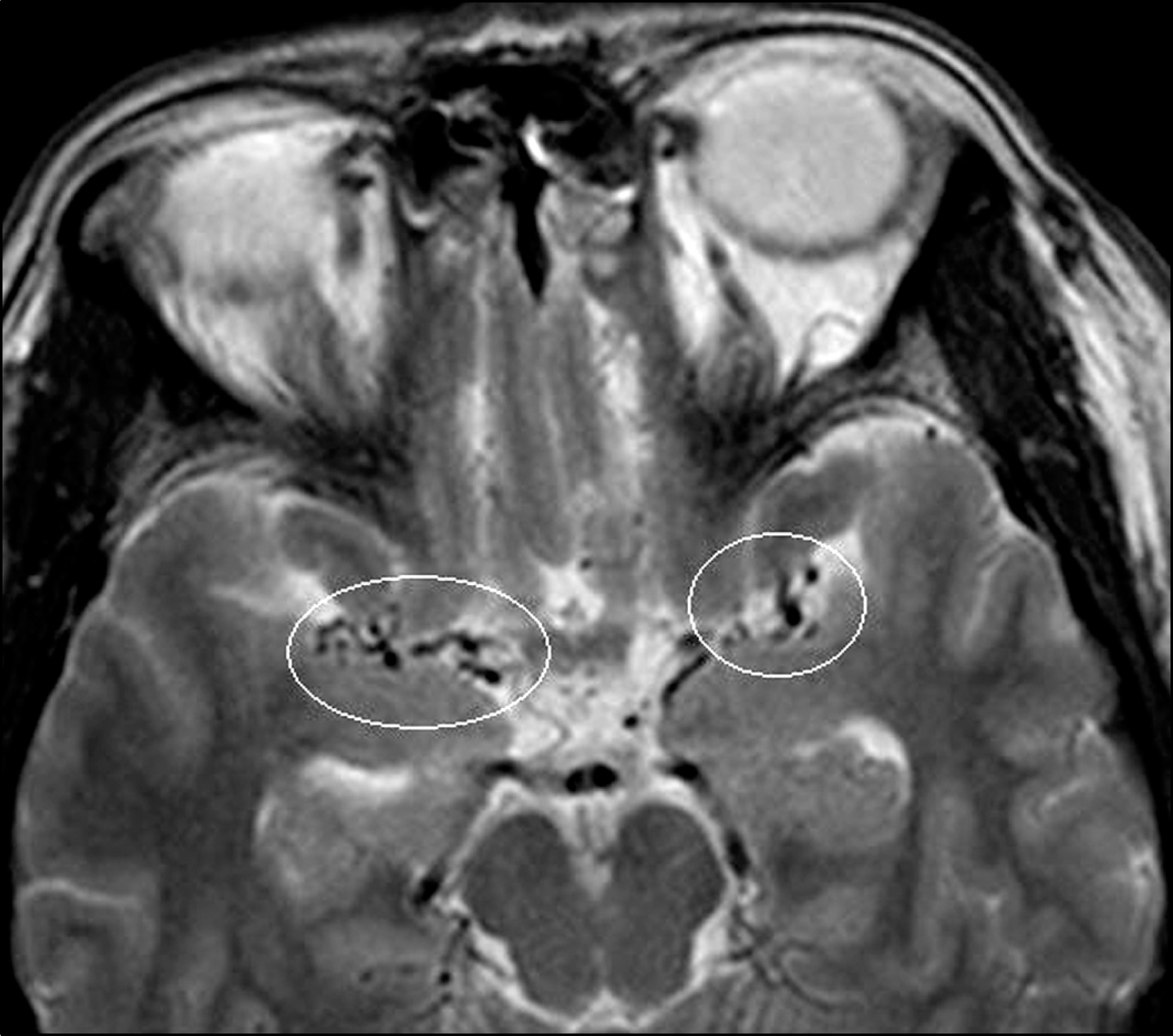 | Figure 4.T2-weighted brain MRI shows increased tiny signal voids in both middle cerebral artery trunks (R/O Moyamoya disease). |
 XML Download