Abstract
Purpose
To report the results of performing an Ahmed glaucoma valve implantation without removing the anterior chamber lens to treat secondary glaucoma.
Case summary
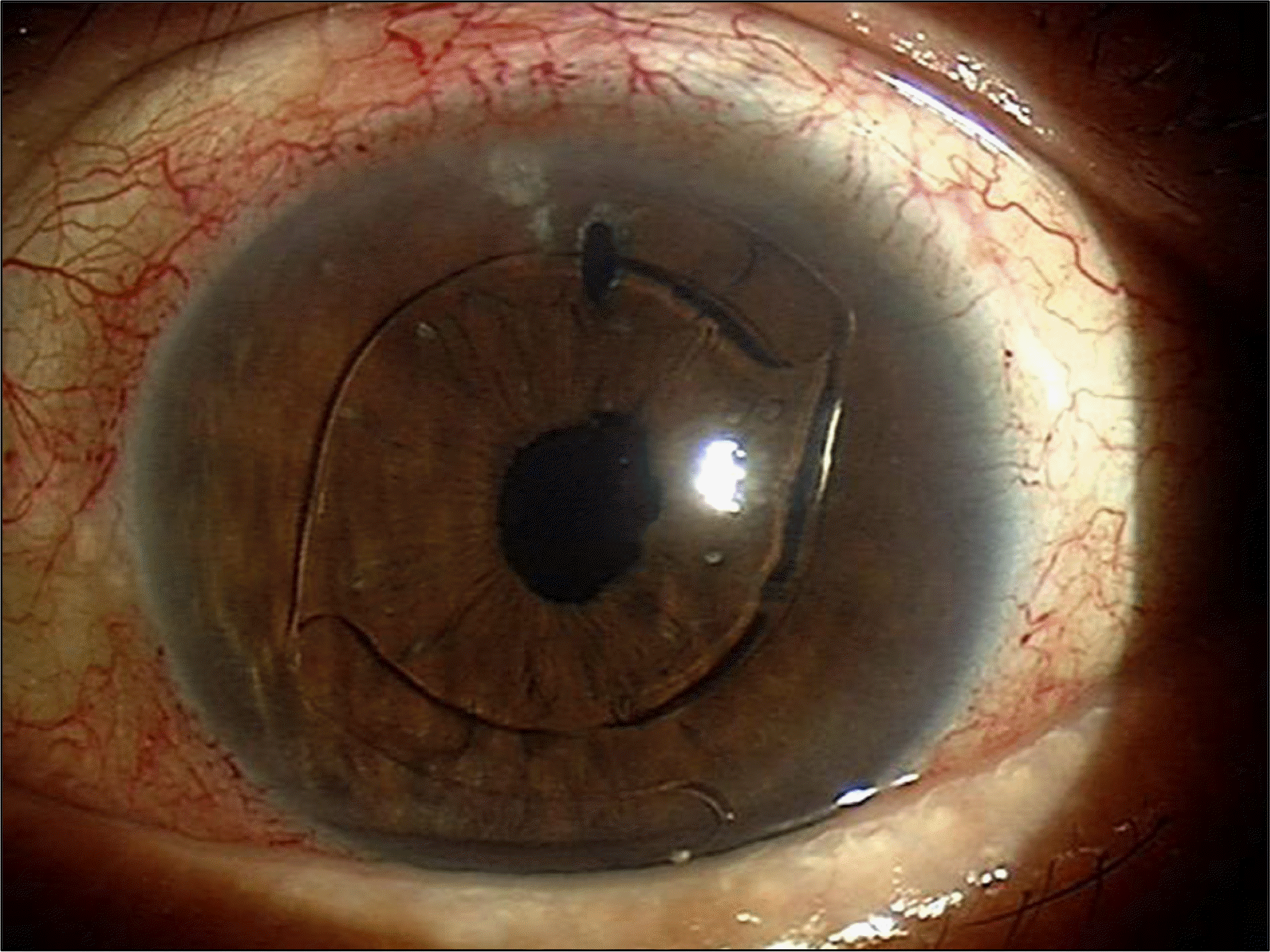
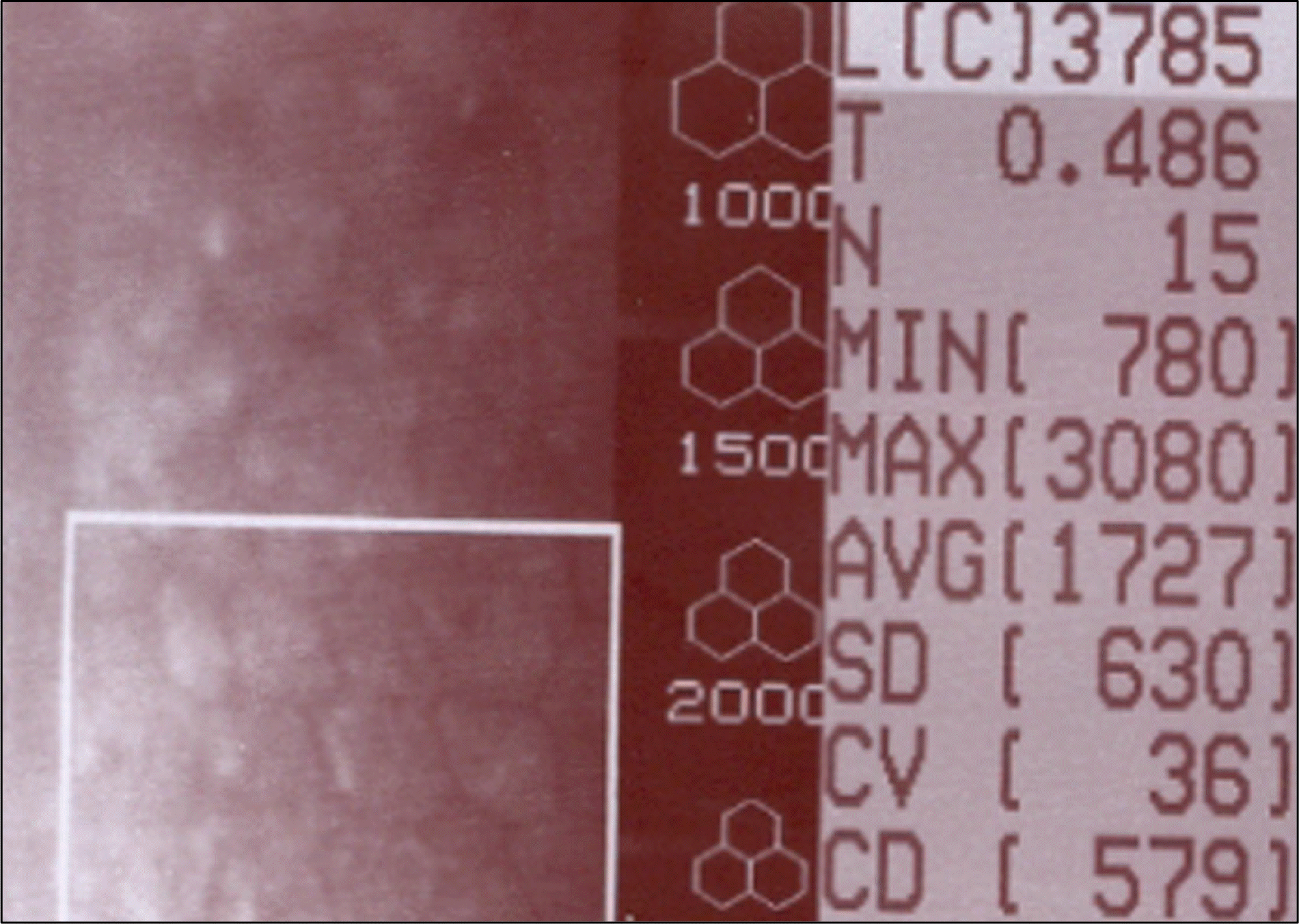
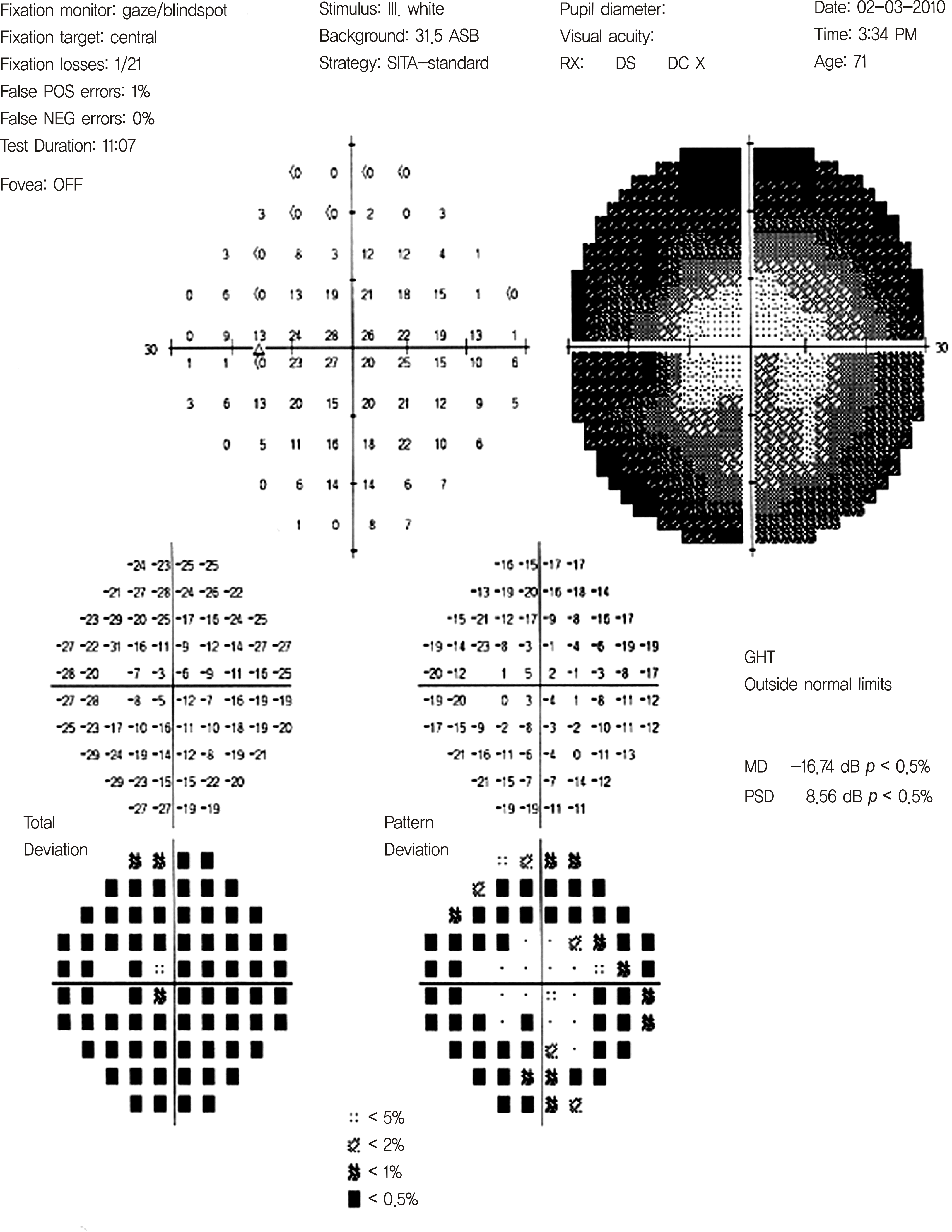
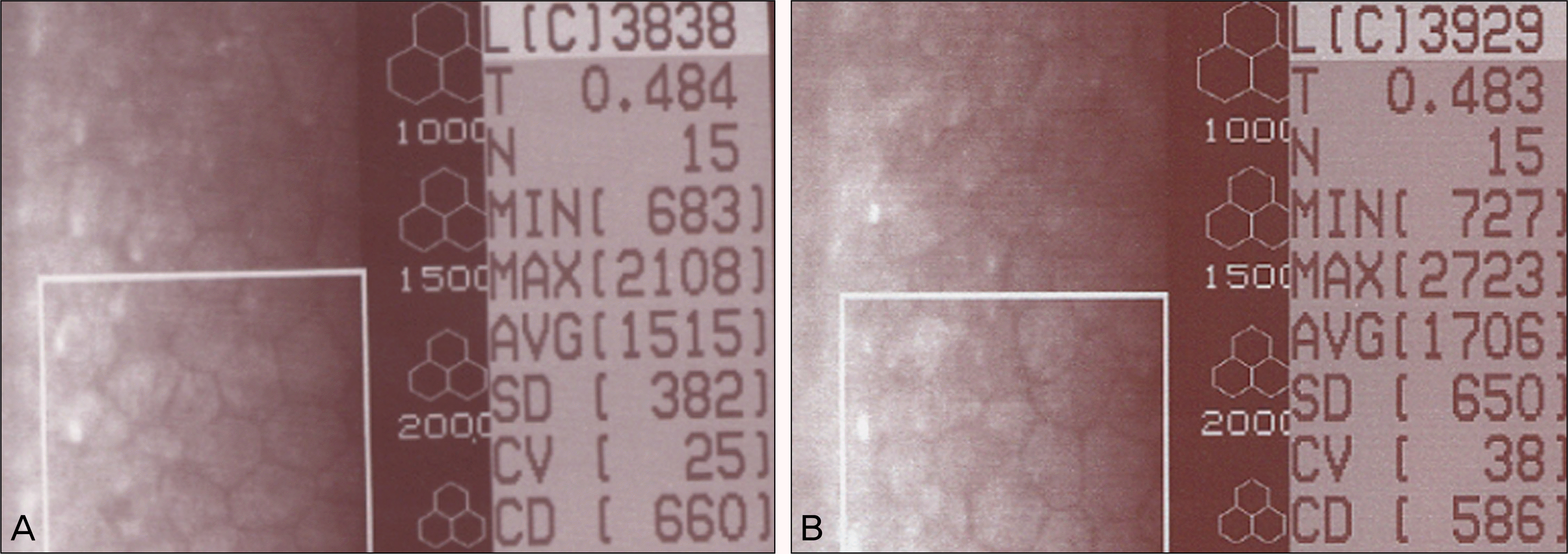
A 72-year-old male visited the hospital for imminent visual field loss in the left eye. At the time of the visit, he had a mild headache, and the intraocular pressure of the left eye was 38 mm Hg. The left eye had received anterior chamber lens insertion with iridectomy and the posterior capsule was ruptured. The vitreous protruded at the two o'clock site and adhered to the backside of the anterior chamber lens tilting it toward the temporal cornea. At the time of the visit, the maximum corrected vision of the left eye was 0.32. The patient was diagnosed with secondary glaucoma caused by the anterior chamber lens. Because the patient did not want to have the anterior chamber lens removed and the supporting area of the anterior chamber lens blocked the trabeculectomy and peripheral iridectomy sites, the authors performed an Ahmed glaucoma valve implantation instead. The outcome of Ahmed glaucoma valve implantation was evaluated through a preoperative and postoperative vision test and periodical corneal endothelial cell analysis. During the six months of post-operative follow-up, the vision was improved, intraocular pressure was stabilized, and corneal endothelial cells were maintained.
Conclusions
When a patient requires glaucoma surgery for secondary glaucoma caused by an anterior chamber lens, an Ahmed implantation without lens removal may be an option which may carefully be considered. However, because of the possibility of subsequent complications, a longer follow-up period is necessary.
Go to : 
References
2. Kook MS, Jeon SK, Kim MJ, Yoon YH. Combined pars plana vitrectomy and ahmed implantation for refractory glaucoma. J Korean Ophthalmol Soc. 1998; 39:559–65.
3. Lee JH, Kim SS, Hong YJ. A clinical study of the ahmed valve implant in refractory glaucoma. J Korean Ophthalmol Soc. 2001; 42:1003–10.
4. Molteno AC. New implant for drainage in glaucoma. Clinical trial. Br J Ophthalmol. 1969; 53:606–15.

5. Yeom IY, Chang JH, Jung YC. A clinical study on implantation of anterior chamber intraocular lens and posterior chamber intra-ocular lens by scleral fixation in eyes without capsular or zonular support. J Korean Ophthalmol Soc. 1993; 34:950–5.
6. Yi TK, Kwon JS, Shin DE. Visual outcome and complications of modern kelman style anterior chamber intraocular lens implantation. J Korean Ophthalmol Soc. 1999; 40:2773–84.
7. Rummelt V, Lang GK, Yanoff M, Naumann GO. A 32-year follow-up of the rigid Schreck anterior chamber lens. A clinicopathological correlation. Arch Ophthalmol. 1990; 108:401–4.
8. Drews RC. The Barraquer experience with intraocular lenses. 20 years later. Ophthalmology. 1982; 89:386–93.
9. Wolter JR. Foreign body giant cells selectively covering haptics of intraocular lens implants: indicators of poor toleration? Ophthalmic Surg. 1983; 14:839–44.

10. Yeo JH, Jakobiec FA, Pokorny K, et al. The ultrastructure of an IOL “cocoon membrane”. Ophthalmology. 1983; 90:410–9.

11. McDermott ML, Swendris RP, Shin DH, et al. Corneal endothelial cell counts after Molteno implantation. Am J Ophthalmol. 1993; 115:93–6.

12. Edelhauser HF, Holley GP, Geroski DH, Williams KK. A comparison of 12(R) HETE and ouabain reversibility and additive effects on corneal endothelial Na/K atpase and swelling. Investigative Ophthalmol Vis Sci. 1991; 32:1177.
13. Setälä K. Corneal endothelial cell density after an attack of acute glaucoma. Acta Ophthalmol. 1979; 57:1004–13.
14. Han GH, Jeon SL. The change of the corneal endothelial cell after acute angle closure glaucoma. J Korean Ophthalmol Soc. 2003; 44:16–21.
15. Gagnon MM, Boisjoly HM, Brunette I, et al. Corneal endothelial cell density in glaucoma. Cornea. 1997; 16:314–8.

16. Bigar F, Witmer R. Corneal endothelial changes in primary acute angle-closure glaucoma. Ophthalmology. 1982; 89:596–9.

17. Fiore PM, Richter CU, Arzeno G, et al. The effect of anterior chamber depth on endothelial cell count after filtration surgery. Arch Ophthalmol. 1989; 107:1609–11.

18. Kim JH, Kim CS. The change in corneal endothelial cells after ahmed glaucoma valve implantation. J Korean Ophthalmol Soc. 2006; 47:1972–80.
Go to : 
 | Figure 2.The left eye on the visit. The left eye had received anterior chamber lens insertion and iridectomy and the posterior capsule had already ruptured. In addition, the vitreous body escaped in the direction of two o'clock and stuck on the backside of the anterior chamber lens, and the anterior chamber lens was tilted toward the temporal cornea. |
 | Figure 3.In the preoperative corneal endothelial cell analysis of the left eye, the corneal endothelial cell density was 579 cells/mm2. |


 PDF
PDF ePub
ePub Citation
 Print
Print


 XML Download