

 PDF
PDF ePub
ePub Citation
 Print
Print


Abstract
Purpose
To report the first domestic case of choroidal neovascularization in a choroideremia patient treated with intra-vitreal bevacizumab injection.
Case summary
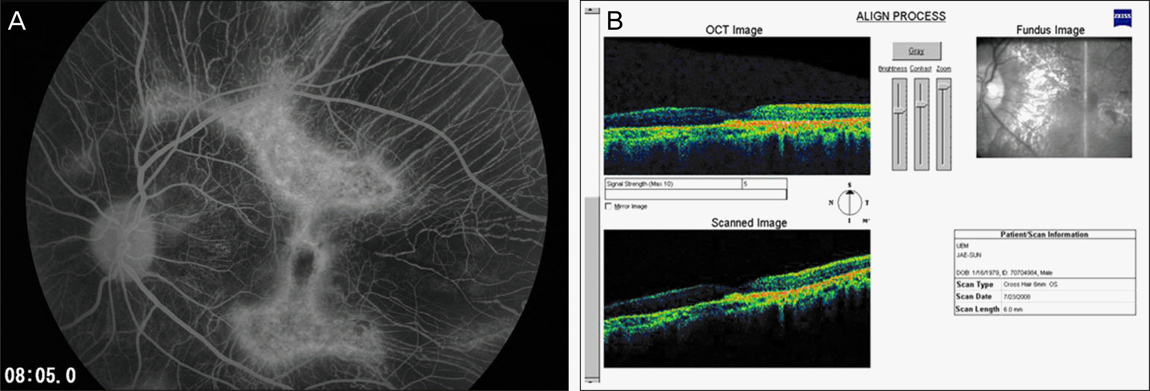
A 29-year-old male presented with a sudden decline in vision in the left eye. Fundus examination revealed areas of choriocapillaries and retinal pigment epithelium atrophy with macular hemorrhage. Fluorescein angiogram revealed vascular hyperfluorescence in the juxtafoveal area. Neurosensory detachment around the macula and increased central macular thickness was also observed using optical coherence tomography. Upon the diagnosis of choroideremia with choroidal neovascularization, the patient was treated with 1.25 mg intravitreal bevacizumab. Visual acuity improved after four injections of intravitreal Bevacizumab with improvement in both detachment and fluorescein leakage.
Conclusions
In patients with choroideremia presenting sudden decline in vision, ophthalmologists should detect for possible choroidal neovascularization. The results from the present study show that judicious use of intravitreal Bevacizumab may be effective in such cases. Further studies with a large sample size and sufficiently long follow-up periods are required.
References
1. Roberts MF, Fishman GA, Roberts DK, et al. Retrospective, longitudinal, and cross sectional study of visual acuity impairment in choroideraemia. Br J Ophthalmol. 2002; 86:658–62.

2. Wakabayashi K, Yoneura D, Kawasaki K, Uyama M. New approach to electrodiagnosis in diseases of the retina choroideremia. Folia Ophthalmol Japan. 1984; 35:775–9.
3. Robinson D, Tiedeman J. Choroideremia associated with a subretinal neovascular membrane. Case report. Retina. 1987; 7:70–4.
4. Noro Y, Yamaguchi K, Tamai M. A case of choroideremia associated with subretinal proliferation. Jap J Clin Ophthalmol. 1992; 46:962–3.
5. Endo K, Yuzawa M, Ohba N. Choroideremia associated with subretinal neovascular membrane. Acta Ophthalmol Scand. 2000; 78:483–6.
6. Sawa M, Tamaki Y, Klancnik JM Jr, Yannuzzi LA. Intraretinal foveal neovascularization in choroideremia. Retina. 2006; 26:585–8.
7. Mauthner H. Ein fall von choroideremia. Ber Nat Med Ver Innsbruck. 1872; 2:191.
8. Krill AE, Archer D. Classification of the choroidal atrophies. Am J Ophthalmol. 1971; 72:562–85.
Figure 1.
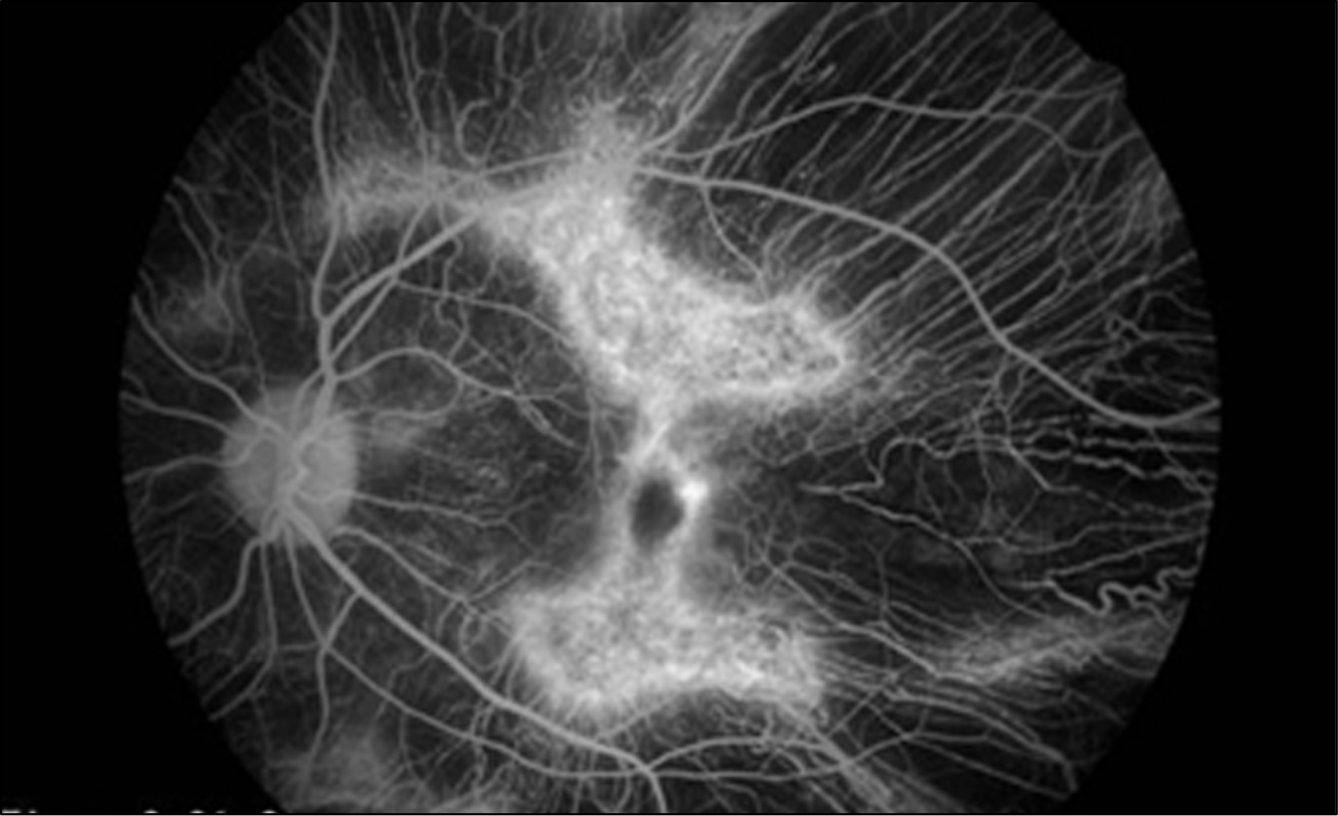
Fundus photographs of right eye (A) and left eye (B) at presentation show areas of choriocapillaries and retinal pigment epithelium atrophy in the mid-periphery and posterior pole relatively sparing the macula. Note the macular hemorrhage in the left eye (B). Fluorescein angiogram of right eye (C) and left eye (D) at presentation show symmetrical areas of retinal pigment epithelium and choriocapillary loss. The fovea is spared with surrounding areas of hyperfluorescence. Note the leakage from a juxtafo-veal choroidal neovascularization (CNV) in the left eye (D).

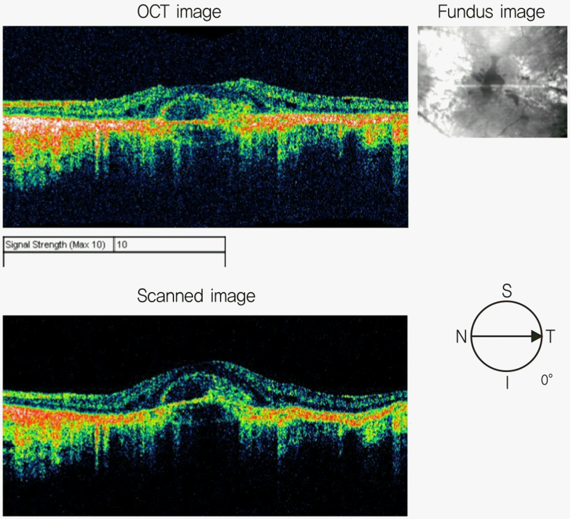
Figure 2.
The optical coherence tomographs (OCT) of the patient's left eye at initial visit shows increased central retinal thickness owing to neurosensory detachment and shallow elevation of the pigmented epithelium.
 XML Download