Abstract
Purpose
To evaluate the clinical features and treatment results of optic nerve sheath meningioma (ONSM) in Korean patients.
Methods
The present retrospective noncomparative case series was comprised of 10 eyes of 10 patients with a diagnosis of ONSM who were treated between 1997 and 2010 at the Seoul National University Hospital.
Results
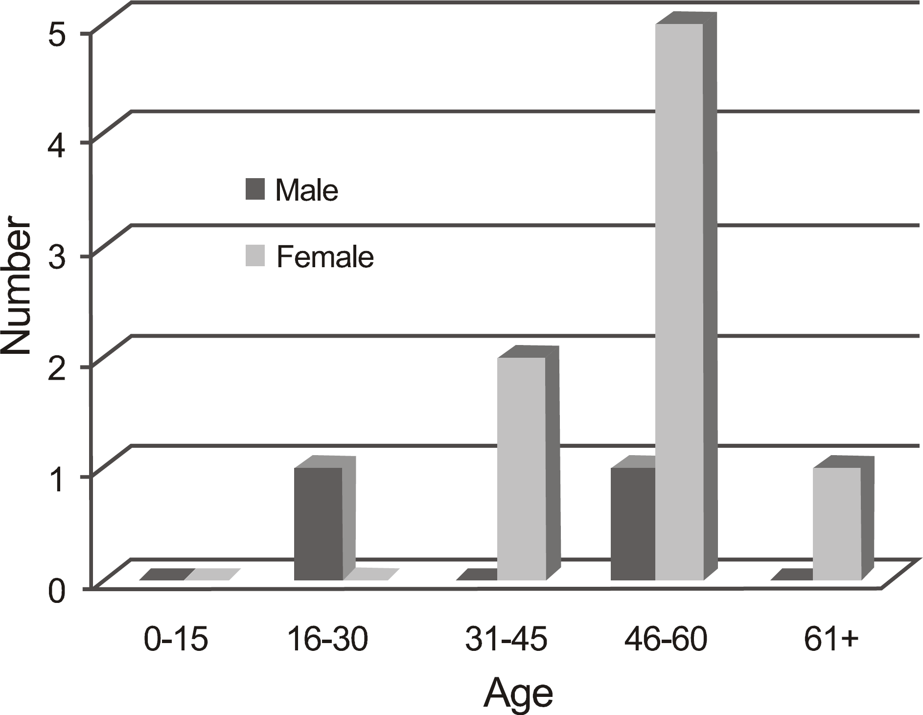
The mean age at presentation was 47.9 years. Two males and 8 females participated in the study. Decreased vision and proptosis were the most common presenting symptoms. On imaging, the most common pattern was tubular. The most common histopathologic pattern was a meningothelial type. Two patients who were observed maintained good vision during the follow-up period and showed a slow progression of the tumor. Two patients out of 3 who underwent surgical management presented significant visual loss and complications. Three patients in the gamma-knife surgery (GKS) group showed no significant changes in visual acuity after treatment; however, tumor growth was halted. Two patients who underwent 3-dimensional conformal radiotherapy (3D-CRT) presented improvement in their visual acuity or visual field, and tumor growth was halted.
Conclusions
Management should be conservative in most cases because of the slow and indolent growth pattern of ONSM. However, 3D-CRT can be considered as an initial treatment in patients with progressive visual deterioration and having still higher possibility of vision maintenance. GKS can be indicated in patients with progressive visual deterioration and a rare possibility of visual recovery. Surgical indications for ONSM are limited and must be carefully considered for each patient.
References
2. Saeed P, Rootman J, Nugent RA, et al. Optic nerve sheath meningiomas. Ophthalmology. 2003; 110:2019–30.

3. Wright JE, McNab AA, McDonald WI. Primary optic nerve sheath meningioma. Br J Ophthalmol. 1989; 73:960–6.

4. Sibony PA, Krauss HR, Kennerdell JS, et al. Optic nerve sheath meningiomas: clinical manifestations. Ophthalmology. 1984; 91:1313–26.
5. Berman D, Miller NR. New concepts in the management of optic nerve sheath meningiomas. Ann Acad Med Singapore. 2006; 35:168–74.
6. Miller NR. New concepts in the diagnosis and management of optic nerve sheath meningiomas. J Neuroophthalmol. 2006; 26:200–8.
7. Probst C, Gessaga E, Leuenberger AE. Primary meningioma of the optic nerve sheaths: case report. Ophthalmologica. 1985; 190:83–90.

8. Alper MG. Management of primary optic meningiomas. Current status-therapy in controversy. J Clin Neuroophthalmol. 1981; 1:101–17.
9. Hollenhorst RW Jr, Hollenhorst RW Sr, MacCarty CS. Visual prognosis of optic nerve sheath meningiomas producing shunt vessels on the optic disk: the Hoyt-Spencer syndrome. Trans Am Ophthalmol Soc. 1977; 75:141–63.
10. Jakobiec FA, Depot MJ, Kennerdell JS, et al. Combined clinical and computed tomographic diagnosis of orbital glioma and meningioma. Ophthalmology. 1984; 91:137–55.
11. Rootman J. Diseases of the orbit: A multidisciplinary approach. Philadelphia: JB Lippincott;1988. p. 281–5.
12. Marquardt MD, Zimmerman LE. Histopathology of meningiomas and gliomas of the optic nerve. Hum Pathol. 1982; 13:226–35.

13. Kennerdell JS, Maroon JC, Malton M, Warren FA. The management of optic nerve sheath meningiomas. Am J Ophthalmol. 1988; 106:450–7.

14. Rose GE. Orbital meningiomas: surgery, radiotherapy, or hormones? Br J Ophthalmol. 1993; 77:313–4.

15. Samples JR, Robertson DM, Taylor JZ, Waller RR. Optic nerve meningioma. Ophthalmology. 1983; 90:1591–4.

16. Lee AG, Woo SY, Miller NR, et al. Improvement in visual function in an eye with a presumed optic nerve sheath meningioma after treatment with three-dimensional conformal radiation therapy. J Neuroophthalmol. 1996; 16:247–51.

17. Moyer PD, Golnik KC, Breneman J. Treatment of optic nerve sheath meningioma with three-dimensional conformal radiation. Am J Ophthalmol. 2000; 129:694–6.

Figure 2.
Eyelid photos of two patients showing proptosis resulting from ONSM. One patient (case 1) presented with proptosis of the left eye (A, white arrow), and the other patient (case 10) showed proptosis of the right eye (B, white arrow).
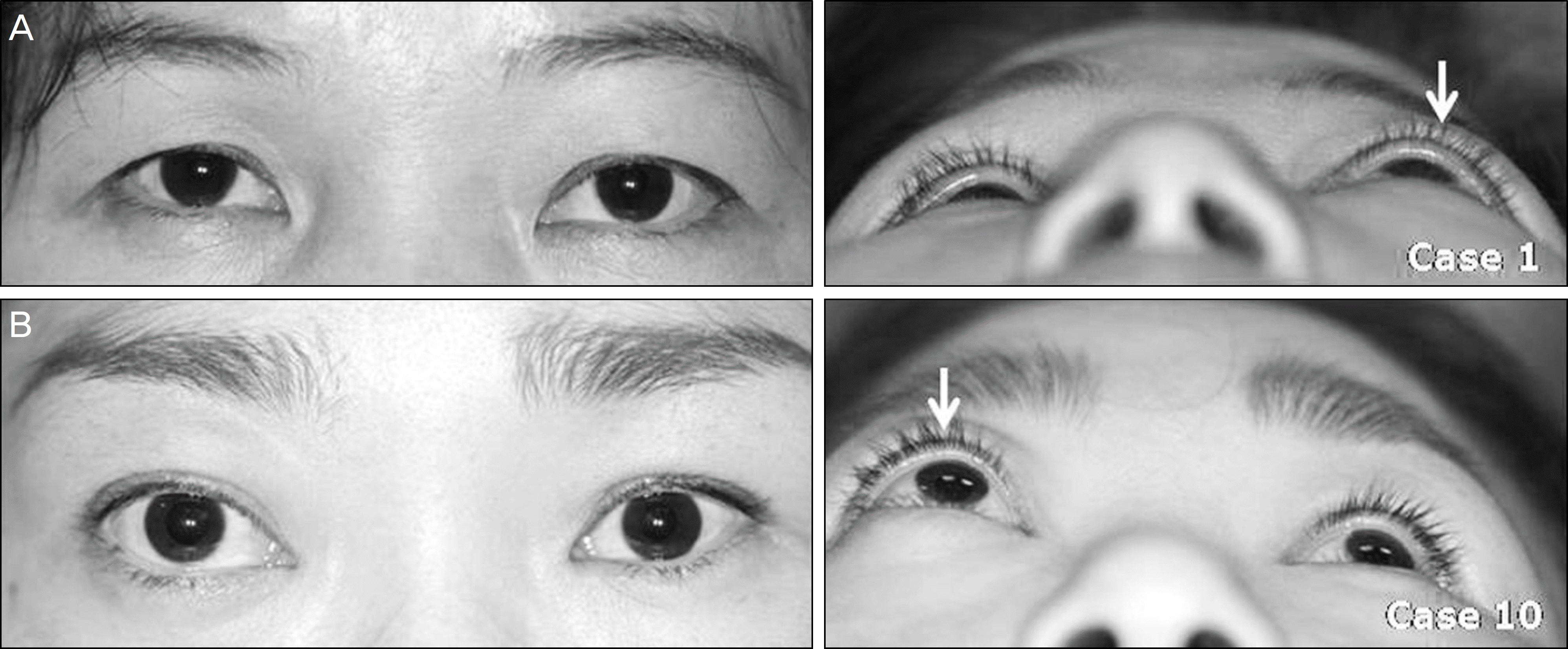
Figure 3.
Fundus photographs of patients with ONSM. (A) Fundus photograph of case 1 with globular ONSM demonstrates blurring of optic nerve disc margin (white arrow) and choroidal folds (black arrow). (B) Fundus photograph of case 9 with tubular-diffuse pattern ONSM also showed optic disc swelling (white arrow). (C) Fundus photograph of case 10 with globular ONSM also demonstrates optic disc swelling (white arrow) and choroidal folds (black arrow) radiating superonasally and inferonasally from the disc.
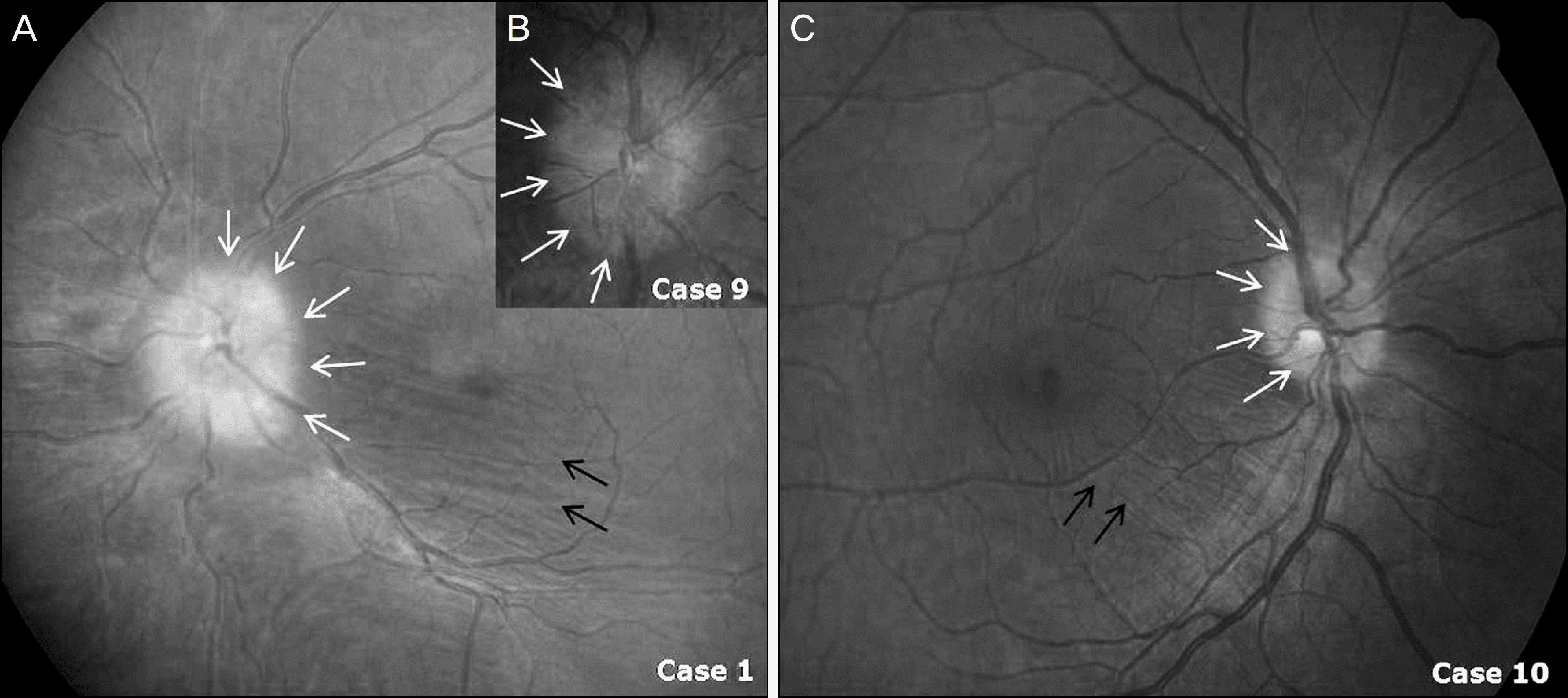
Figure 4.
(A) The tubular-apical expansion ONSM (white arrow) is seen in a 57-year-old woman. Her tumor showed intracranial involvement. (B) The globular pattern ONSM (white arrow) is seen in a 50-year-old woman with a history of decreased vision on the left. (C) The fusiform pattern ONSM (white arrow) occurred in a 52-year-old man who presented with proptosis of his right eye. Note tram tracking in this meningioma.
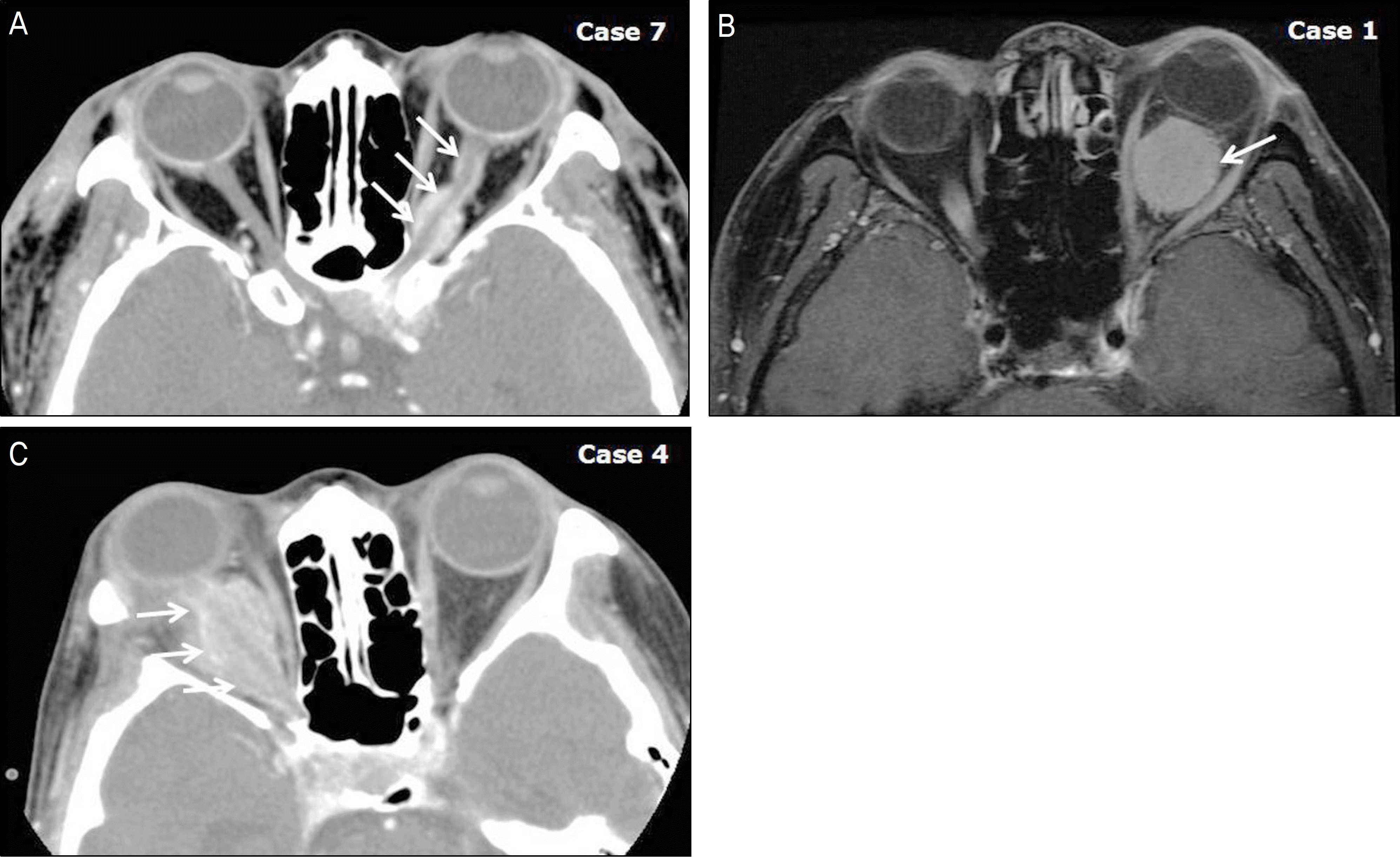
Figure 5.
(A) Histologic findings of case 3 showed a meningothelial ONMS, in which polygonal cells are arranged in sheets separated by vascular trabeculae. (B) Histologic findings of transitional pattern of ONSM in a 19-year-old man, case 5, in which spindle cells are arranged in whorls and result in deposition of calcium salts (psammomma bodies, white arrow).
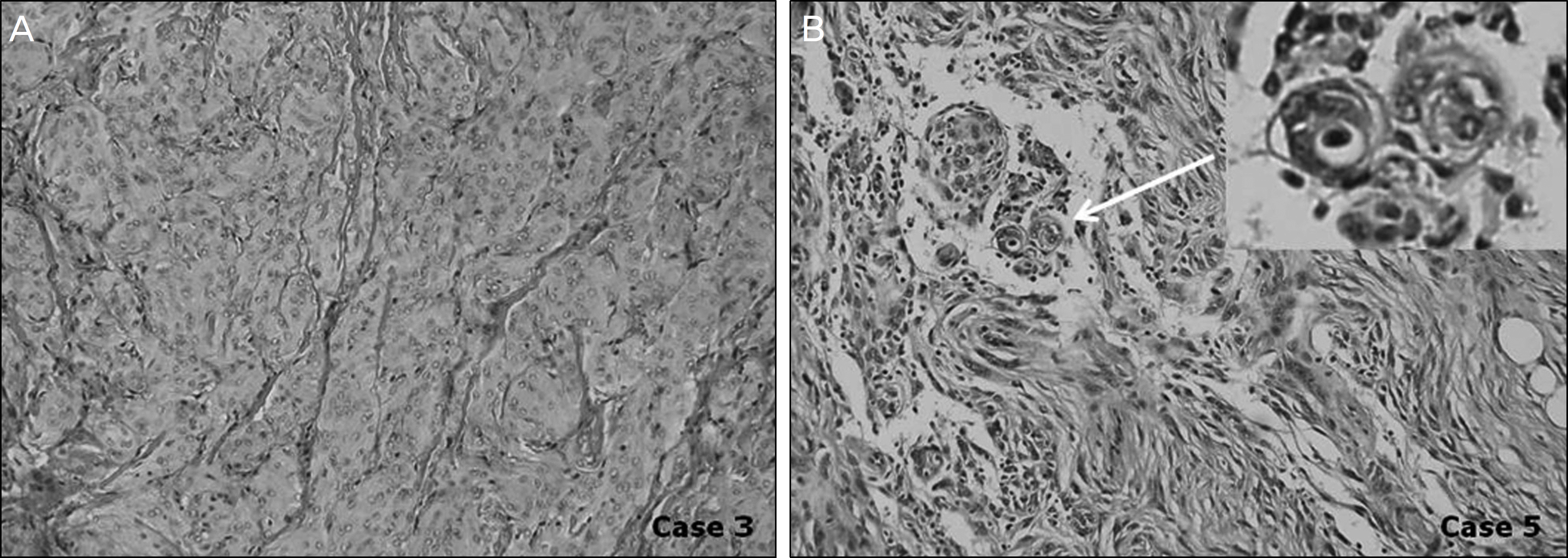
Figure 6.
(A) This 50-year-old woman (Case 1) presented with decreased vision and proptosis of the left eye. Under diagnosis of globular ONSM of the left eye, her visual field examinations were shown to have an unchanged enlarged blind spot and inferonasal field defect for 2 years and 7 months. (B) This 52-year-old man (Case 2) presented with proptosis of the right eye. He was diagnosed with tubular-diffuse ONSM of the right eye. His visual field examination was normal and unchanged for 1 year and 1 month.
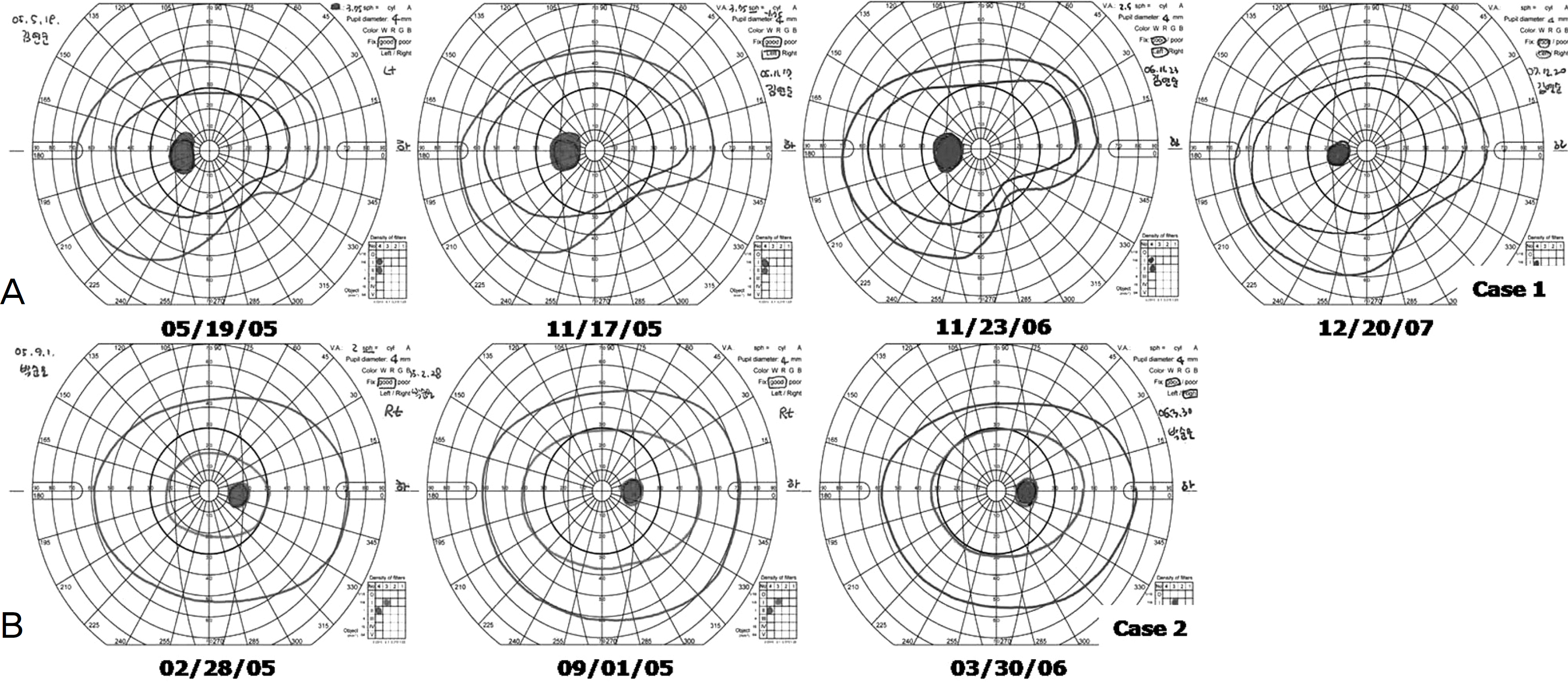
Figure 7.
T1-weighted transverse MRI imagings of Case 1 patient with globular ONSM (white arrow) of the left eye. (A) MRI imaging taken January 12 th, 2005. (B) MRI imaging taken December 28 th, 2007. These MRI scans of the same patient 2 years and 11 months later demonstrates only a slight change in tumor size and shows slowly progressive pattern of the ONSM.

Figure 8.
(A) This 46-year-old woman (Case 6) presented with decreased vision of the left eye. She was diagnosed with ONSM (white arrow) of the left eye. (B) This MRI scan of the same patient 5 years and 1 month later demonstrates a dramatically increase in size of tumor with marked proptosis. (C) Gamma-knife surgery was performed. (Volume 15.7 cc, Dose 12 Gy at 50% isodose line, Shot 16x) (D) After 5 years' follow-up, tumor decreased in volume slightly and she remains with decreased exophthalmos.

Figure 9.
(A) This tubular-diffuse ONSM (white arrow) was seen in a 64-year-old woman (Case 9). Optic disc swelling was found accidentallyon regular medical checkup. On physical examination, her vision was 0.9, with evidence of a temporal constriction (C). After 2 years' follow-up, her visual field examinations were shown to have an enlarged blind spot (D). Over a 1-year period, her tumor showed an increase in size and thickness (B). Her vision deteriorated to 0.4–1 and her visual field revealed central island of vision only (E). She underwent a fractionated stereotactic radiotherapy and maintained stable vision with a recovery of visual field (F∼H). She has no more increase in tumor size on MRI (I∼J).
Table 1.
Demographics of patients
Table 2.
Presenting symptoms and signs
Table 3.
Configuration of tumor on imaging
Table 4.
Treatment results


 PDF
PDF ePub
ePub Citation
 Print
Print


 XML Download