

 PDF
PDF ePub
ePub Citation
 Print
Print


Abstract
Purpose
To evaluate the long-term clinical outcomes of femtosecond LASER-assisted Descemet's stripping endothelial keratoplasty (DSEK).
Methods
The clinical results of endothelial keratopathy from 11 consecutive patients who were followed up for at least 12 months after femtosecond LASER-assisted DSEK were retrospectively analyzed. The best corrected visual acuities (BCVA), manifest refractions, intraocular pressures, and perioperative complications were evaluated preoperatively and up to 24 months after the femtosecond LASER-assisted DSEK.
Results
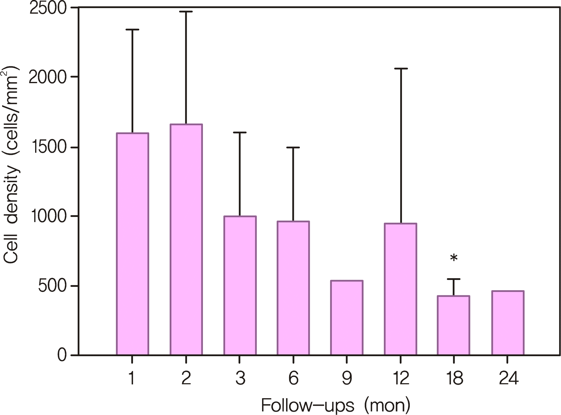
The average follow-up period was 18 months. Postoperative visual acuity had significantly improved from 1.26 (logMAR) to 0.80 (logMAR) at 3 months (p < 0.05) and this change was maintained during postoperative follow-up. All eyes underwent successful transplantation and the donor discs were well-attached. The mean endothelial cell density continued to decrease during the follow-up period. The donor-recipient stromal interface was the area where varying degree of haziness and birefringent particles were found.
Conclusions
The femtosecond LASER-assisted DSEK was effective in creating an endothelial donor disc which resulted in rapid visual recovery and low surgically-induced astigmatism. However, the operation caused rapid decrease in endothelial cell density which requires continuing further consideration by the physician.
Go to : 
References
1. Price FW Jr, Price MO. Descemet's stripping with endothelial keratoplasty in 200 eyes: Early challenges and techniques to enhance donor adherence. J Cataract Refract Surg. 2006; 32:411–8.
2. Terry MA, Ousley PJ. Replacing the endothelium without corneal surface incisions or sutures: the first United States clinical series using the deep lamellar endothelial keratoplasty procedure. Ophthalmology. 2003; 110:755–64.
3. Terry MA, Ousley PJ. Deep lamellar endothelial keratoplasty visual acuity, astigmatism, and endothelial survival in a large prospective series. Ophthalmology. 2005; 112:1541–8.
4. Melles GR, Wijdh RH, Nieuwendaal CP. A technique to excise the descemet membrane from a recipient cornea (descemetorhexis). Cornea. 2004; 23:286–8.

5. Cheng YY, Pels E, Nuijts RM. Femtosecond-laser-assisted Descemet's stripping endothelial keratoplasty. J Cataract Refract Surg. 2007; 33:152–5.
6. Espana EM, Huang B. Confocal microscopy study of donor-recipient interface after Descemet's stripping with endothelial keratoplasty. Br J Ophthalmol. 2010; 94:903–8.
7. Price FW Jr, Price MO. Descemet's stripping with endothelial keratoplasty in 50 eyes: a refractive neutral corneal transplant. J Refract Surg. 2005; 21:339–45.
8. Seo WM, Kim HK. Early result of femtosecond laser assisted descemet's membrane stripping endothelial keratoplasty. J Korean Ophthalmol Soc. 2008; 49:40–7.
9. Durrie DS, Kezirian GM. Femtosecond laser versus mechanical keratome flaps in wavefront-guided laser in situ keratomileusis: prospective contralateral eye study. J Cataract Refract Surg. 2005; 31:120–6.
10. Cheng YY, Schouten JS, Tahzib NG, et al. Efficacy and safety of femtosecond laser-assisted corneal endothelial keratoplasty: a randomized multicenter clinical trial. Transplantation. 2009; 88:1294–302.
11. Cheng YY, Pels E, Cleutjens JP, et al. Corneal endothelial viability after femtosecond laser preparation of posterior lamellar discs for Descemet-stripping endothelial keratoplasty. Cornea. 2007; 26:1118–22.
12. Cheng YY, Kang SJ, Grossniklaus HE, et al. Histologic evaluation of human posterior lamellar discs for femtosecond laser Descemet's stripping endothelial keratoplasty. Cornea. 2009; 28:73–9.
13. Cheng YY, Hendrikse F, Pels E, et al. Preliminary results of femtosecond laser-assisted descemet stripping endothelial keratoplasty. Arch Ophthalmol. 2008; 126:1351–6.
14. Seitz B, Langenbucher A, Hofmann-Rummelt C, et al. Nonmechanical posterior lamellar keratoplasty using the femtosecond laser (femto- plak) for corneal endothelial decompensation. Am J Ophthalmol. 2003; 136:769–72.
15. Lee DH, Chung TY, Chung ES, et al. Case report: Femtosecond laser-assisted small incision deep lamellar endothelial keratoplasty. Korean J Ophthalmol. 2008; 22:43–8.
16. Sarayba MA, Juhasz T, Chuck RS, et al. Femtosecond laser posterior lamellar keratoplasty: a laboratory model. Cornea. 2005; 24:328–33.
17. Mehta JS, Shilbayeh R, Por YM, et al. Femtosecond laser creation of donor cornea buttons for Descemet-stripping endothelial keratoplasty. J Cataract Refract Surg. 2008; 34:1970–5.
18. Lee JS, Park YG, Yoon KC. Long-term results of Descemet's stripping automated endothelial keratoplasty in Korea. J Korean Ophthalmol Soc. 2010; 51:1431–7.
19. Pineros O, Cohen EJ, Rapuano CJ, Laibson PR. Long-term results after penetrating keratoplasty for Fuchs' endothelial dystrophy. Arch Ophthalmol. 1996; 114:15–8.
20. Kim KE, Joo CK. Changes in astigmatism after suture removal in penetrating keratoplasty. J Korean Ophthalmol Soc. 2003; 44:284–8.
21. Ousley PJ, Terry MA. Stability of vision, topography, and endothelial cell density from 1 year to 2 years after deep lamellar endothelial keratoplasty surgery. Ophthalmology. 2005; 112:50–7.
22. Fogla R, Padmanabhan P. Initial results of small incision deep lamellar endothelial keratoplasty (DLEK). Am J Ophthalmol. 2006; 141:346–51.
23. Yepes N, Segev F, Hyams M, et al. Five-millimeter-incision deep lamellar endothelial keratoplasty: one-year results. Cornea. 2007; 26:530–3.
24. Patel SV, Hodge DO, Bourne WM. Corneal endothelium and postoperative outcomes 15 years after penetrating keratoplasty. Am J Ophthalmol. 2005; 139:311–9.
25. Ing JJ, Ing HH, Nelson LR, et al. Ten-year postoperative results of penetrating keratoplasty. Ophthalmology. 1998; 105:1855–65.
26. Bahar I, Kaiserman I, McAllum P, et al. Comparison of posterior lamellar keratoplasty techniques to penetrating keratoplasty. Ophthalmology. 2008; 115:1525–33.
27. Mearza AA, Qureshi MA, Rostron CK. Experience and 12-month results of descemet-stripping endothelial keratoplasty (DSEK) with a small-incision technique. Cornea. 2007; 26:1292–3.
28. Koenig SB, Covert DJ, Dupps WJ Jr, Meisler DM. Visual acuity, refractive error, and endothelial cell density six months after Descemet stripping and automated endothelial keratoplasty (DSAEK). Cornea. 2007; 26:670–4.
29. Terry MA, Chen ES, Shamie N, et al. Endothelial cell loss after Descemet's stripping endothelial keratoplasty in a large prospective series. Ophthalmology. 2008; 115:488–96.
30. Price MO, Price FW Jr. Endothelial cell loss after descemet stripping with endothelial keratoplasty influencing factors and 2-year trend. Ophthalmology. 2008; 115:857–65.
31. Flanagan GW, Binder PS. Precision of flap measurements for laser in situ keratomileusis in 4428 eyes. J Refract Surg. 2003; 19:113–23.
32. Thomas J, Wang J, Rollins AM, Sturm J. Comparison of corneal thickness measured with optical coherence tomography, ultrasonic pachymetry, and a scanning slit method. J Refract Surg. 2006; 22:671–8.
33. Thompson RW Jr, Choi DM, Price MO, et al. Noncontact optical coherence tomography for measurement of corneal flap and residual stromal bed thickness after laser in situ keratomileusis. J Refract Surg. 2003; 19:507–15.
34. Eisner RA, Binder PS. Technique for measuring laser in situ keratomileusis flap thickness using the IntraLase laser. J Cataract Refract Surg. 2006; 32:556–8.
35. Koenig SB, Covert DJ. Early results of small-incision Descemet's stripping and automated endothelial keratoplasty. Ophthalmology. 2007; 114:221–6.
36. Covert DJ, Koenig SB. New triple procedure: Descemet's stripping and automated endothelial keratoplasty combined with phacoe-mulsification and intraocular lens implantation. Ophthalmology. 2007; 114:1272–7.
37. Sonigo B, Iordanidou V, Chong-Sit D, et al. In vivo corneal confocal microscopy comparison of intralase femtosecond laser and mechanical microkeratome for laser in situ keratomileusis. Invest Ophthalmol Vis Sci. 2006; 47:2803–11.
38. Carlson EC, Wang IJ, Liu CY, et al. Altered KSPG expression by keratocytes following corneal injury. Mol Vis. 2003; 9:615–23.
39. Sundarraj N, Fite D, Belak R, et al. Proteoglycan distribution during healing of corneal stromal wounds in chick. Exp Eye Res. 1998; 67:433–42.
40. Toti P, Tosi GM, Traversi C, et al. CD-34 stromal expression pattern in normal and altered human corneas. Ophthalmology. 2002; 109:1167–71.
41. Espana EM, Kawakita T, Liu CY, Tseng SC. CD-34 expression by cultured human keratocytes is downregulated during myofibroblast differentiation induced by TGF-beta1. Invest Ophthalmol Vis Sci. 2004; 45:2985–91.
42. Ljubimov AV, Burgeson RE, Butkowski RJ, et al. Extracellular matrix alterations in human corneas with bullous keratopathy. Invest Ophthalmol Vis Sci. 1996; 37:997–1007.
43. Funderburgh JL, Hevelone ND, Roth MR, et al. Decorin and biglycan of normal and pathologic human corneas. Invest Ophthalmol Vis Sci. 1998; 39:1957–64.
44. Ivarsen A, Thøgersen J, Keiding SR, et al. Plastic particles at the LASIK interface. Ophthalmology. 2004; 111:18–23.
Go to :
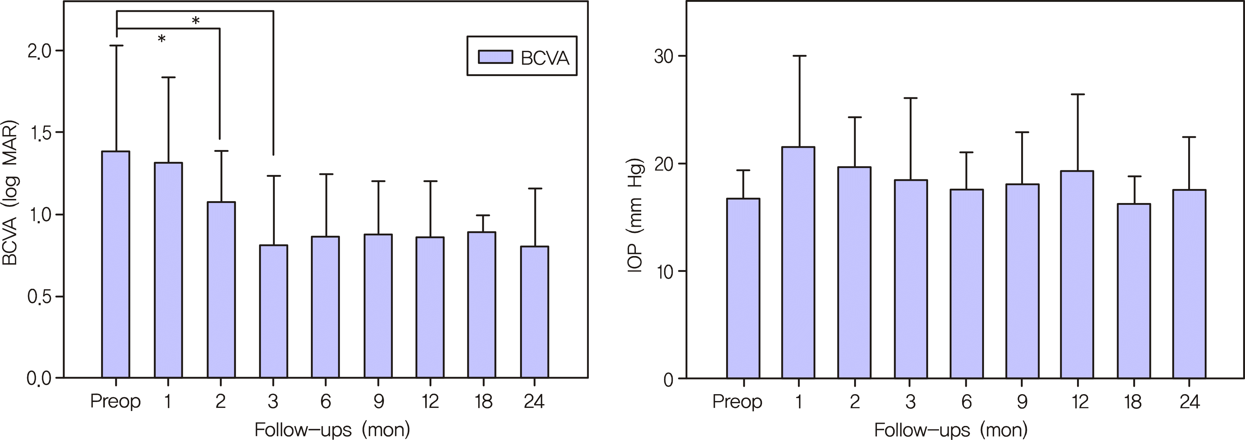 | Figure 1.The mean pre-operative and postoperative best corrected visual acuity (BCVA) expressed as the logarithm of the minimum angle of resolution (logMAR) (A). The mean values of pre-operative and postoperative intraocular pressure measured by Tonopen (B). (* p<0.05). |
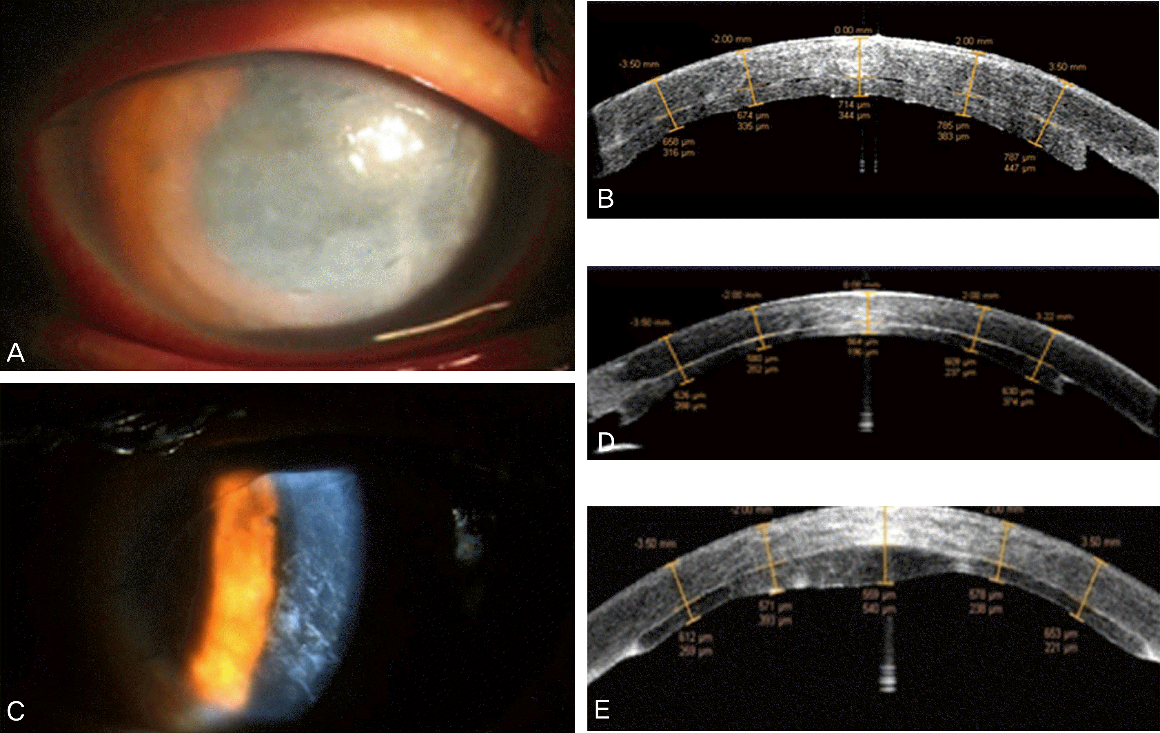 | Figure 3.Slit lamp photograph after air injection into the anterior chamber of patient 6 showing corneal edema and previous stromal puncture sites (A). The high resolution anterior segment optical coherence tomography showed that interface space still remained in this patient (B). Slit lamp photograph of patient 7 showing the white interface opacity between the recipient's cornea and the donor disc (C). The anterior segment OCT of the same patient showed the thickness at the vertex and at 2.0 mm and 3.5 mm on each side of the vertex (D). The high resolution anterior segment optical coherence tomography of patient 5 showed thickened posterior corneal disc center which was suspected to be caused by the donor disc being cut unevenly at the time of operation (E). |
 | Figure 5.The grading of donor-recipient stromal interface haziness: Grade 1 (very mild haze) (A), Grade 2 (mild haze) (B), Grade 3 (moderate haze) (C), and Grade 4 (severe haze) (D). |
Table 1.
The preoperative patient data, femtosecond LASER settings and follow-up periods
| Patient | t Sex | Age (yr) | Diagnosis | Operation | Femtosecond laser setting (Full lamellar cut) | F/U (mon) | ||
|---|---|---|---|---|---|---|---|---|
| Depth (μ m) | Diameter (mm) | Energy (μ J) | ||||||
| 1 | M | 78 | PBK* | FS-DSEK† | 360 | 8.7 | 2.0 | 24 |
| 2 | M | 69 | Fuchs dystrophy | FS-DSEK, CE IOL‡ | 380 | 8.7 | 1.8 | 18 |
| 3 | M | 66 | PBK | FS-DSEK | 380 | 8.7 | 1.8 | 24 |
| 4 | M | 49 | ABKΠ | FS-DSEK secondary IOL | 400 | 8.7 | 1.8 | 24 |
| 5 | M | 46 | Traumatic bullous keratopathy | FS-DSEK | 390 | 8.7 | 1.8 | 18 |
| 6 | M | 59 | PBK | FS-DSEK | 360 | 8.7 | 1.8 | 24 |
| 7 | F | 27 | Fuchs dystrophy | FS-DSEK | 380 | 8.7 | 2.0 | 12 |
| 8 | M | 78 | PBK | FS-DSEK | 380 | 8.7 | 1.8 | 18 |
| 9 | M | 68 | PBK | FS-DSEK | 380 | 8.75 | 1.8 | 12 |
| 10 | F | 35 | Fuchs dystrophy | FS-DSEK | 360 | 8.75 | 1.8 | 12 |
| 11 | M | 66 | ACIOL§ PBK | FS-DSEK IOL exchange | 380 | 8.7 | 2.0 | 12 |
| Mean | 58.27 | 377.3 | 8.7 | 1.85 | 18.00 | |||
Table 2.
Patient's preoperative and postoperative clinical data
| Patient |
Preoperative exam |
3 months |
6 months |
12 months |
||||||||
|---|---|---|---|---|---|---|---|---|---|---|---|---|
| BCVA*(logMAR) | IOP†(mm Hg) | Astig‡(D§) | BCVA (logMAR) | IOP (mm Hg) | Astig (D) | BCVA (logMAR) | IOP(mm Hg) | Astig (D) | BCVA (logMAR) | IOP (mm Hg) | Astig (D) | |
| 1 | 1.52 | 15 | 2.75 | 0.52 | 14 | 2.25 | 0.70 | 11 | NA | 0.80 | 11 | 3.12 |
| 2 | 1.00 | 16 | 4.5 | 0.70 | 18 | NA | 0.70 | 12 | NA | 0.80 | 14 | 1.75 |
| 3 | 2.20 | 16 | 1.75 | 0.52 | 12 | 0.5 | 0.49 | 13 | NA | 0.52 | 34 | 1 |
| 4 | 0.80 | 21 | 1.5 | 1.00 | 20 | 0 | 1.15 | 19 | NA | 1.30 | 20.5 | NA |
| 5 | 2.30 | 19 | 2.5 | 0.49 | 17 | 2.5 | NA | NA | NA | NA | NA | NA |
| 6 | 2.20 | 17 | 2.6 | 0.90 | 18 | 0 | 1.00 | 17 | 2 | 0.70 | 19 | 1.37 |
| 7 | 0.70 | 18 | 1 | 0.10 | 12 | 1 | 0.70 | 13 | NA | 0.52 | 11 | 0.25 |
| 8 | 0.52 | 18 | NA | 1.52 | 15 | NA | 1.00 | 20 | 0.5 | 0.90 | 22 | 1.75 |
| 9 | 1.00 | 11 | NA | 0.80 | 17.5 | NA | 0.70 | 20 | 0.75 | 0.70 | 22 | NA |
| 10 | 1.52 | 17 | 1 | 0.80 | 19 | NA | 0.49 | 17 | 1.5 | NA | NA | NA |
| 11 | 1.52 | 16 | NA | 1.52 | 40 | 2 | 1.52 | 22 | NA | 1.52 | 20 | NA |
Table 3.
Postoperative complications and management for each case
| Case | Complication | Time | Management |
|---|---|---|---|
| 2 | Graft dehiscence | POD2 | Air injection & reposition |
| Rejection | PO12 mon | SC* dexamethasone injection | |
| Rejection | PO18 mon | SC dexamethasone injection | |
| 3 | IIOP† | PO2 wk | IOP lowering agents |
| Interface opacity | PO2 wk | Frequent topical steroids | |
| Rejection | PO5.5 mon | SC dexamethasone injection | |
| 4 | Uneven donor corneal thickness | PO1 mon | Observation |
| IIOP | PO1 mon | A/C‡ tapping, IOP lowering agents | |
| Rejection | PO2 mon | SC dexamethasone inj, IV§ methylprednisolone | |
| 5 | IIOP | POD1 | IOP lowering agents |
| Graft dehiscence | POD3 | Air injection | |
| 6 | IIOP | PO1 mon | IOP lowering agents |
| Corectopia | PO24 mon | Observation | |
| 7 | Interface opacity | PO3 wk | Frequent topical steroids |
| 8 | Interface opacity | PO2 wk | Observation |
| IIOP | PO2 wk | IOP lowering agents | |
| Diffuse interface opacity | PO9 mon | Frequent topical steroids | |
| 9 | IIOP | PO2 mon | IOP lowering agents |
| 10 | Anterior uveitis | PO1 mon | A/C irrigation |
| Graft dehiscence | PO1 mon | Air injection | |
| Interface opacity | PO3 mon | Frequent topical steroids | |
| 11 | Graft dehiscence | PO1 wk | Air injection |
Table 4.
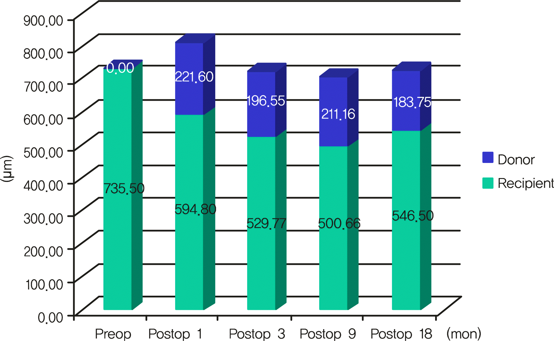
The thickness of donor disc measured at vertex and 2 mm/3.5 mm at nasal and temporal side
Table 5.
The grading of donor-recipient stromal interface haze, other interface findings, and best-corrected visual acuity in all patients
| Patient | PostDSEK (mon) | Donor-recipient stromal interface | Other findings | BCVA* (logMAR†) |
|---|---|---|---|---|
| 1 | 3 | 3 | Mild birefringent bodies | 0.52 |
| 12 | 4 | Mild birefringent bodies | 0.80 | |
| 2 | 1 | 2 | Mild birefringent bodies | 0.70 |
| 8 | 1 | Mild birefringent bodies | 0.80 | |
| 3 | 2 | 4 | Severe birefringent bodies | 0.90 |
| 9 | 2 | Mild birefringent bodies | 0.52 | |
| 4 | 6 | 1 | No birefringent bodies | 1.15 |
| 12 | 1 | Mild birefringent bodies | 1.30 | |
| 5 | 2 | 3 | Mild birefringent bodies | 1.30 |
| 20 | 3 | Mild birefringent bodies | 1.30 | |
| 6 | 1 | 2 | Mild birefringent bodies | 1.52 |
| 18 | 2 | Moderate birefringent bodies | 0.90 | |
| 7 | 3 | 2 | Mild birefringent bodies | 0.10 |
| 18 | 2 | Mild birefringent bodies | 0.30 | |
| 8 | 3 | 4 | Severe birefringent bodies | 1.52 |
| 9 | 3 | Moderate birefringent bodies | 1.00 | |
| 9 | 1 | 4 | Severe birefringent bodies | 1.00 |
| 3 | 2 | Moderate birefringent bodies | 0.80 | |
| 10 | 1 | 3 | Mild birefringent bodies | 1.52 |
| 2 | 3 | Moderate birefringent bodies | 1.0 | |
| 11 | 1 | 3 | Moderate birefringent bodies | 1.30 |
| 3 | 1 | Mild birefringent bodies | 1.52 |
 XML Download