

 PDF
PDF ePub
ePub Citation
 Print
Print


Abstract
Case summary
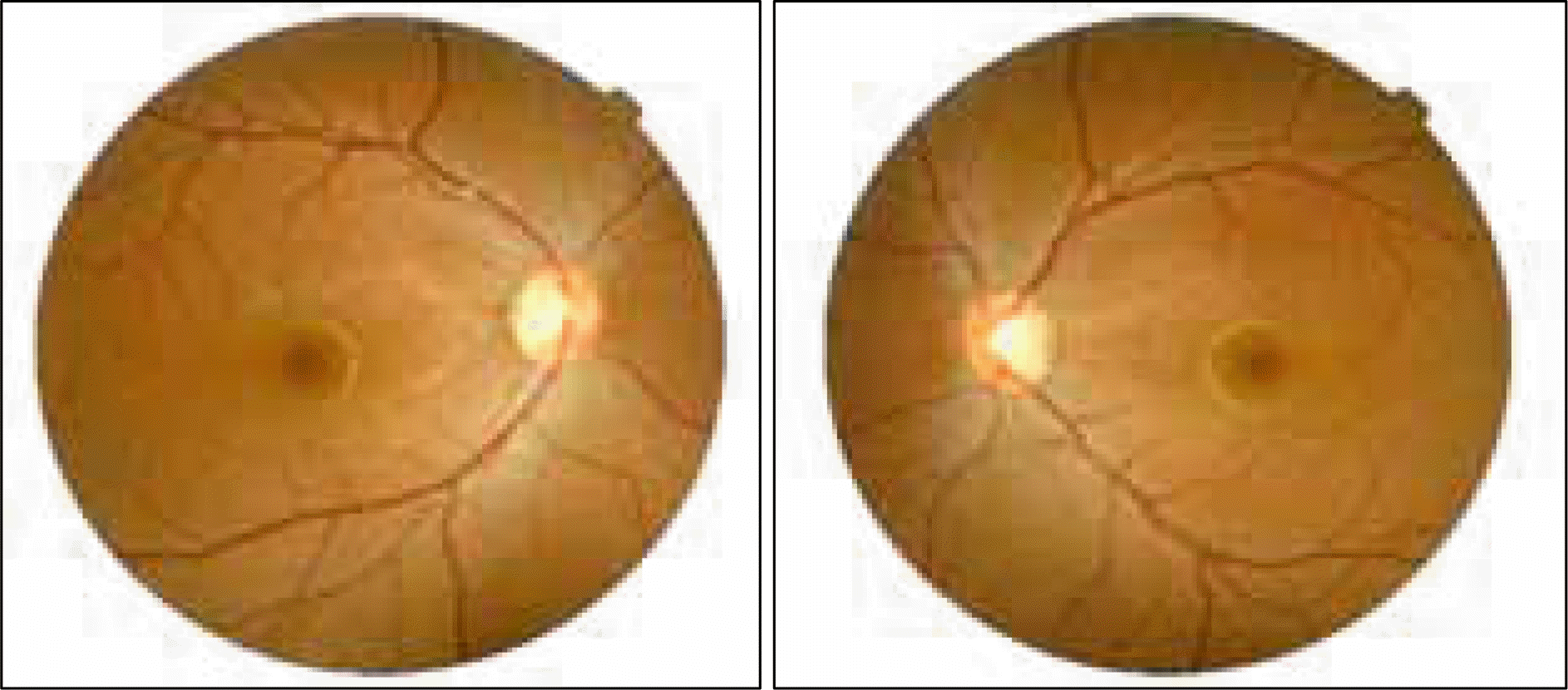
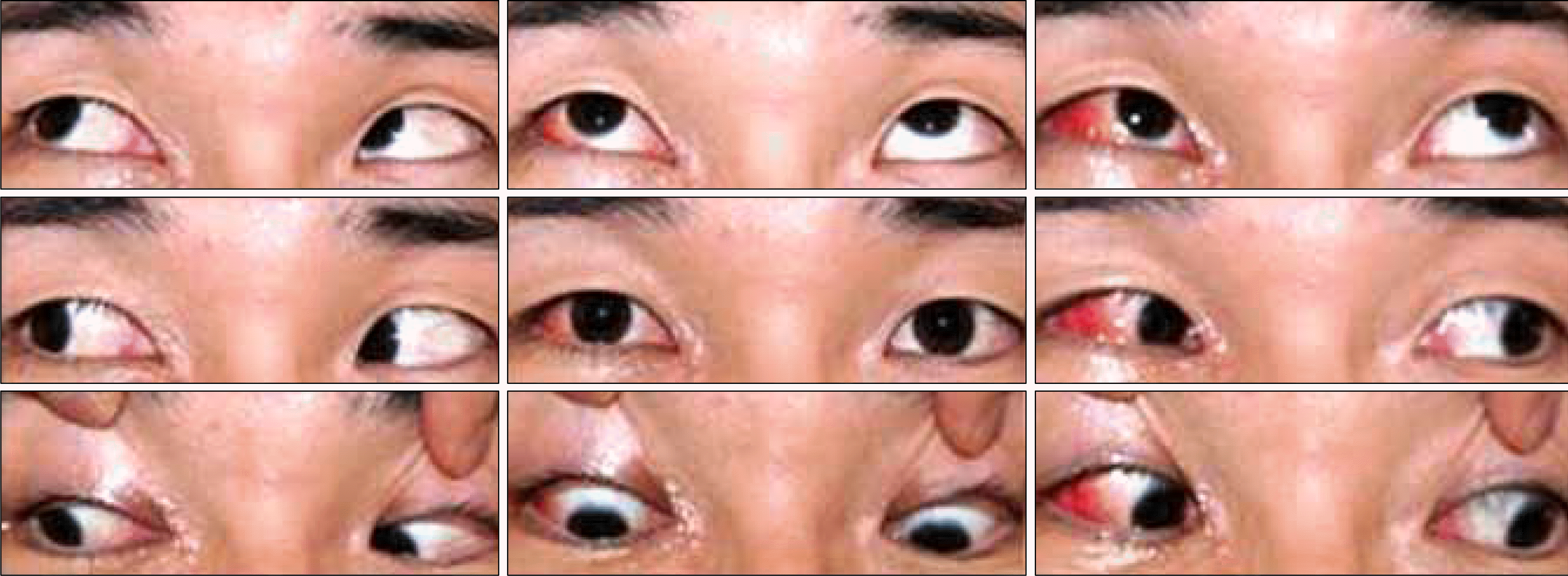
A 19-year-old male presented with a blowout fracture of the right inferior orbital wall. The patient had no history of facial asymmetry, head tilt, strabismus or diplopia. The day after the reduction operation, the patient complained of diplopia on the down-gaze. The patient had orhthophoria in the primary position. However, the right eye showed a limitation on infraduction. Six weeks later, the limitation of infraduction resolved, however the diplopia worsened. The right eye showed excessive elevation in adduction and hypertropia in the primary position. The hypertropia increased on left gaze, and decreased on right gaze. Bielschowsky’s head test revealed a negative result. The fundus photographs showed a mild excyclotorsion of the right eye. Five months later, the symptoms and signs were stable and surgery was performed. Under general anesthesia, the exaggerated forced duction test and traction with strabismus hook on the right inferior oblique muscle revealed tightness. The inferior oblique muscle was found to be recessed. After the operation, the diplopia, hypertropia and the excessive elevation on adduction of the right eye resolved.
References
1. Hammer B, Killer HE, Wieser D. Ophthalmic aspects. Hammer B, editor. Orbital Fractures— Diagnosis, Operative Treatment, Secondary Corrections. Seattle: Hogrefe & Huber;1995. p. 18–28.
2. Biesman BS, Hornblass A, Lisman R, et al. Diplopia after surgical repair of orbital floor fractures. Ophthal Plast Reconstr Surg. 1996; 12:9–17.

3. Lyon DB, Newman SA. Evidence of direct damage to extraocular muscles as a cause of diplopia following orbital trauma. Ophthal Plast Reconstr Surg. 1989; 5:81–91.
4. Koornneef L. Current concepts on the management of orbital blow-out fractures. Ann Plast Surg. 1982; 9:185–200.
5. Converse JM, Smith B, Obear MF, Wood-Smith D. Orbital blow-out fractures: a ten-year survey. Plast Reconstr Surg. 1967; 39:20–36.
6. Duane A. Binocular movements. Arch Ophthalmol. 1933; 9:579.
7. Guyton D. Exaggerated traction test for the oblique muscles. Ophthalmology. 1981; 88:1035–40.
8. Hawes MJ, Dortzbach RK. Surgery on orbital floor fracture: influence of time of repair and fracture size. Ophthalmology. 1983; 90:1066–70.
9. Greenwald HS, Keeney AH, Shannon GM. A review of 128 patients with orbital fractures. Am J Ophthalmol. 1974; 78:655–64.
10. Emery JM, von Noorden GK, Schlernitzauer DA. Orbital floor fractures: long-term follow-up of cases with and without surgical repair. Trans Am Acad Ophthalmol Otolaryngol. 1971; 75:802–12.
11. Smith B, Lisman RD, Simonton J, Della Rocca R. Volkmann's contracture of the extraocular muscles following blowout fracture. Plast Reconstr Surg. 1984; 74:200–16.
12. Awadein A, Pesheva M, Guyton DL. “Inverted Brown pattern”: A tight inferior oblique muscle masquerading as a superior oblique muscle underaction— clinical characteristics and surgical management. J AAPOS. 2006; 10:565–72.
13. Hunter DG, Lam GC, Guyton DL. Inferior oblique muscle injury from local anesthesia for cataract surgery. Ophthalmology. 1995; 102:501–9.
14. Kim JH, Hwang JM. Imaging of the superior rectus in superior rectus overaction after retrobulbar anesthesia. Ophthalmology. 2006; 113:1681–4.
15. Capo H, Guyton DL. Ipsilateral hypertropia after cataract surgery. Ophthalmology. 1996; 103:721–30.
16. Grimmett MR, Lambert SR. Superior rectus muscle overaction after cataract extraction. Am J Ophthalmol. 1992; 114:72–80.
17. Kushner BJ. Extraocular muscle contracture and overaction syndrome occurring after periocular anesthesia. JAAPOS. 2004; 8:182–3.

 XML Download