Abstract
Purpose
To report a case of temporal hemianopsia of a healthy eye occurring in the contralateral silicone oil-filled eye due to migration of silicone oil into the optic chiasm and lateral ventricle.
Case summary
A 56-year-old man visited our clinic with temporal hemianopsia for 10 days in the left eye. Three months before, the patient had presented with decreased vision and ocular pain in the right eye as well as a headache. The patient underwent vitrectomy at another hospital for the management of retinal detachment occurring in the right eye 8 years earlier. In addition, for recurred retinal detachment, reoperations were performed twice with silicone oil injection. Funduscopy revealed findings such as glaucomatous optic disc and an intraocular pressure of 54 mmHg in the right eye. On visual field examination, the temporal hemianopsia was detected in the left eye. Under the suspicion of cerebral le-sions, a magnetic resonance imaging (MRI) examination was performed. On the right side of the optic chiasm and the suprasellar region, materials were present whose signal intensity was identical to silicone oil in the right vitreal cavity. During a follow-up, the migration of silicone oil into the lateral ventricle and the alteration of its location with the positional change were observed.
References
1. Cibis PA, Becker B, Okun E, Canaan S. The use of liquid silicone in retinal detachment surgery. Arch Ophthalmol. 1962; 68:590–9.

2. Scott JD. The treatment of massive vitreous retraction by the separation of preretinal membranes using liquid silicone. Mod Probl Ophthalmol. 1975; 15:185–90.
3. Zivojnović R, Mertens DA, Baarsma GS. Fluid silicon in detachment for surgery (author’s transl). Klin Monbl Augenheilkd. 1981; 179:17–22.
4. Bodanowitz S, Kir N, Hesse L. Silicone oil for recurrent vitreous hemorrhage in previously vitrectomized diabetic eyes. Ophthalmologica. 1997; 211:219–22.

5. Heimann K, Dahl B, Dimopoulos S, Lemmen KD. Pars plana vitrectomy and silicone oil injection in proliferative diabetic retinopathy. Graefes Arch Clin Exp Ophthalmol. 1989; 227:152–6.

6. The Silicone Study Group. Vitrectomy with silicone oil or sulfur hexafluoride gas in eyes with severe proliferative vitreoretinopathy: results of a randomized clinical trial. Silicone Study Report 1. Arch Ophthalmol. 1992; 110:770–9.
7. The Silicone Study Group. Vitrectomy with silicone oil or perfluoropropane gas in eyes with severe proliferative vitreoretinopathy: results of a randomized clinical trial. Silicone Study Report 2. Arch Ophthalmol. 1992; 110:780–92.
8. Yeo JH, Glaser BM, Michels RG. Silicone oil in the treatment of complicated retinal detachments. Ophthalmology. 1987; 94:1109–13.

9. Seong MC, Chung HW, Lee SY, et al. The clinical results of silicone oil tamponade in pars plana vitrectomy for various vitreoretinal diseases. J Korean Ophthalmol Soc. 2007; 48:1057–66.

10. Gallemore RP, McCune II BW. Silicone oil in vitreoretinal surgery. Ryan SJ, editor. Retina. 4th ed.St. Louis: Elsevier Mosby;2006. 3:chap 130.

11. Gonvers M. Temporary silicone oil tamponade in the management of retinal detachment with proliferative vitreoretinopathy. Am J Ophthalmol. 1985; 100:239–45.

12. Nguyen QH, Lloyd MA, Heuer DK, et al. Incidence and management of glaucoma after intravitreal silicone oil injection for complicated retinal detachments. Ophthalmology. 1992; 99:1520–6.

13. Beekhuis WH, van Rij G, Zivojnović R. Silicone oil keratopathy: indications for keratoplasty. Br J Ophthalmol. 1985; 69:247–53.

14. Abrams GW, Azen SP, Barr CC, et al. The incidence of corneal abnormalities in the Silicone Study. Silicone Study Report 7. Arch Ophthalmol. 1995; 113:764–9.
15. Valone J Jr, McCarthy M. Emulsified anterior chamber silicone oil and glaucoma. Ophthalmology. 1994; 101:1908–12.

16. Yoon TJ, Oum BS. Factors for epiretinal membrane formation after retinal detachment surgery with silicone oil tamponade. J Korean Ophthalmol Soc. 2004; 45:1681–8.
17. Gonvers M, Andenmatten R. Temporary silicone oil tamponade and intraocular pressure: An 11-year retrospective study. Eur J Ophthalmol. 1996; 6:74–80.

18. Federman JL, Schubert HD. Complications associated with the use of silicone oil in 150 eyes after retina-vitreous surgery. Ophthalmology. 1988; 95:870–6.

19. Riedel KG, Gabel VP, Neubauer L, et al. Intravitreal silicone oil injection: complications and treatment of 415 consecutive patients. Graefes Arch Clin Exp Ophthalmol. 1990; 228:19–23.

20. Oh TS, Kim SY. Complications associated with intravitreal silicone oil injection. J Korean Ophthalmol Soc. 1993; 34:1012–22.
21. Stefánsson E, Anderson MM Jr, Landers MB 3rd, et al. Refractive changes from use of silicone oil in vitreous surgery. Retina. 1988; 8:20–3.

22. Barr CC, Lai MY, Lean JS, et al. Postoperative intraocular pressure abnormalities in the Silicone Study. Silicone Study Report 4. Ophthalmology. 1993; 100:1629–35.
23. Quintyn JC, Genevois O, Ranty ML, Retout A. Silicone oil migration in the eyelid after vitrectomy for retinal detachment. Am J Ophthalmol. 2003; 136:540–2.

24. Scholda C, Egger S, Lakits A, Haddad R. Silicone oil removal: results, risks and complications. Acta Ophthalmol Scand. 1997; 75:695–9.

25. Jonas JB, Budde WM, Knorr HL. Timing of retinal redetachment after removal of intraocular silicone oil tamponade. Am J Ophthalmol. 1999; 128:628–31.

26. Kampik A, Höing C, Heidenkummer HP. Problems and timing in the removal of silicone oil. Retina. 1992; 12:S11–6.
27. Halberstadt M, Domig D, Kodjikian L, et al. PVR recurrence and the timing of silicon oil removal. Klin Monbl Augenheilkd. 2006; 223:361–6.
28. Yoon JS, Lee SY, Lee SC, Kwon OW. Clinical outcomes after silicone oil removal. J Korean Ophthalmol Soc. 2003; 44:642–8.
29. Eckle D, Kampik A, Hintschich C, et al. Visual field defect in association with chiasmal migration of intraocular silicone oil. Br J Ophthalmol. 2005; 89:918–20.

30. Yu JT, Apte RS. A case of intravitreal silicone oil migration to the central nervous system. Retina. 2005; 25:791–3.

31. Eller AW, Friberg TR, Mah F. Migration of silicone oil into the brain: a complication of intraocular silicone oil for retinal tamponade. Am J Ophthalmol. 2000; 129:685–8.

32. Parel JM, Milne P, Gautier S, et al. Silicone oils: Physicochemical properties. Ryan SJ, editor. Retina. 4th ed.St. Louis: Elsevier Mosby;2006. 3:chap. 129.

33. Friberg TR, McLellan TG. Vitreous pulsations, relative hypotony, and retrobulbar cyst associated with a congenital optic pit. Am J Ophthalmol. 1992; 114:767–9.

34. Shields CL, Eagle RC Jr. Pseudo-Schnabel’s cavernous degeneration of the optic nerve secondary to intraocular silicone oil. Arch Ophthalmol. 1989; 107:714–7.

35. Manschot WA. Intravitreal silicone injection. Adv Ophthalmol. 1978; 36:197–207.
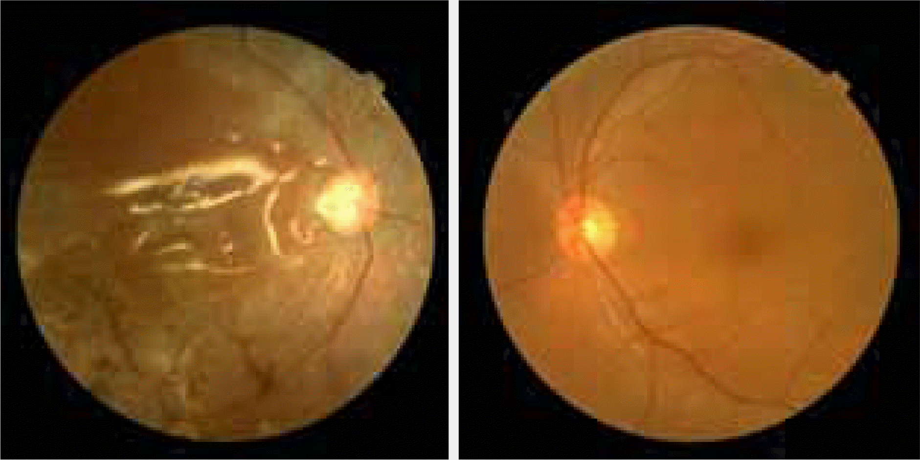
Figure 1.
Color fundus photograph shows elevated retina and the cup-disc ratio is 1.0 in the right eye. There is the reflex on the retinal surface by the silicone oil. There were no abnormal findings in the left eye.

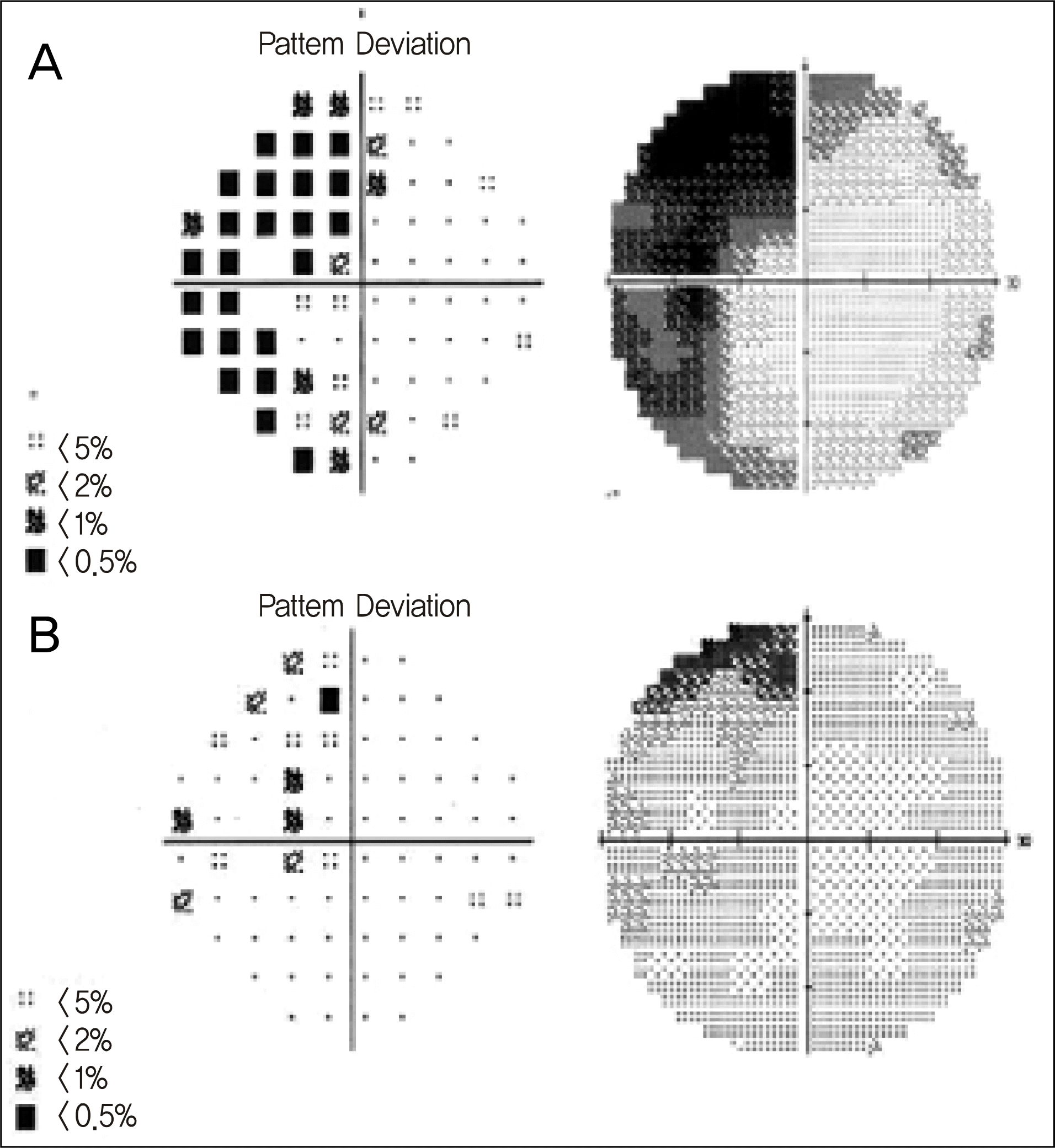
Figure 2.
(A) Left temporal hemianopia of the left eye. Visual acuity was 1.0. (B) 1 month later, after migration of silicone oil into lateral ventricle, the visual field defect improved.

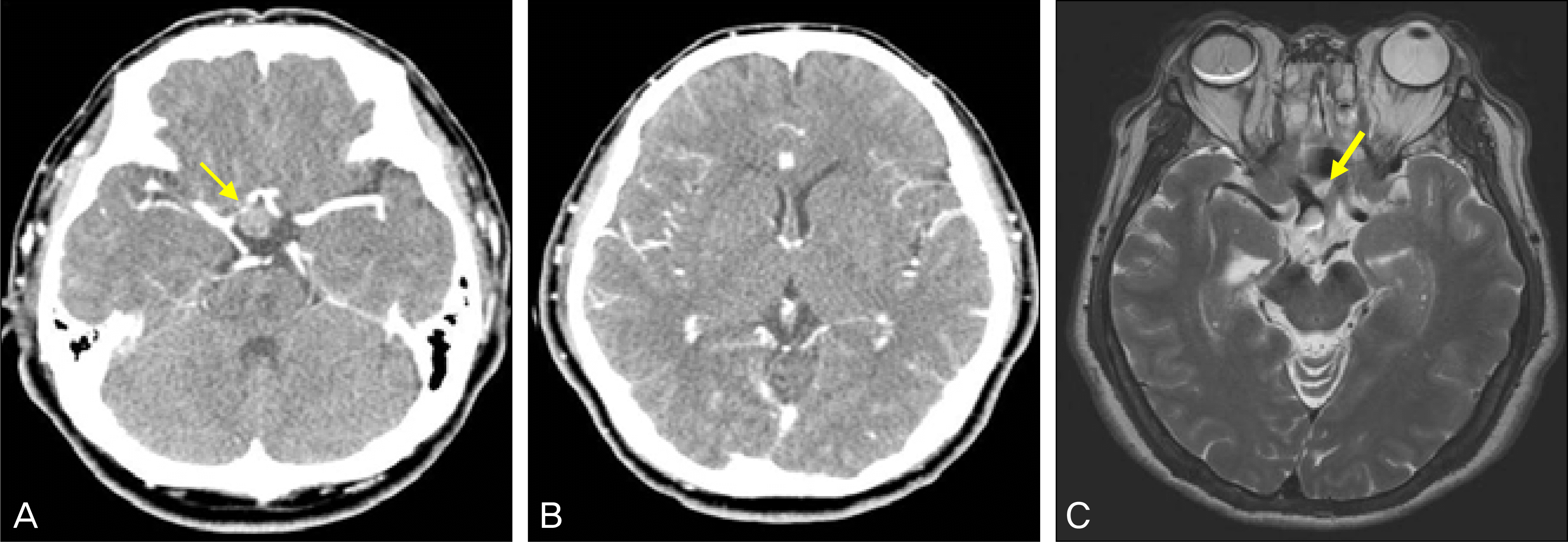
Figure 3.
(A) Computed tomography (CT) of the head shows 1.4 cm sized no enhanced mass in the suprasellar area (arrow). (B) There was no mass in the ventricle. (C) A axial T2-weighted magnetic resonance imaging (MRI) scan of the head shows hetero-genous signal intensity in the silicone filled right eye as well as in the right side of the optic chiasm (arrow).

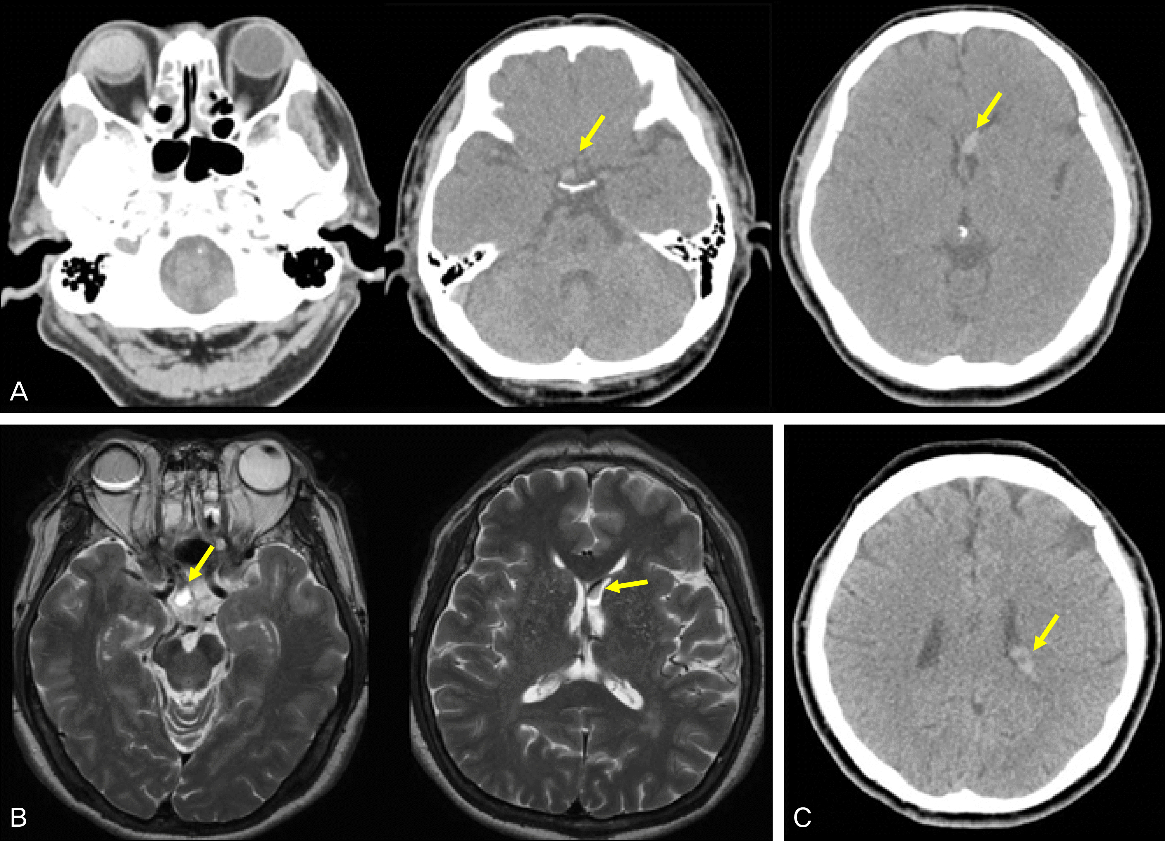
Figure 4.
(A) Computed tomography (CT) scan of the head, about 1 cm sized hyperdense mass is newly seen in the left lateral ventricle. The material filling the right eyeball, suprasellar area and the new lesion in the frontal horn of the left lateral ventricle showed identical CT density (arrow). (B) T2-weighted magnetic resonance imaging (MRI) scan shows a new lesion (arrow). (C) Computed tomography (CT) scan of the head, axial section, with the patient in a prone position. Silicone oil shifts to the posterior portion of the lateral ventricle (arrow).


 PDF
PDF ePub
ePub Citation
 Print
Print


 XML Download