Abstract
Purpose
To compare the changes in strabismus angle and deviation between two groups: a bilateral lateral rectus recession (Group 1) and a unilateral lateral rectus recession in exotropia (Group 2).
Methods
A retrospective survey was conducted on 198 patients who had received exotropia surgery in our ophthalmology clinic from September 2003 to April 2007. A total of 116 patients were in Group 1, and 82 patients were in Group 2.
Unilateral lateral rectus recession was performed on patients under 25 PD, and bilateral lateral rectus recession was performed on patients above 25 PD. A case of less than 4 PD esodeviation and less than 10 PD exodeviaton was defined as a success and any other case as a failure at the 1-year follow-up visit.
Results
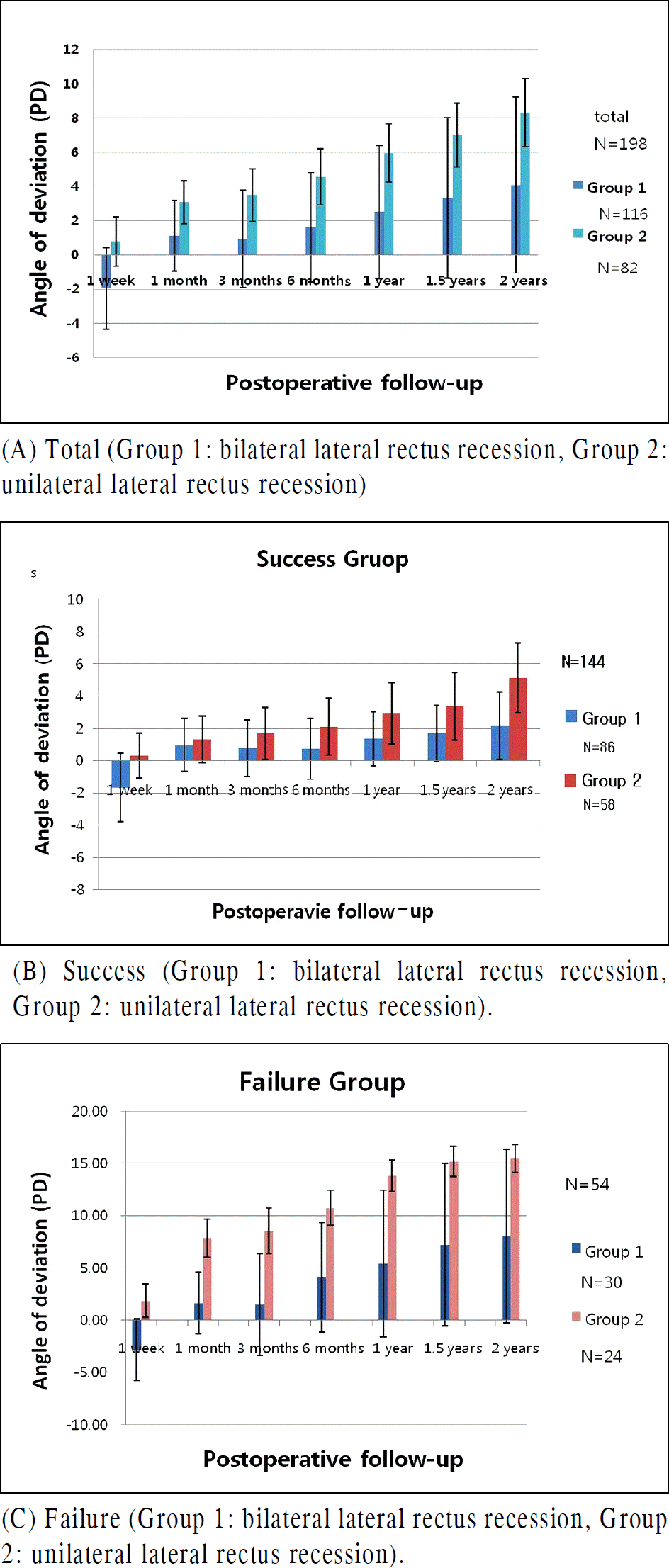
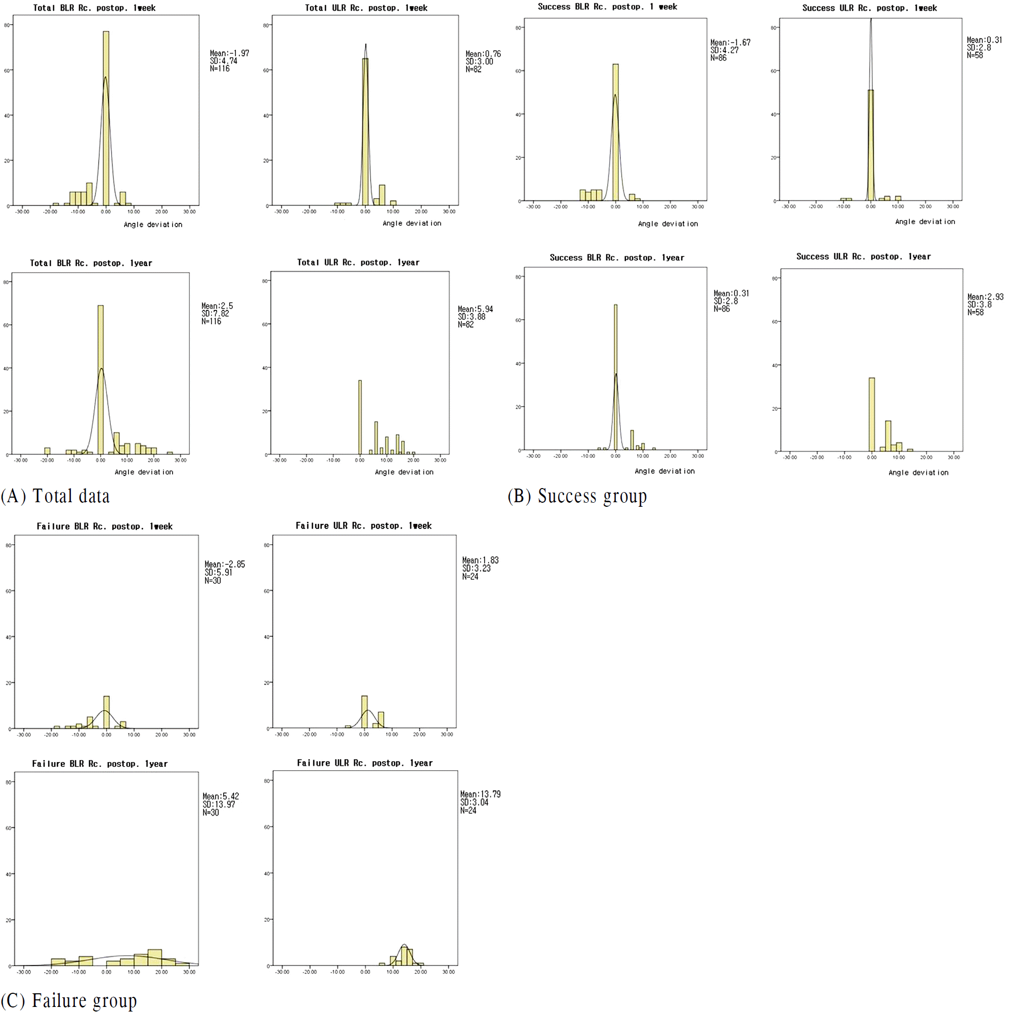
The average deviations of the first postoperative month and the first postoperative year were −1.96 D ± 4.75, 2.5 D ± 7.82 for Group 1 and 0.77 D ± 2.87, 5.94 D ± 3.38 for Group 2. Revealing statistical significance between the 2 Groups: Group 1 had 30 failure cases (25.9%) and their 1 postoperative year average deviation was 5.42 D ± 13.97, while Group 2 showed 24 failure cases (29.3%) and their 1-postoperative-year average deviation was 13.0–79 ± 3.04. Group 1 had less strabismus angle and a greater standard deviation than Group 2, as Group 1 had more overcorrection. Among the 30 failure cases of Group 1, 9 were overcorrected and 21 were undercorrected, but all 24 failure cases in Group 2 were undercorrected.
References
1. von Noorden GK, Campos EC. Binocular vision and ocular motility: theory and management of strabismus. 6th ed.St. Louis: Mosby;2002. p. 631.
2. Burian HM. Exodeviations. Their classification, diagnosis, and treatment. Am J Ophthalmol. 1966; 62:1161–6.

3. Scott WE, Keech R, Mash J. The postoperative results and stability of exodeviations. Arch Ophthalmol. 1981; 99:1814–8.

4. Ruttum MS. Initial versus subsequent postoperative motor alignment in intermittent exotropia. J AAPOS. 1997; 1:88–91.

5. Scott WE, Keech R, Mash AJ. The postoperative results and stability of exodeviations. Arch Ophthalmol. 1981; 99:1814–8.

6. Souza-Dias C, Uesugui CF. Postoperative evolution of the planned initial overcorrection in intermittent exotropia: 61 cases. Binocular Vis Eye Muscle Surg Q. 1993; 8:141–8.
7. Reynolds JD, Hiles DA. Single lateral rectus muscle recession for small angle exotropia. Reinecke RD, editor. Strabismus 2. New York: Grune & Stratton;1984. p. 247–53.
8. Kushner BJ. Selective surgery for intermittent exotropia based on distance/near differences. Arch Ophthalmol. 1998; 116:324–8.

9. Lee SH, Kim J-Y, Kwon J-Y. The effect of unilateral lateral rectus recession for the treatment of moderate-angle exotropia. J Korean Ophthalmol Soc. 2005; 46:2045–9.
10. Parks MM. Clinical opththalmoloty. 2nd ed.New York: Harper & Row;1978. p. 7.
11. Moon KJ, Choi WC, Park C. The long term effect of unilateral lateral rectus muscle recession for moderate angle exotropia. J Korean Ophthalmol Soc. 1998; 39:1885–90.
12. Stavis MI. Large recession of one lateral rectus muscle for moderate angle exotropia. Ophthalmology. 1986; 93:88.
13. Nelson LB, Bacal DA, Burke MJ. An alternative approach to the surgical management of exotropia: the unilateral lateral rectus recession. J Pediatr Ophthalmol Strabismus. 1992; 29:357–60.
14. Weakley DR, Stager DR. Unilateral lateral rectus recessions in exotropia. Ophthalmic Surg. 1993; 24:458–60.

15. Dunlap EA, Gaffney RB. Surgical management of intermittent exotropia. Am Orthopt J. 1963; 18:20–33.

16. Lee SY. The effect of unilateral lateral rectus muscle recession over 11 mm in the treatment of intermittent exotropia of 15–20PD. J Korean Ophthalmol Soc. 1999; 40:550–4.
17. Harley RD. Pediatric Ophthalmology. 2nd ed.Philadelphia: W.B. Saunders;1983. p. 238–9.
18. Feretis D, Mela E, Vasilopoulos G. Excessive single lateral rectus muscle recession in the treatment of intermittent exotropia. J Pediatr Ophthalmol Strabismus. 1990; 27:315–6.

19. Sul CY, Park C. Large recession of one lateral rectus muscle for moderate angle exotropia. J Korean Ophthalmol Soc. 1988; 29:125–9.
20. Moon KJ, Choi WC, Park C. The long term effect of unilateral lateral rectus muscle recession for moderate angle exotropia. J Korean Ophthalmol Soc. 1998; 39:1885–90.
21. Reynolds JD, Hiles DA. Single lateral rectus muscle recession for small angle exotropia. Reinecke RD, editor. Strabismus II. Orlando: Grune & Stratton;1984. p. 247–53.
22. Deutsch JA, Nelson LB, Sheppard RW, Burke MJ. Unilateral lateral rectus recession for the treatment of exotropia. Ann Ophthalmol. 1992; 24:111–3.
23. Olitsky SE. Early and late postoperative alignment following unilateral lateral rectus recession for intermittent exotropia. J Pediatr Ophthalmol Strabismus. 1998; 35:146–8.

24. Roh YB, Choi HY. Surgical results of unilateral and bilateral lateral rectus recessions in exotropia under 25 prism diopter. J Korean Ophthalmol Soc. 1997; 38:474–8.
25. Choi SJ, Kim IC. The effect of recession of single lateral rectus muscle. J Korean Ophthalmol Soc. 1995; 36:1273–7.
26. Cho HJ, Ma YR, Park YG. Comparison of surgical results between bilateral and unilateral lateral rectus recession in 20–25 prism diopters intermittent exotropia. J Korean Ophthalmol Soc. 2002; 43:1993–9.
27. Lee SN, Shin DB, Xu YG, Min BM. Effect of unilateral recession for intermittent exotropia under 25PD. J Korean Ophthalmol Soc. 2002; 43:1469–73.
28. Dadeya S, Kamlesh . Long term results of unilateral lateral rectus recession in intermittent exotropia. J Pediatr Ophthalmol Strabismus. 2003; 40:283–7.
29. Sheppard RW, Panton CM, Smith DR. The single horizontal muscle recession operation, a survey. Can J Ophthalmol. 1973; 8:68–74.
30. Keech RV, Stewart SA. The surgical overcorrection of intermittent exotropia. J Pediatr Ophthalmol Strabismus. 1990; 27:218–20.

31. Park HS, Kim JB, Seo MS, Park YG. A study on the consecutive esotropia after intermittent exotropia surgery. J Korean Ophthalmol Soc. 1994; 35:1327–34.
Table 1.
Clinical characteristics of patients
| | Group 1* | Group 2† | p value |
|---|---|---|---|
| Number | 116 | 82 | |
| Sex | | | |
| male | 45 | 35 | |
| Female | 71 | 47 | |
| Age | | | |
| Mean ± SD | 6.44 ± 2.33 | 6.98 ± 2.19 | 0.104 |
| Range | 3–14 | 3–13 | |
| Preop deviation (PD‡) | | | |
| Mean ± SD | 29.18 ± 4.71 | 20.95 ± 3.26 | 0.000 |
| Range | 25–40 | 16–24 | |
Table 2.
Comparison of angle of deviation
| Group (N*) | Postoperative Follow up |
Angle of deviation (Mean±SD) (PD†) |
||
|---|---|---|---|---|
| Group 1‡ | Group 2§ | p value | ||
| | 1 week | −1.97 ± 4.75 | 0.77 ± 2.87 | 0.000 |
| | 1 year | 2.5 ± 7.82 | 5.94 ± 3.38 | 0.000 |
| Success (144) | | | | |
| | 1 week | −1.67 ± 4.27 | 0.31 ± 2.80 | 0.002 |
| | 1 year | 1.36 ± 3.32 | 2.93 ± 3.80 | 0.010 |
| Failure (54) | | | | |
| | 1 week | −2.85 ± 5.91 | 1.83 ± 3.23 | 0.001 |
| | 1 year | 5.42 ± 13.97 | 13.79 ± 3.04 | 0.008 |
Table 3.
Comparison of success rate between Group 1 and Group 2 in postoperative 1 year
| Group (N*) | Deviation angle (PD†) |
No. of patient (%) |
|
|---|---|---|---|
| Group 1‡ (N = 116) | Group 2§ (N = 82) | ||
| Success (144) | | 86 (74.1%) | 58 (70.7%) |
| Failure | | 30 (25.9%) | 24 (29.3%) |
| Overcorrection (10) | >10 PD esodeviation | 8 (6.9%) | 0 |
| | 4–10 PD esodeviation | 2 (1.7%) | 0 |
| Undercorrection (44) | 10–19 PD exodeviation | 7 (6.0%) | 20 (24.4%) |
| | >20 PD exodeviation | 13 (11.2%) | 4 (4.9%) |
Table 4.
Coefficient of variation at each follow-up period
| Group (N*) | 1 week | 1 month | 3 month | 6 month | 1 year | 1.5 year | 2 year |
|---|---|---|---|---|---|---|---|
| Group 1† | 241.5 | 372.9 | 605.4 | 393.7 | 312.7 | 282.5 | 253.1 |
| Group 2‡ | 375.5 | 81.9 | 86.8 | 72.1 | 57.0 | 53.1 | 48.1 |
| Success (144) | | | | | | | |
| Group 1† (86) | 255.1 | 336.5 | 444.6 | 513.9 | 244.2 | 204.7 | 194.7 |
| Group 2‡ (58) | 901.7 | 222.4 | 188.8 | 167.2 | 129.8 | 124.32 | 83.95 |
| Failure (54) | | | | | | | |
| Group 1† (30) | 207.6 | 367.8 | 655.2 | 255.3 | 257.6 | 216.0 | 206.9 |
| Group 2‡ (24) | 176.0 | 46.8 | 51.6 | 31.4 | 22.0 | 19.2 | 17.3 |


 PDF
PDF ePub
ePub Citation
 Print
Print


 XML Download