

 PDF
PDF ePub
ePub Citation
 Print
Print


Abstract
Purpose
To evaluate histopathologic properties of eyelid skin and conjunctiva and the association between clinical mani-festation and histopathologic findings.
Methods
A prospective study was conducted on the histologic findings of the upper eyelid skin and conjunctiva performed from November 2009 to February 2010 in 27 patients for upper eyelid blepharoplasty procedures. Histopathologic studies were performed for specimens collected from the upper eyelid skin and inferotemporal bulbar conjunctiva. Preoperative photographs of the upper eyelid and conjunctiva were taken to grade clinical severity of dermatochalasis and conjunctivochalasis.
Results
Decrease of collagen density and elastic degeneration in the eyelid skin and conjunctiva were significantly associated with aging (p < 0.001, p = 0.001, p = 0.001, p < 0.001). Clinical severity of dermatochalasis was correlated with a decrease of collagen density and elastic degeneration in the eyelid skin, and clinical severity of conjunctivochalasis was associated with a decrease of collagen density, elastic degeneration and lymphangiectasia in conjunctiva (p < 0.001).
Go to : 
References
1. DeAngelis DD, Carter SR, Seiff SR. Dermatochalasis. Int Ophthalmol Clin. 2002; 42:89–101.

2. Hughes WL. Conjunctivochalasis. Am J Ophthalmol. 1942; 25:48–51.
3. Meller D, Tseng SC. Conjunctivochalasis: literature review and possible pathophysiology. Surv Ophthalmol. 1998; 43:225–32.
4. Francis IC, Chan DG, Kim P, et al. Case-controlled clinical and histopathological study of conjunctivochalasis. Br J Ophthalmol. 2005; 89:302–5.
5. Watanabe A, Yokoi N, Kinoshita S, et al. Clinicopathologic study of conjunctivochalasis. Cornea. 2004; 23:294–8.
6. Höh H, Schirra F, Kienecker C, Ruprecht KW. Lid-parallel conjunctival folds are a sure diagnostic sign of dry eye. Ophthalmologe. 1995; 92:802–8.
7. Baumann L. Skin ageing and its treatment. J Pathol. 2007; 211:241–51.
8. Makrantonaki E, Zouboulis CC, William J. Cunliffe Scientific Awards. Characteristics and pathomechanisms of endogenously aged skin. Dermatology. 2007; 214:352–60.
9. Hwang K, Kim DJ, Kim SK. Does the upper eyelid skin become thinner with age? J Craniofac Surg. 2006; 17:474–6.
10. Kocaoglu FA, Katircioglu YA, Tok OY, et al. The histopathology of involutional ectropion and entropion. Can J Ophthalmol. 2009; 44:677–9.
Go to :
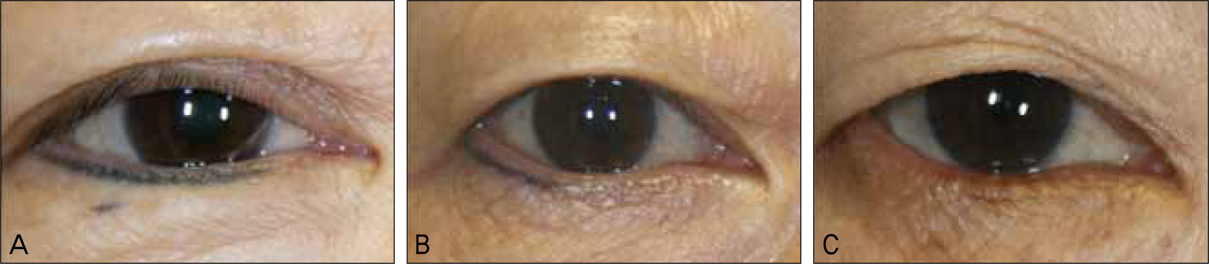 | Figure 1.Clinical severity score of dermatochalasis. (A) Grade 1, hanging skin margin above the upper eyelid skin margin. (B) Grade 2, hanging skin margin on the lid margin. (C) Grade 3, hanging skin margin below the lid margin. |
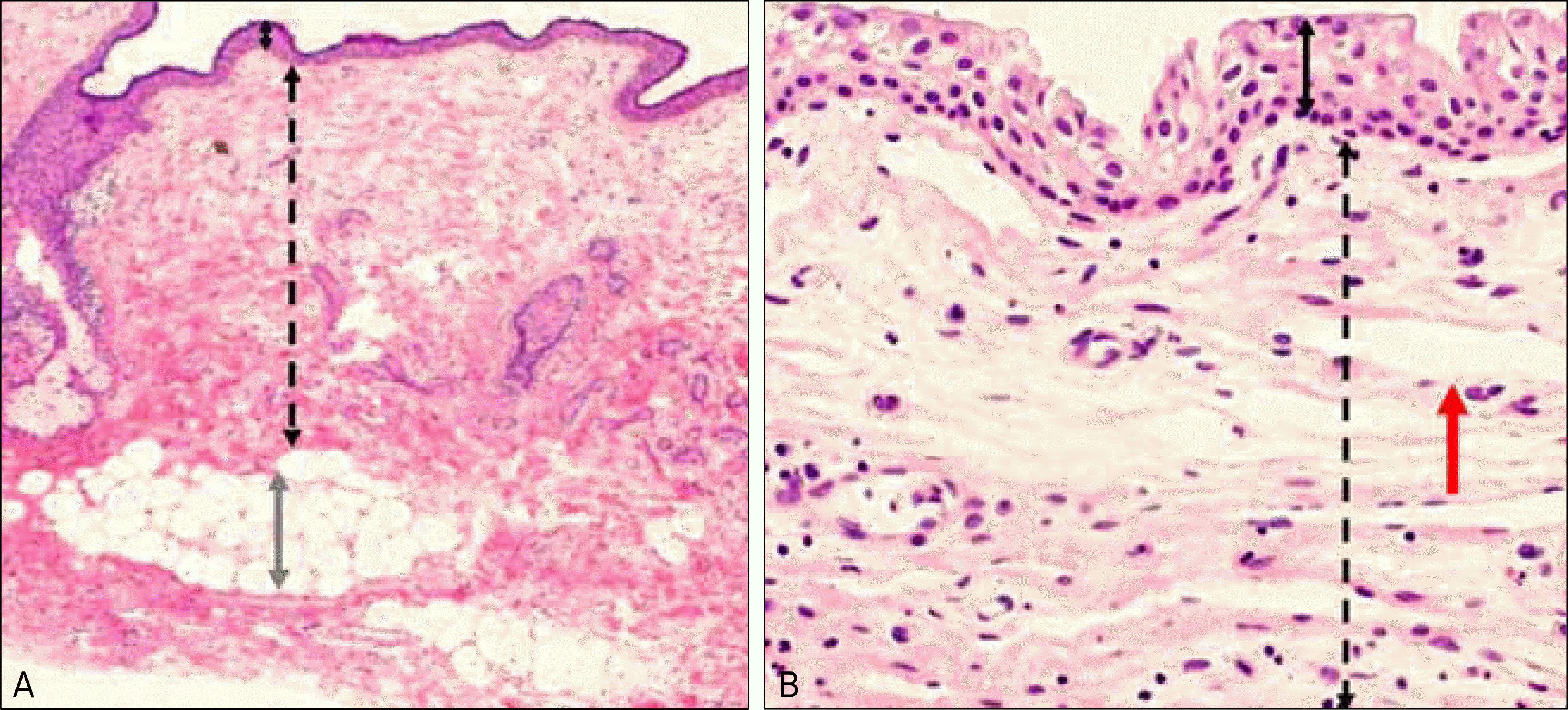 | Figure 2.Histopathologic findings of the eyelid and conjunctiva. (A) Histologic findings of the upper eyelid skin (Hematoxylin-eo-sin stain, ×50). Black arrow indicates epidermis, dotted arrow indicates dermis and gray arrow is subcutaneous fat. (B) Histologic findings of the conjunctiva (Hematoxylin-eosin stain, ×100). Black arrow indicates epithelium, dotted arrow indicates substantia propria and red arrow denotes lymphangiectasia. |
 | Figure 3.Collagen density of the upper eyelid skin and conjunctiva. (A, B, C) Collagen density of upper eyelid skin (Trichrome stain, ×400). (A) Grade 3, high collagen density. (B) Grade 2, moderate collagen density. (C) Grade 1, low collagen density. (D, E, F) Collagen density of conjunctiva (Trichrome stain, ×400). (D) Grade 3, high collagen density. (E) Grade 2, moderate collagen density. (F) Grade 1, low collagen density. |
 | Figure 4.Elastic degeneration of upper eyelid skin and conjunctiva. (A, B, C) Elastic degeneration of the upper eyelid skin (Verhoeff Van Gieson stain, ×400). (A) Grade 1, mild elastic degeneration. (B) Grade 2, moderate elastic degeneration. (C) Grade 3, severe elastic degeneration. (D, E, F) Elastic degeneration of the conjunctiva (Verhoeff Van Gieson stain, ×400). (D) Grade 1, mild elastic degeneration. (E) Grade 2, moderate elastic degeneration. (F) Grade 3, severe elastic degeneration. |
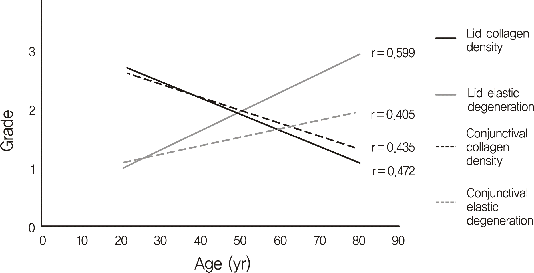 | Figure 5.Association between age and histologic findings of the eyelid and conjunctiva in dermatochalasis patients. Collagen density & elastic degeneration were significantly correlated with age regarding histologic findings of the eyelid skin and conjunctiva. Lid collagen density (p < 0.001) and conjunctival collagen density (p = 0.001) noted to decrease according to age. Lid elastic degeneration (p = 0.001) and conjunctival elastic degeneration (p < 0.001) were noted to decrease according to age. |
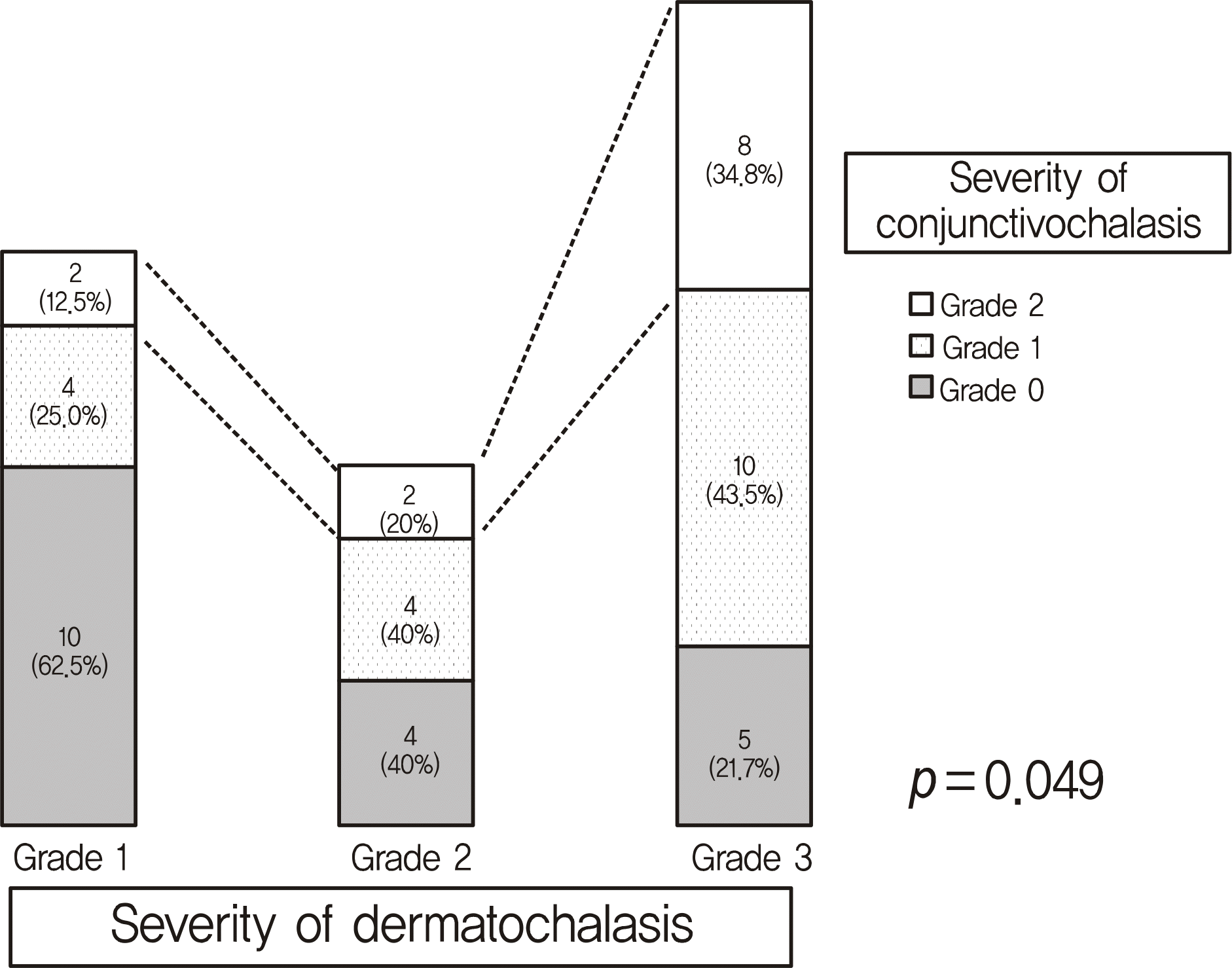 | Figure 6.Association between severity of dermatochalasis and conjunctivochalasis. Clinical severity of dermatochalasis tends to be related proportionally with the severity of conjunctivochalasis. |
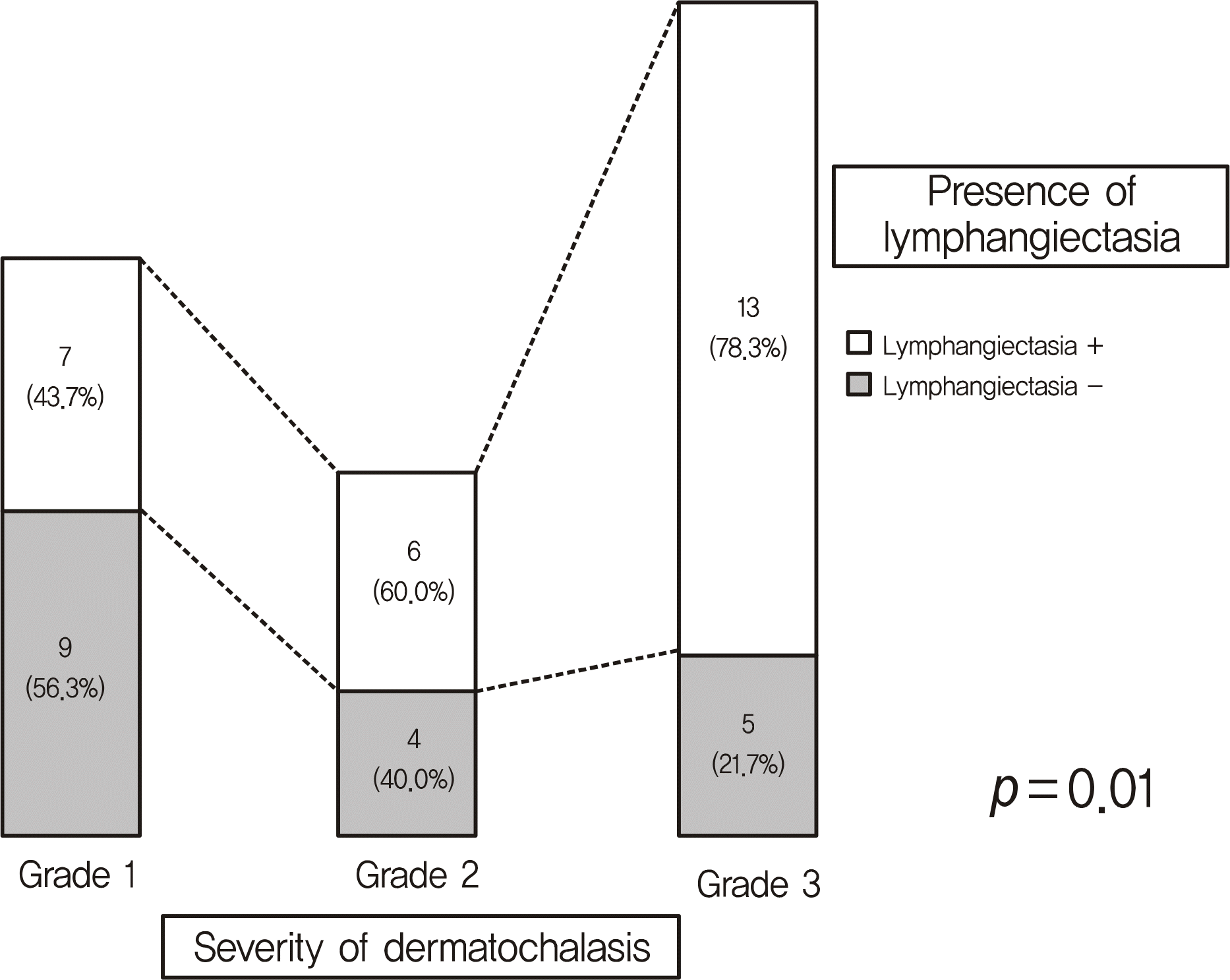 | Figure 7.Association between clinical severity of dermatochalasis and conjunctival lymphangiectasia. Clinical severity of dermatochalasis is significantly associated with presence of conjunctival lymphangiectasia (p = 0.01). |
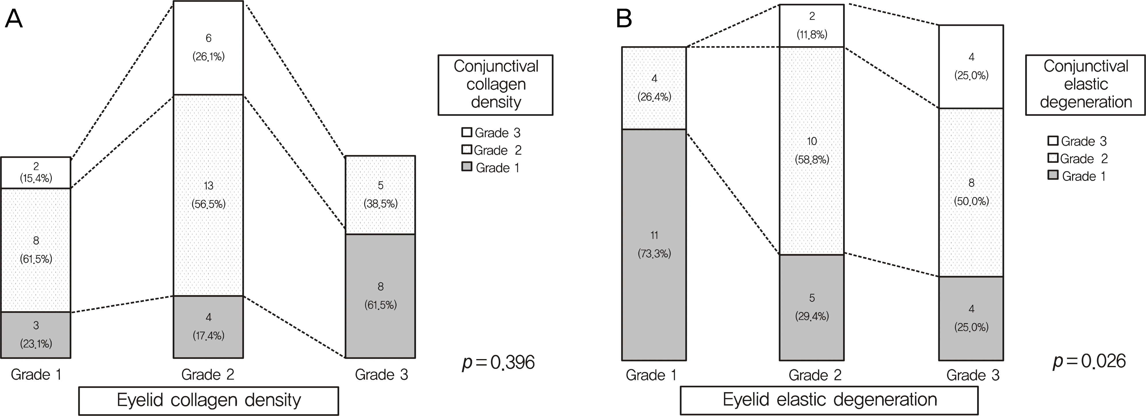 | Figure 8.Associations of histologic findings between eyelid skin and conjunctiva in the dermatochalasis patients. (A) Collagen density between the eyelid skin and conjunctiva is not significantly associated (p = 0.396), (B) eyelid skin elastic degeneration is significantly related to conjunctival elastic degeneration (p = 0.026). |
Table 1.
Grading of conjunctivochalasis
| Grade | |
|---|---|
| 0 | No persistent fold |
| 1 | Single, small fold |
| 2 | More than two folds and not higher than the tear meniscus |
| 3 | Multiple folds and higher than the tear meniscus |
Table 2.
Demographic features
| Factors | |
|---|---|
| Age (mean ± SD, yr) Distribution of age | 54.9 ± 20.5 |
| <40 | 7 |
| 41-50 | 2 |
| 51-60 | 2 |
| 61-70 | 11 |
| >71 | 5 |
| Sex | |
| Male | 5 (18.5%) |
| Female | 22 (72.5%) |
| Laterality | |
| OD | 25 (51.0%) |
| OS | 24 (49.0%) |
Table 3.
Thickness of upper eyelid skin
| Age (yr) | Epidermis (mean ± SD, μm) | Dermis (mean ± SD, μm) |
|---|---|---|
| <40 | 48.72 ± 14.37 | 512.54 ± 137.63 |
| 41-50 | 48.50 ± 8.10 | 632.50 ± 98.78 |
| 51-60 | 35.00 ± 4.08 | 690.00 ± 27.08 |
| 61-70 | 45.68 ± 9.54 | 673.86 ± 164.38 |
| >71 | 44.50 ± 6.71 | 605.00 ± 157.11 |
| Total | 45.50 ± 10.40 | 624.3 ± 156.30 |
| p-value* | 0.274 | 0.154 |
Table 4.
Association between clinical severity and histologic grading in the patients with dermatochalasis
| Site | Clinical severity score |
Histologic grading |
||
|---|---|---|---|---|
| Collagen density‡ | Elastic degeneration§ | Lymphangiectasia∏ | ||
| Eyelid* | 1 (n = 16) | 2.4 | 1.2 | |
| 2 (n = 10) | 1.9 | 1.8 | ||
| 3 (n = 23) | 1.7 | 2.7 | ||
| p-value | p = 0.013 | p < 0.001 | ||
| Conjunctiva† | 0 (n = 20) | 1.0 | 2.4 | 0.5 |
| 1 (n = 22) | 0.8 | 2.1 | 0.6 | |
| 2 (n = 7) | 0.3 | 1.2 | 0.7 | |
| p-value | p < 0.001 | p < 0.001 | p < 0.001 | |
* Clinical severity of dermatochalasis were classified from 1 to 3: grade 1, hanging skin margin above the upper eyelid skin margin; grade 2, hanging skin margin on the lid margin; grade 3, hanging skin margin below the lid margin.;
† Clinical severity of conjunctivochalasis were classified according to conjunctival fold and tear meniscus height: grade 0, no persistent fold; grade 1, single small fold; grade 2, more than two folds and not higher than the tear meniscus.;
‡ Collagen density was scored from 1 to 3: grade 1, low collagen density; grade 2, moderate collagen density; grade 3, high collagen density.;
 XML Download