Journal List > J Korean Ophthalmol Soc > v.52(5) > 1009037

Shin, Cho, Lee, and Seong: The Long-Term Observation of Intraocular Pressure after Intravitreal Injections of Bevacizumab, Triamcinolone, and Combination of Both

Abstract

Purpose

The present study investigates the onset and frequency of increased intraocular pressure (IOP) after injections of intravitreal Bevacizumab, Triamcinolone, and a combination of both drugs.

Methods

Patients were classified into three groups: Bevacizumab group (group A), Triamcinolone group (group B), and combined drug group (group C), irrespective of the underlying causes. The IOP was measured 30 minutes prior to followed by one day, one week, one month, two months, three months, six months, and one year after injection. The measured IOP at each time point was compared with the pre-injection IOP and the differences in IOP among the three groups were statistically analyzed. The relationships between various factors possible of increasing IOP were also analyzed.

Results

A total of 259 subjects were enrolled in the present study. An IOP increase of more than 5 mm Hg was observed in 25 eyes (15%) in group A, 34 eyes (40%) in group B, and 40 eyes (50%) in group C. There was no statistically significant mean IOP change after injection in group A, but in groups B and C there was an increase in mean IOP up until two and three months after injection, respectively. However, eyes in group A with a history of glaucoma showed statistically significant increases in IOP.

Conclusions

The onsets and frequencies of increased IOP were different with the different drugs. Proper follow-up for increased IOP after injection is necessary based on the type of drug used.

References

1. Chang MW, Kim SW, Oh IK, et al. Intravitreal triamcinolone injection with or without bevacizumab for diabetic macular edema. J Korean Ophthalmol Soc. 2008; 49:1269–74.
crossref
2. Ahmadieh H, Ramezani A, Shoeibi N, et al. Intravitreal bevacizumab with or without triamcinolone for refractory diabetic macular edema; a placebo-controlled, randomized clinical trial. Graefes Arch Clin Exp Ophthalmol. 2008; 246:483–9.
crossref
3. Ehrlich R, Ciulla TA, Moss AM, Harris A. Combined treatment of intravitreal bevacizumab and intravitreal triamcinolone in patients with retinal vein occlusion: 6 months of follow-up. Graefes Arch Clin Exp Ophthalmol. 2010; 248:375–80.
crossref
4. Faghihi H, Roohipoor R, Mohammadi SF, et al. Intravitreal bevacizumab versus combined bevacizumab-triamcinolone versus macular laser photocoagulation in diabetic macular edema. Eur J Ophthalmol. 2008; 18:941–8.
crossref
5. Folgosa MS, Messias A, Takata C, et al. Single intravitreal injection of triamcinolone combined with bevacizumab for the treatment of diffuse diabetic macular oedema refractory to grid photocoagulation. Acta Ophthalmol. 2010; 88:e297–8.
crossref
6. Jonas JB, Hayler JK, Söfker A, Panda-Jonas S. Intravitreal injection of crystalline cortisone as adjunctive treatment of proliferative diabetic retinopathy. Am J Ophthalmol. 2001; 131:468–71.
crossref
7. Park CH, Jaffe GJ, Fekrat S. Intravitreal triamcinolone acetonide in eyes with cystoid macular edema associated with central retinal vein occlusion. Am J Ophthalmol. 2003; 136:419–25.
crossref
8. Antcliff RJ, Spalton DJ, Stanford MR, et al. Intravitreal triamcinolone for uveitic cystoid macular edema: an optical coherence tomography study. Ophthalmology. 2001; 108:765–72.
crossref
9. Danis RP, Ciulla TA, Pratt LM, Anliker W. Intravitreal triamcinolone acetonide in exudative age-related macular degeneration. Retina. 2000; 20:244–50.
crossref
10. Jonas JB, Kreissig I, Degenring R. Intraocular pressure after intravitreal injection of triamcinolone acetonide. Br J Ophthalmol. 2003; 87:24–7.
crossref
11. Jonas JB, Kreissig I, Degenring R, et al. Secondary chronic open-angle glaucoma after intravitreal triamcinolone acetonide. Arch Ophthalmol. 2003; 121:729–30.
crossref
12. Roth DB, Realini T, Feuer WJ, et al. Short-term complications of intravitreal injection of triamcinolone acetonide. Retina. 2008; 28:66–70.
crossref
13. Smithen LM, Ober MD, Maranan L, Spaide RF. Intravitreal triamcinolone acetonide and intraocular pressure. Am J Ophthalmol. 2004; 138:740–3.
crossref
14. Jonas JB, Degenring RF, Kreissig I, et al. Intraocular pressure elevation after intravitreal triamcinolone acetonide injection. Ophthalmology. 2005; 112:593–8.
crossref
15. Roth DB, Verma V, Realini T, et al. Long-term incidence and timing of intraocular hypertension after intravitreal triamcinolone acetonide injection. Ophthalmology. 2009; 116:455–60.
crossref
16. Jung JW, Nam DH, Shyn KH. The complications after intravitreal injection of triamcinolone acetonide. J Korean Ophthalmol Soc. 2007; 48:55–62.
17. Kim SJ, Park YM, Lee SU, et al. Short-term safety and efficacy of intravitreal bavacizumab injection. J Korean Ophthalmol Soc. 2009; 50:219–26.
crossref
18. Falkenstein IA, Cheng L, Freeman WR. Changes of intraocular pressure after intravitreal injection of bevacizumab (avastin). Retina. 2007; 27:1044–7.
crossref
19. Bakri SJ, Larson TA, Edwards AO. Intraocular inflammation following intravitreal injection of bevacizumab. Graefes Arch Clin Exp Ophthalmol. 2008; 246:779–81.
crossref
20. Shima C, Sakaguchi H, Gomi F, et al. Complications in patients after intravitreal injection of bevacizumab. Acta Ophthalmologica. 2008; 86:372–6.
crossref
21. Ozkiriş A, Erkiliç K. Complications of intravitreal injection of triamcinolone acetonide. Can J Ophthalmol. 2005; 40:63–8.
22. Yang YH, Kim KR, Yang SW, Yim HB. The effect of intravitreal triamcinolone acetonide on intraocular pressure. J Korean Ophthalmol Soc. 2004; 45:1081–5.
23. Smithen LM, Ober MD, Maranan L, Spaide RF. Intravitreal triamcinolone acetonide and intraocular pressure. Am J Ophthalmol. 2004; 138:740–3.
crossref
24. Bakri SJ, Beer PM. The effect of intravitreal triamcinolone acetonide on intraocular pressure. Ophthalmic Surg Lasers Imaging. 2003; 34:386–90.
crossref
25. Park HY, Yi K, Kim HK. Intraocular pressure elevation after intravitreal triamcinolone acetonide injection. Korean J Ophthalmol. 2005; 19:122–7.
crossref
26. Hattenbach LO, Klais C, Koch FH, Gümbel HO. Intravitreous injection of tissue plasminogen activator and gas in the treatment of submacular hemorrhage under various conditions. Ophthalmology. 2001; 108:1485–92.
crossref
27. Shin JH, Lee SW, Kim DK, et al. Intravitreal injection of triamcinolone acetonide for macular edema: long-term safety and efficacy after two years. J Korean Ophthalmol Soc. 2007; 48:1670–4.
28. Chung MS, Chang WH. Efficacy of anterior chamber paracentesis in intravitreal triamcinolone injection. J Korean Ophthalmol Soc. 2005; 46:1328–32.
29. Lee MW, Kyung SE, Chang MH. Prophylactic effect of brimoni-dine 0.15% on IOP elevation after intravitreal triamcinolone acetonide injection. J Korean Ophthalmol Soc. 2008; 49:743–52.
crossref
30. Seong HK, Lee JM, Park YS, Lee BR. Immediate natural course of IOP after IVTA and the effect of preoperative ocular massage. J Korean Ophthalmol Soc. 2007; 48:808–14.
31. Kahook MY, Kimura AE, Wong LJ, et al. Sustained elevation in intraocular pressure associated with intravitreal bevacizumab injections. Ophthalmic Surg Lasers Imaging. 2009; 40:293–5.
crossref
32. Jalil A, Fenerty C, Charles S. Intravitreal bevacizumab (Avastin) causing acute glaucoma: an unreported complication. Eye. 2007; 21:1541.
crossref
33. Kubota T, Okabe H, Hisatomi T, et al. Ultrastructure of the trabecular meshwork in secondary glaucoma eyes after intravitreal triamcinolone acetonide. J Glaucoma. 2006; 15:117–9.
crossref
34. Cho P, Lui T. Comparison of the performance of the Nidek NT-2000 noncontact tonometer with the Keeler Pulsair 2000 and the Goldmann applanation tonometer. Optom Vis Sci. 1997; 74:51–8.
crossref
35. Audren F, Lecleire-Collet A, Erginay A, et al. Intravitreal triamcinolone acetonide for diffuse diabetic macular edema: phase 2 trial comparing 4 mg vs 2 mg. Am J Ophthalmol. 2006; 142:794–9.
crossref
36. Lee JM, Kim SJ, Yi KY, Kim HK. Intraocular pressure change after secondary intravitreal triamcinolone acetonide injection. J Korean Ophthalmol Soc. 2007; 48:97–102.
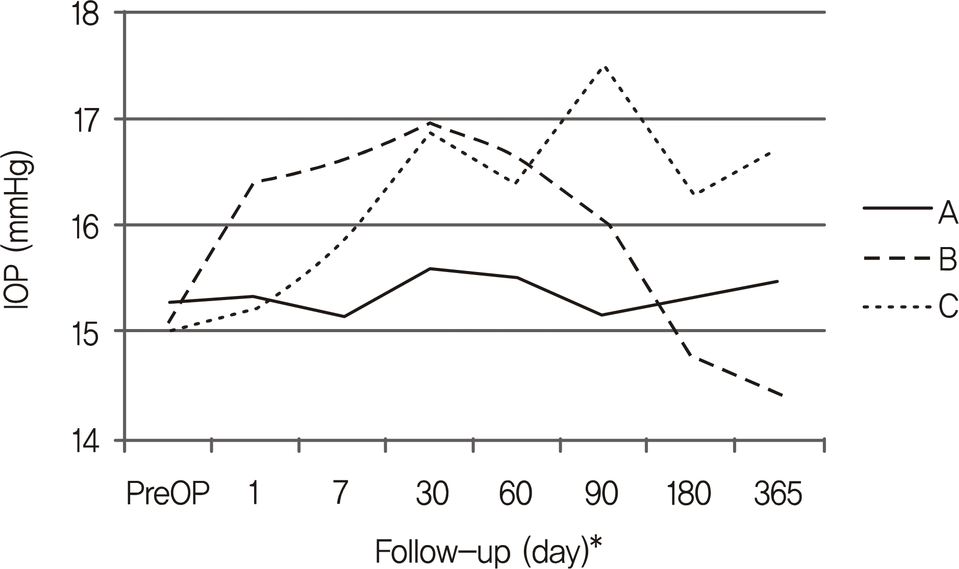
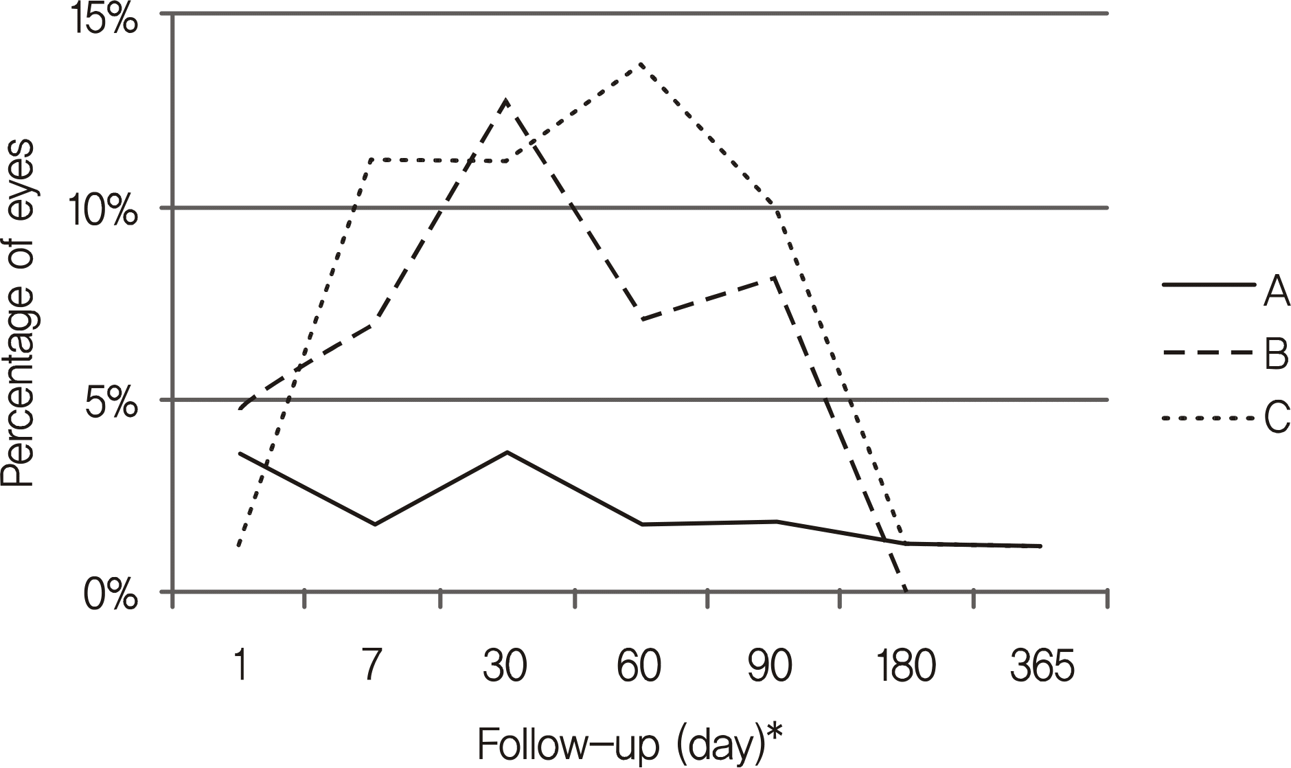
Figure 1.
The change of mean intraocular pressure (IOP) during follow up period. Note that the mean IOP of group B increased during post injection 7 to 60 days. The mean IOP of group C increased during post injection 30 to 90 days. The mean IOP of Group A showed no statistically significant change. Group A: Bevacizumab, Group B: Triamcinolone, Group C: Bevacizumab + Triamcinolone. * Post injection days.
jkos-52-574f1.tif
Figure 2.
The percentage of eyes showing increased IOP of more than 5 mmHg at each follow up time. Group B and C showed higher percentages of increased IOP than Group A during post injection 7 to 90 days. There were no statistically significant differences between Group B and C (p > 0.05) during the entire follow up period. Group A: Bevacizumab, Group B: Triamcinolone, Group C: Bevacizumab + Triamcinolone. * Post injection days.
jkos-52-574f2.tif
Table 1.
Clinical characteristics of the eyes enrolled in the current study
Bevacizumab (A) Triamcinolone (B) B Bevacizumab + Triamcinolone (C) p-value
Number of eyes 166 85 80
Age (mean ± SD, yr) 59.46 ± 12.36 61.2 ± 11.29 58.90 ± 10.60 0.405
Mean follow up (day) 197.80 221.41 210.38 0.071
1 year follow up* (n) 75 38 30 0.496
Lens (phakic : pseudophakic) 96:70 52:33 59:21 0.098
Past injection history (n) 28 (16.8%) 30 (35.3%) 26 (32.5%) 0.002
History of glaucoma (n) 10 (6%) 2 (2.4%) 7 (8.8%) 0.205
Diagnosis
PDR 86 (51.5%) 16 (18.8%) 36 (45%) 0.003
NPDR with ME 26 (15.6%) 39 (45.9%) 21 (26.3%) 0.001
RVO with ME 13 (7.8%) 25 (29.4%) 22 (27.5%) 0.000
ARMD 32 (19.23%) 4 (4.71%) 1 (1.3%) 0.000
Others 9 (5.42%) 1 (1.18%) 0 (0%) 0.041

DR = proliferative diabetic retinopathy; NPDR = non-proliferative diabetic retinopathy; ME = macular edema; RVO = retinal vein occlusion; ARMD = age-related macular degeneration.

* The number of eyes followed up for one year;

ANOVA test;

Chi-square test.

Table 2.
Intraocular pressure of each group during follow-up period
Follow up (day) A
B
C
p-value*
Mean IOP ± SD (mmHg) p-value Mean IOP ± SD (mmHg) p-value Mean IOP ± SD (mmHg) p-value
PreOP 15.28 ± 3.10 15.09 ± 3.18 15.01 ± 1.98 0.543
1 15.33 ± 3.43 0.393 16.41 ± 3.32 0.013 15.23 ± 3.15 0.422 0.265
7 15.15 ± 3.26 0.146 16.62 ± 4.11 0.008 15.82 ± 4.00 0.049 0.030
30 15.62 ± 3.96 0.189 16.97 ± 4.10 0.002 16.88 ± 5.98 0.005 0.091
60 15.53 ± 3.19 0.666 16.67 ± 4.82 0.020 16.39 ± 4.99 0.024 0.328
90 15.16 ± 3.56 0.240 16.09 ± 4.50 0.128 17.48 ± 7.32 0.006 0.026
180 15.36 ± 3.45 0.454 14.81 ± 4.16 0.287 16.30 ± 5.37 0.056 0.175
365 15.49 ± 3.14 0.659 14.45 ± 3.90 0.335 16.73 ± 5.81 0.097 0.053

A = Bevacizumab group; B = Triamcinolone group; C = Bevacizumab + Triamcinolone group; preOP = 30 minutes before injection; IOP = intraocular pressure; IIOP = increased intraocular pressure.

* Statistical difference among three groups in each follow up day by ANOVA test;

Statistical difference of intraocular pressure between preinjection and each follow up day by independent t-test.

Table 3.
The mean IOP of eyes with increased IOP of more than 5 mmHg during follow up in each group
Follow up (day) A (n = 25)
B (n = 34)
C (n = 40)
p-value*
Mean IOP ± SD (mmHg) N. of onset Mean IOP ± SD (mmHg) N. of onset Mean IOP ± SD (mmHg) N. of onset
PreOP 14.44 ± 3.07 14.18 ± 3.08 15.00 ± 1.90 0.406
1 16.36 ± 4.38 6 16.73 ± 3.76 4 16.27 ± 2.69 1 0.900
7 15.27 ± 3.71 3 18.38 ± 5.20 6 17.32 ± 4.45 9 0.053
30 17.66 ± 5.50 6 18.78 ± 5.20 11 19.41 ± 7.22 9 0.657
60 16.91 ± 4.00 3 18.57 ± 6.55 6 19.10 ± 4.99 11 0.324
90 17.13 ± 4.57 3 17.40 ± 5.56 7 20.56 ± 8.53 8 0.140
180 17.33 ± 4.69 2 15.31 ± 5.35 0 18.46 ± 5.95 1 0.111
365 16.77 ± 3.86 2 14.86 ± 4.57 0 18.69 ± 6.95 1 0.167
Average IIOP (mean ± SD, mmHg) 6.22 ± 1.50 8.76 ± 3.81 9.83 ± 7.31 0.022

A = bevacizumab group, B = triamcinolone group; C = bevacizumab + triamcinolone group; preOP = 30 minutes before injection; IOP = intraocular pressure; IIOP = increased intraocular pressure; N. of onset = the number of eyes which begin to increase intraocular pressure at follow-up day; Average IIOP = the average value of increased intraocular pressure from baseline study at the time of the beginning of IIOP.

* Statistical significance was tested by one-way ANOVA among groups;

Statistical significance appeared only between group A and C by Turkey’s multiple comparison test (p = 0.017). The comparison of group A and B or B and C show non-significant difference (p = 0.142, 0.655).

Table 4.
The comparison of various factors that influence IOP between non-IIOP and IIOP groups
Group Non-IIOP IIOP p-value
Preop IOP (mmHg)* A 15.53 14.44 0.086
B 15.61 14.18 0.040
C 14.70 15.00 0.503
Psedophakic:Phakic A 59:82 11:14 0.830
B 23:28 10:24 0.177
C 10:30 11:29 1.000
History of glaucoma A 136:5 20:5 0.030
G(-):G(+) B 50:1 33:1 1.00
C 39:1 34:6 0.108
History of injection A 119:15:7 19:3:3 0.474
(N:Beva:Tri) B 34:5:12 21:3:10 0.807
C 27:3:10 27:2:11 0.884

Preop IOP = intraocular pressure before injection; IOP = intraocular pressure; IIOP = increased intraocular pressure; A = Bevacizumab group; B = Triamcinolone group; C = Bevacizumab + Triamcinolone group; G(-) = no history of glaucoma; G(+) = history of glaucoma; N = no previous intravitreal injection history; Beva = history of intravitreal Bevacizumab injection; Tri = history of intravitreal Triamcinolone injection.

* p-value by independent t-test;

p-value by Chi-square test.

TOOLS
Similar articles