

 PDF
PDF ePub
ePub Citation
 Print
Print


Abstract
Purpose
To generate a map relating visual field (VF) test points to corresponding areas of the retinal nerve fiber layer (RNFL) measured with optical coherence tomography (OCT) in patients with localized RNFL defects.
Methods
Twenty-four patients with preperimetric glaucoma and 173 patients with perimetric glaucoma, all with localized RNFL defects, underwent standard automated perimetry (SAP) and OCT measurements. To define zones of related point, factor analysis of the mean thresholds for the SAP test points was performed, independently for each hemifield. A map relating the VF zones to the 12 OCT sectors was plotted based on the strongest correlations between both techniques.
Results
Factor analysis divided the VF points into five VF zones for each hemifield. Distribution of the VF zones for the superior and inferior hemifields was slightly asymmetric. Linear regression results showed that superior VF zones corresponding to the superior arcuate and nasal step regions were best correlated with 6- and 7-o’clock RNFL sectors (inferior and inferior temporal) of thickness (r = 0.51-0.59). RNFL thinning (defined by abnormal sector at p < 5%) and regional de-creases in SAP sensitivity (defined by abnormal pattern deviation at p < 5%) were topographically related.
Go to : 
References
1. Sommer A, Miller NR, Pollack I, et al. The nerve fiber layer in the diagnosis of glaucoma. Arch Ophthalmol. 1977; 95:2149–56.

2. Quigley HA, Addicks EM, Green WR. Optic nerve damage in human glaucoma. III. Quantitative correlation of nerve fiber loss and visual field defect in glaucoma, ischemic neuropathy, papilledema, and toxic neuropathy. Arch Ophthalmol. 1982; 100:135–46.
3. Mikelberg FS, Yidegiligne HM, Schulzer M. Optic nerve axon count and axon diameter in patients with ocular hypertension and normal visual fields. Ophthalmology. 1995; 102:342–8.
4. Yamagishi N, Anton A, Sample PA, et al. Mapping structural damage of the optic disk to visual field defect in glaucoma. Am J Ophthalmol. 1997; 123:667–76.
5. Anton A, Yamagishi N, Zangwill L, et al. Mapping structural to functional damage in glaucoma with standard automated perimetry and confocal scanning laser ophthalmoscopy. Am J Ophthalmol. 1998; 125:436–46.
6. Garway-Heath DF, Poinoosawmy D, Fitzke FW, Hitchings RA. Mapping the visual field to the optic disc in normal tension glaucoma eyes. Ophthalmology. 2000; 107:1809–15.
7. Gardiner SK, Johnson CA, Cioffi GA. Evaluation of the struc-ture-function relationship in glaucoma. Invest Ophthalmol Vis Sci. 2005; 46:3712–7.
8. Mai TA, Reus NJ, Lemij HG. Structure-function relationship is stronger with enhanced corneal compensation than with variable corneal compensation in scanning laser polarimetry. Invest Ophthalmol Vis Sci. 2007; 48:1651–8.
9. Zangwill LM, Williams J, Berry CC, et al. A comparison of optical coherence tomography and retinal nerve fiber layer photography for detection of nerve fiber layer damage in glaucoma. Ophthalmology. 2000; 107:1309–15.
10. El Beltagi TA, Bowd C, Boden C, et al. Retinal nerve fiber layer thickness measured with optical coherence tomography is related to visual function in glaucomatous eyes. Ophthalmology. 2003; 110:2185–91.
11. Kanamori A, Nakamura M, Escano MF, et al. Evaluation of the glaucomatous damage on retinal nerve fiber layer thickness measured by optical coherence tomography. Am J Ophthalmol. 2003; 135:513–20.
12. Leung CK, Yung WH, Ng AC, et al. Evaluation of scanning resolution on retinal nerve fiber layer measurement using optical coherence tomography in normal and glaucomatous eyes. J Glaucoma. 2004; 13:479–85.
13. Weber J, Dannheim F, Dannheim D. The topographical relationship between optic disc and visual field in glaucoma. Acta Ophthalmol (Copenh). 1990; 68:568–74.
14. Wirtschafter JD, Becker WL, Howe JB, Younge BR. Glaucoma visual field analysis by computed profile of nerve fiber function in optic disc sectors. Ophthalmology. 1982; 89:255–67.
15. Kanamori A, Naka M, Nagai-Kusuhara A, et al. Regional relationship between retinal nerve fiber layer thickness and corresponding visual field sensitivity in glaucomatous eyes. Arch Ophthalmol. 2008; 126:1500–6.
16. Ferreras A, Pablo LE, Garway-Heath DF, et al. Mapping standard automated perimetry to the peripapillary retinal nerve fiber layer in glaucoma. Invest Ophthalmol Vis Sci. 2008; 49:3018–25.
17. Bowd C, Zangwill LM, Berry CC, et al. Detecting early glaucoma by assessment of retinal nerve fiber layer thickness and visual function. Invest Ophthalmol Vis Sci. 2001; 42:1993–2003.
18. Zangwill LM, Bowd C, Berry CC, et al. Discriminating between normal and glaucomatous eyes using the Heidelberg Retina Tomograph, GDx Nerve Fiber Analyzer, and Optical Coherence Tomograph. Arch Ophthalmol. 2001; 119:985–93.
19. Kamal DS, Garway-Heath DF, Hitchings RA, Fitzke FW. Use of sequential Heidelberg retina tomograph images to identify changes at the optic disc in ocular hypertensive patients at risk of developing glaucoma. Br J Ophthalmol. 2000; 84:993–8.
20. Parisi V, Manni G, Centofanti M, et al. Correlation between optical coherence tomography, pattern electroretinogram, and visual evoked potentials in open-angle glaucoma patients. Ophthalmology. 2001; 108:905–12.
21. Woo SW, Choi HW, Kim JS, Lee JH. Correlation between retinal nerve fiber layer thickness and visual field in normal tension glaucoma patients. J Korean Ophthalmol Soc. 2006; 47:1613–22.
22. Ahn YK, Uhm KB, Hong C. Correlation of the intrapapillary parameters to visual field defects in primary open-angle glaucoma. J Korean Ophthalmol Soc. 1997; 38:1027–36.
23. Airaksinen PJ, Mustonen E, Alanko HI. Optic disc haemorrhages precede retinal nerve fibre layer defects in ocular hypertension. Acta Ophthalmol (Copenh). 1981; 59:627–41.
24. Ogden TE, Duggan J, Danley K, et al. Morphometry of nerve fiber bundle pores in the optic nerve head of the human. Exp Eye Res. 1988; 46:559–68.
25. Quigley HA, Addicks EM. Regional differences in the structure of the lamina cribrosa and their relation to glaucomatous optic nerve damage. Arch Ophthalmol. 1981; 99:137–43.
26. Horn FK, Mardin CY, Viestenz A, Jünemann AG. Association between localized visual field losses and thickness deviation of the nerve fiber layer in glaucoma. J Glaucoma. 2005; 14:419–25.
27. Jonas JB, Fernández MC, Stürmer J. Pattern of glaucomatous neu-roretinal rim loss. Ophthalmology. 1993; 100:63–8.
28. Kim JH, Baek CE, Ahn YK, et al. Pattern of glaucomatous optic disc damage in primary open-angle glaucoma. J Korean Ophthalmol Soc. 1997; 38:1037–43.
Go to :
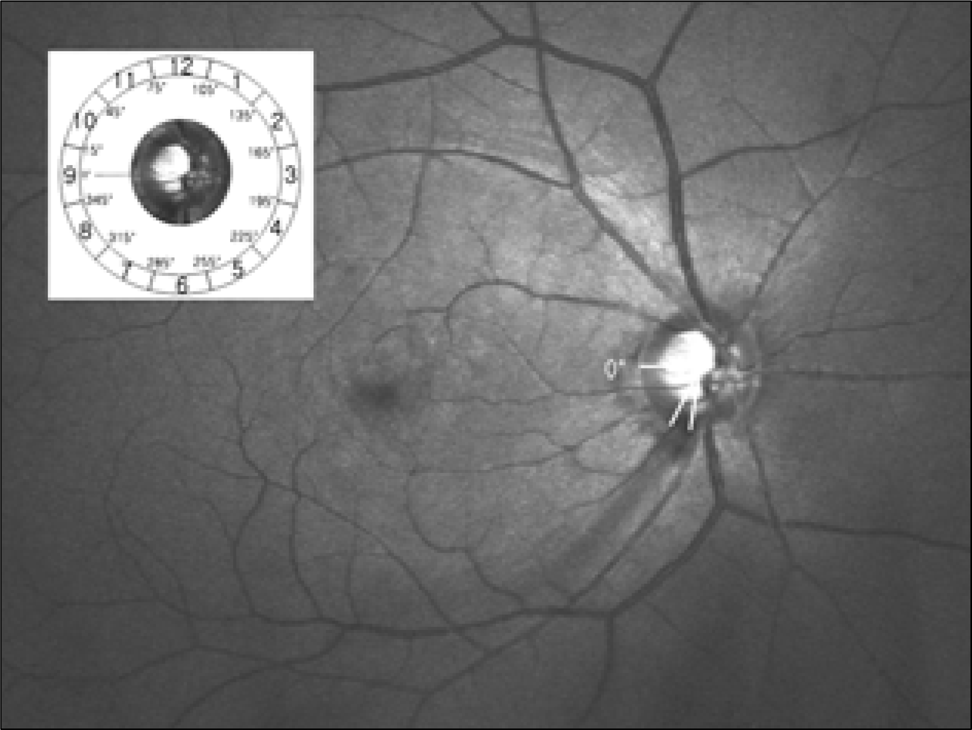 | Figure 1.The angular locations of photographic localized retinal nerve fiber layer (RNFL) defects in the right eye. |
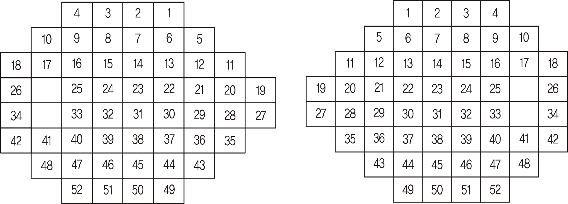
 | Figure 3.Factor analysis generated the visual field points into five visual field zones for each hemifield. The diagram is for the right eye. |
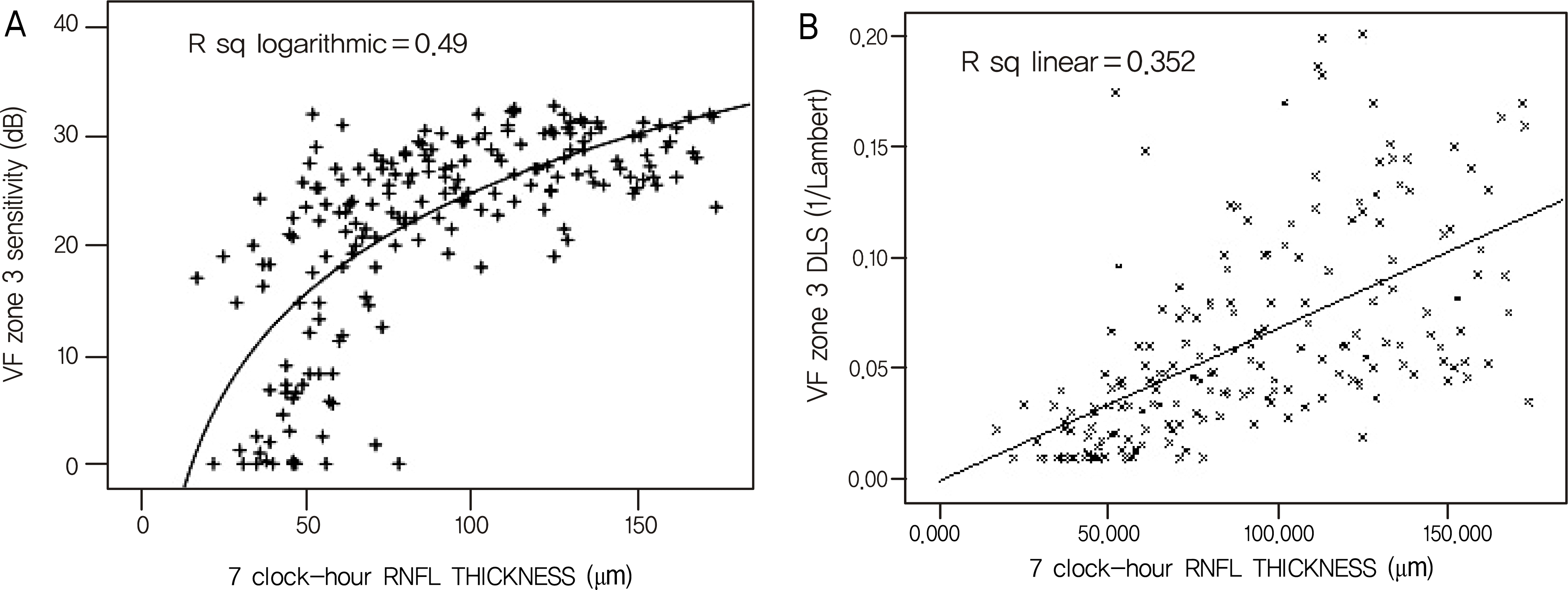 | Figure 4.Scatterplots of 7 clock-hour (inferior temporal) peripapillary retinal nerve fiber layer (RNFL) thickness measured with optical coherence tomography against visual field zone 3 (superior paracentral) sensitivity (or differential light sensitivity, DLS), expressed in the decibel scale (dB) (A) and in the antilog (1/Lambert) scale (B) in all eyes. VF = visual field. |
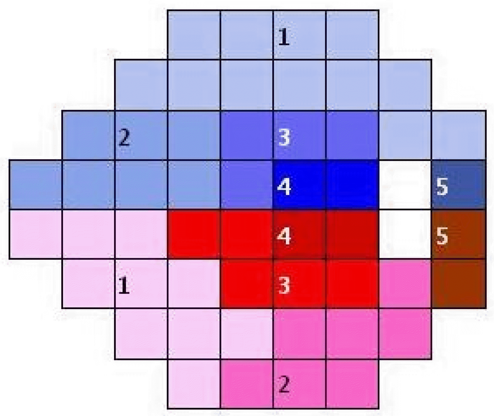
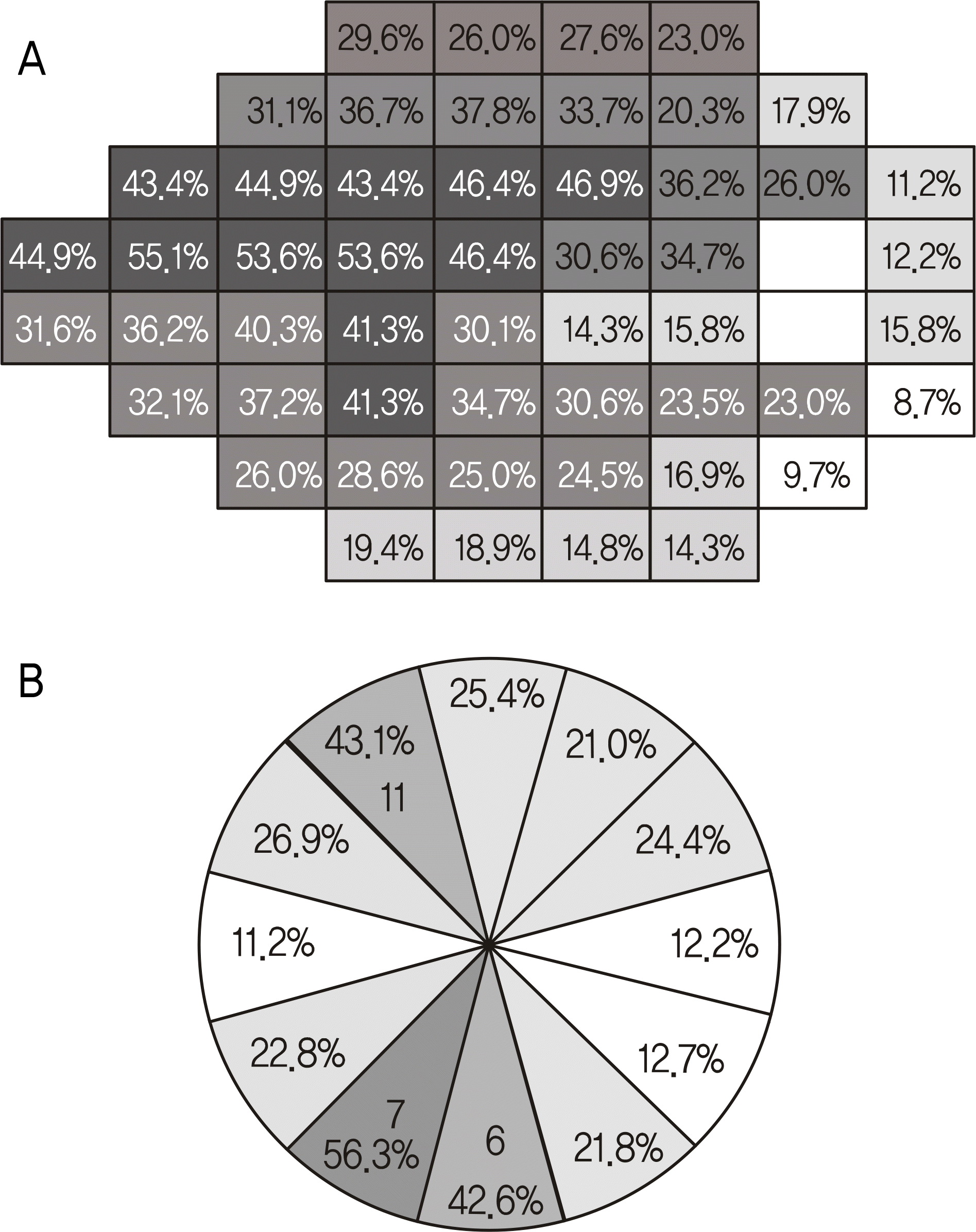 | Figure 5.Diagram represent the percentage of the depressed points of p < 5% in the pattern deviation plot (A) and the percentage of optical coherence tomography retinal nerve fiber layer thickness p < 5% for each clock hour (B). The diagram is for the right eye. |
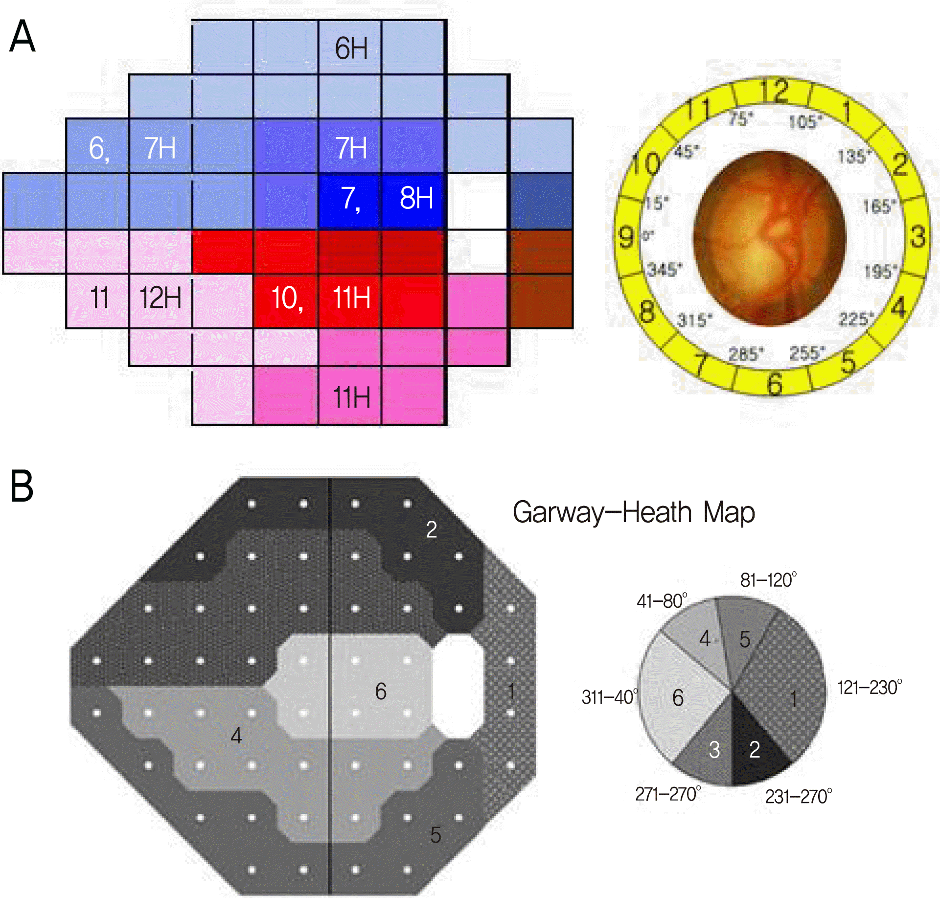 | Figure 6.A new topographic visual field map generated from data in this study. Test points of the visual field were divided by the factor analysis. Visual field zones to corresponding clock-hour sectors measured with optical coherence tomography are shown (A). The corresponding sectors of the optic nerve head for visual field test points according to the results of the Garway-Heath et al study (B). The diagram is for the right eye. |
Table 1.
Demographic and clinical characteristics of study subjects
| Characteristics | |
|---|---|
| Age (mean ± SD, yr) | 56.6 ± 12.4 |
| Gender (M/F) | 113:84 |
| Laterality (right/left) | 80:117 |
| Spherical equivalent (mean ± SD, diopter) | -0.86 ± 2.52 |
| IOP (mean ± SD, mmHg) | 14.03 ± 2.39 |
| Central corneal thickness (mean ± SD, µm) | 536.4 ± 36.4 |
| Type of glaucoma | |
| Preperimetric | 24 |
| Perimetric | 173 |
| SAP mean deviation (mean ± SD, dB) | -6.85 ± 6.04 |
| SAP pattern standard deviation (mean ± SD, dB) | 6.19 ± 4.11 |
| Stage of glaucoma* | |
| No defect | 24 |
| Early defect | 88 |
| Moderate defect | 42 |
| Severe defect | 43 |
| Vertical cup-to-disc ratio (mean ± SD) | 0.79 ± 0.09 |
Table 2.
Rotated component matrix for the superior visual field (VF) points
Table 3.
Rotated component matrix for the inferior visual field (VF) points
Table 4.
Pearson correlation coefficients between the mean threshold for each zones of the visual field (VF) and the RNFL thickness at each of the 12 clock-hour sectors measured with optical coherence tomography (OCT)
| VF zone |
OCT O’clock hour sector |
|||||||||||
|---|---|---|---|---|---|---|---|---|---|---|---|---|
| 9 | 10 | 11 | 12 | 1 | 2 | 3 | 4 | 5 | 6 | 7 | 8 | |
| Superior | ||||||||||||
| 1 | 0.223† | 0.099 | 0.135 | 0.311† | 0.442† | 0.428† | 0.376† | |||||
| 2 | 0.153* | 0.097 | 0.150 | 0.312† | 0.514† | 0.564† | 0.387† | |||||
| 3 | 0.195† | 0.102 | 0.151* | 0.328† | 0.511† | 0.593† | 0.460† | |||||
| 4 | 0.178* | 0.097 | 0.115 | 0.287† | 0.400† | 0.485† | 0.446† | |||||
| 5 | 0.190† | 0.046 | 0.050 | 0.158* | 0.155* | 0.152* | 0.164* | |||||
| Inferior | ||||||||||||
| 1 | 0.242† | 0.354† | 0.465† | 0.318† | 0.155* | 0.083 | 0.048 | |||||
| 2 | 0.298† | 0.358† | 0.403† | 0.313† | 0.216† | 0.082 | 0.039 | |||||
| 3 | 0.267† | 0.397† | 0.434† | 0.275† | 0.168* | 0.087 | 0.067 | |||||
| 4 | 0.196† | 0.208† | 0.230† | 0.186† | 0.111 | 0.073 | 0.046 | |||||
| 5 | 0.097 | 0.122 | 0.176* | 0.220† | 0.173* | 0.076 | 0.052 | |||||
 XML Download