

 PDF
PDF ePub
ePub Citation
 Print
Print


Abstract
Purpose
To evaluate the repeatability and comparability of anterior chamber depth (ACD) and central corneal thickness (CCT) measurements obtained by Galilei dual Scheimpflug analyzer (Ziemer, Port, Switzerland) and slit-lamp optical coherence tomography (SL-OCT; Heidelberg Engineering, Dossenheim, Germany).
Methods
ACD and CCT were measured by Galilei and SL-OCT in 68 eyes of 68 healthy young subjects. Each measurement was performed 3 times by a single examiner, and the repeatability of 3 consecutive measurements was analyzed. ACD and CCT measurements were compared between the 2 devices.
Results
Both Galilei and SL-OCT showed high repeatability (ICCs ≥ 0.994) for ACD and CCT measurements. The mean ACD and CCT measured by Galilei were greater than SL-OCT measurements by 0.11 ± 0.09 mm and 14.01 ± 7.38 μ m, respectively. The 95% limit of agreement values for ACD and CCT measurements were 0.36 mm, 27.66 μ m, respectively, and were highly correlated (correlation coefficients ≥ 0.89, p < 0.001).
Go to : 
References
1. Kim YY, Jung HR. Clarifying the nomenclature for primary angle closure glaucoma. Surv Ophthalmol. 1997; 42:125–36.
2. Holladay JT. Standardizing constants for ultrasonic biometry, keratometry, and intraocular lens power calculations. J Cataract Refract Surg. 1997; 23:1356–70.

3. Doughty MJ, Zaman ML. Human corneal thickness and its impact on intraocular pressure measures: a review and meta-analysis approach. Surv Ophthalmol. 2000; 44:367–408.
4. Reddy AR, Pande MV, Finn P, El-Gogary H. Comparative estimation of anterior chamber depth by ultrasonography, Orbscan II, and IOL master. J Cataract Refract Surg. 2004; 30:1268–71.
5. Vetrugno M, Cardascia N, Cardia L. Anterior chamber depth measured by two methods in myopic and hyperopic phakic IOL implant. Br J Ophthalmol. 2000; 84:1113–6.
6. Ryu HW, Kim KR, Chung SK. Comparison of A-scan, scheimpflug camera, and orbscan for measurement of anterior chamber depth. J Korean Ophthalmol Soc. 2006; 47:1287–91.
7. Giers U, Epple C. Comparison of A-scan device accuracy. J Cataract Refract Surg. 1990; 16:235–42.
8. Lee YE, Jun RM. Parameters obtained by Galilei in normal subjects. J Korean Ophthalmol Soc. 2009; 50:1611–6.
9. Han KE, Jun RM. Measurement of white-to-white diameter and anterior chamber depth by dual Scheimpflug camera. J Korean Ophthalmol Soc. 2010; 51:169–74.
10. Bland JM, Altman DG. Measurement error. BMJ. 1996; 313:744.
11. Bland JM, Altman DG. Statistical methods for assessing agreement between two methods of clinical measurement. Lancet. 1986; 1:307–10.
12. Olsen T. Sources of error in intraocular lens power calculation. J Cataract Refract Surg. 1992; 18:125–9.
13. Wang L, Shirayama M, Koch DD. Repeatability of corneal power and wavefront aberration measurements with a dual-Scheimpflug placido corneal topographer. J Cataract Refract Surg. 2010; 36:425–30.
14. Menassa N, Kaufmann C, Goggin M, et al. Comparison and reproducibility of corneal thickness and curvature readings obtained by the Galilei and the Orbscan II analysis systems. J Cataract Refract Surg. 2008; 34:1742–7.
15. Sandler SF, Zelefsky JR, Dorairaj S, et al. Intraobserver and interobserver reliability and reproducibility of slit-lamp adapted optical coherence tomography for evaluation of anterior chamber depth and central corneal thickness. Ophthalmic Surg Lasers Imaging. 2008; 39:299–303.
16. Salouti R, Nowroozzadeh MH, Zamani M, et al. Comparison of anterior chamber depth measurements using Galilei, HR Pentacam, and Orbscan II. Optometry. 2010; 81:35–9.
17. Park SH, Choi SK, Lee DH, Kim JH. Central corneal thickness measured by four different methods in normal and post-femto-second laser-assisted LASIK eyes. J Korean Ophthalmol Soc. 2010; 51:320–7.
18. Li H, Leung CK, Wong L, et al. Comparative study of central corneal thickness measurement with slit-lamp optical coherence tomography and Visante optical coherence tomography. Ophthalmology. 2008; 115:796–801.
19. Wirbelauer C, Thannhauser CL, Pham DT. Influence of corneal curvature on central and paracentral pachymetry with optical coherence tomography. Cornea. 2009; 28:254–60.
20. Doors M, Cruysberg L, Berendschot T, et al. Comparison of central corneal thickness and anterior chamber depth measurements using three imaging technologies in normal eyes and after phakic intraocular lens implantation. Graefes Arch Clin Exp Ophthalmol. 2009; 247:1139–46.
21. Yi JH, Lee H, Hong S, et al. Anterior chamber measurements by Pentacam and AS-OCT in eyes with normal open angles. Korean J Ophthalmol. 2008; 22:242–5.
Go to :
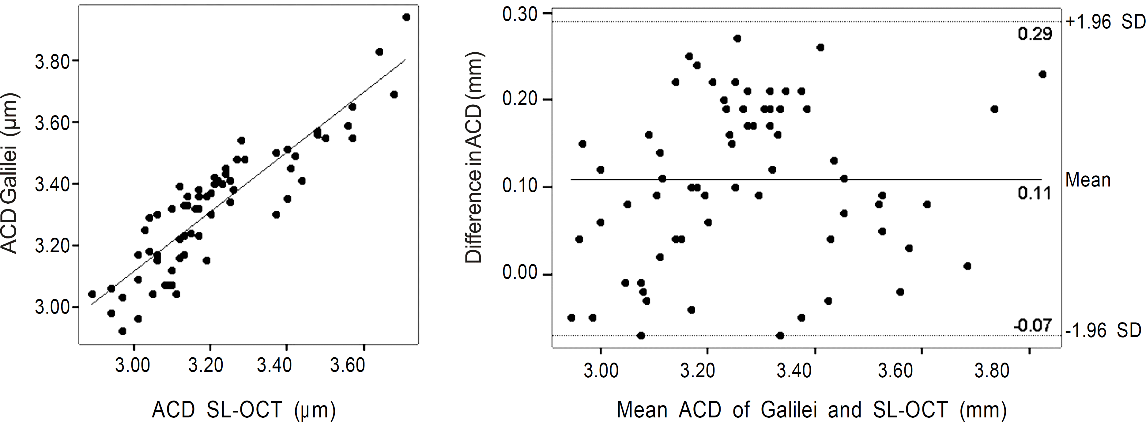 | Figure 1.Scatter plot and Bland-Altman plot of anterior chamber depth (ACD) measurement by Galilei and slit-lamp optical coherence tomography (SL-OCT). |
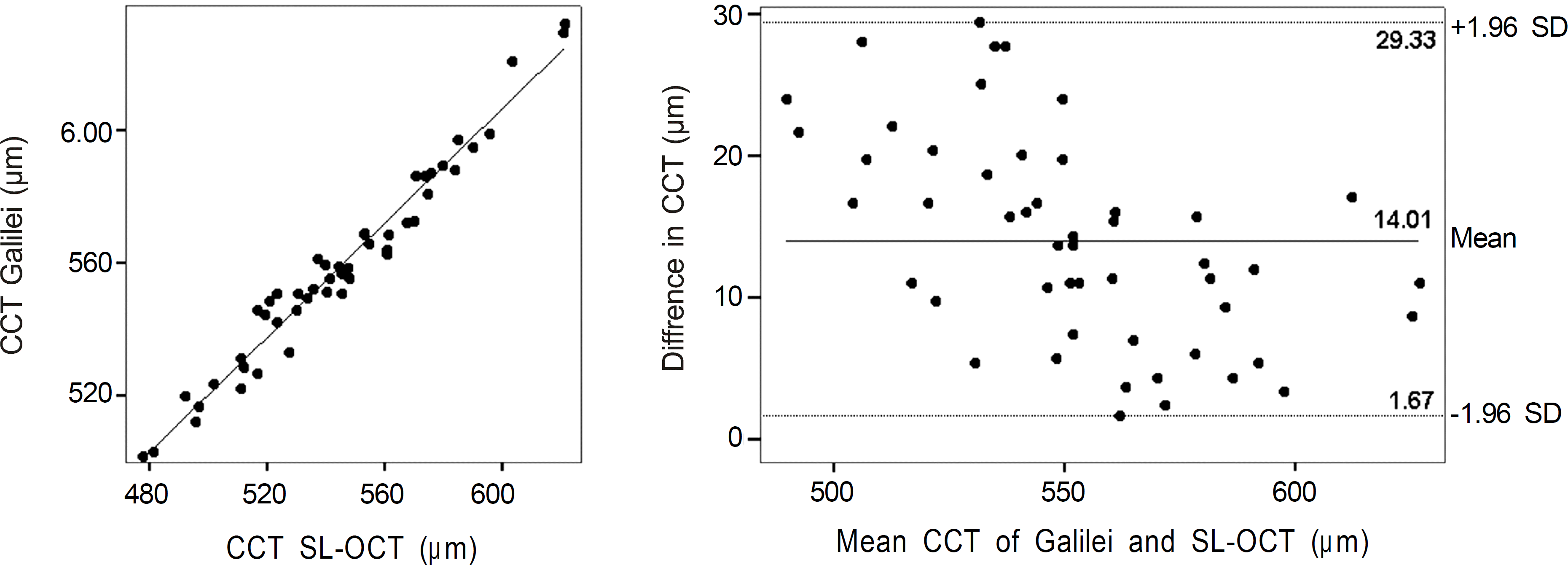 | Figure 2.Scatter plot and Bland-Altman plot of central corneal thickness (CCT) measurement by Galilei and slit-lamp optical coherence tomography (SL-OCT). |
Table 1.
Comparison of anterior chamber depth (ACD) and central corneal thickness (CCT) measured by Galilei and slit-lamp optical coherence tomography (SL-OCT) among the three groups
Table 2.
Anterior chamber depth (ACD) and central corneal thickness (CCT) measurement repeatability of Galilei and slit-lamp optical coherence tomography (SL-OCT)
Table 3.
Pair-wise comparison of anterior chamber depth (ACD) and central corneal thickness (CCT) measurements obtained by Galilei and slit-lamp optical coherence tomography (SL-OCT)
| Pairwise comparison | Mean difference ± S | * P-value P | Pearson correlatio coefficient | † P-value | 95% limit of agreement |
|---|---|---|---|---|---|
| ACD (mm) | | | | | |
| Galilei SL OCT | 0.11 ± 0.09 | < 0.001 | 0.89 | < 0.001 | 0.07 to 0.29 |
| CCT (μ m) | | | | | |
| Galilei SL OCT | 14.01 ± 7.38 | < 0.001 | 0.98 | < 0.001 | 1.67 to 29.33 |
 XML Download