Abstract
Purpose
To report a case of ab interno trabeculotomy with Trabectome® (NeoMedix Corp., CA, USA) conducted on a refractory primary open angle glaucoma (POAG) patient.
Case summary
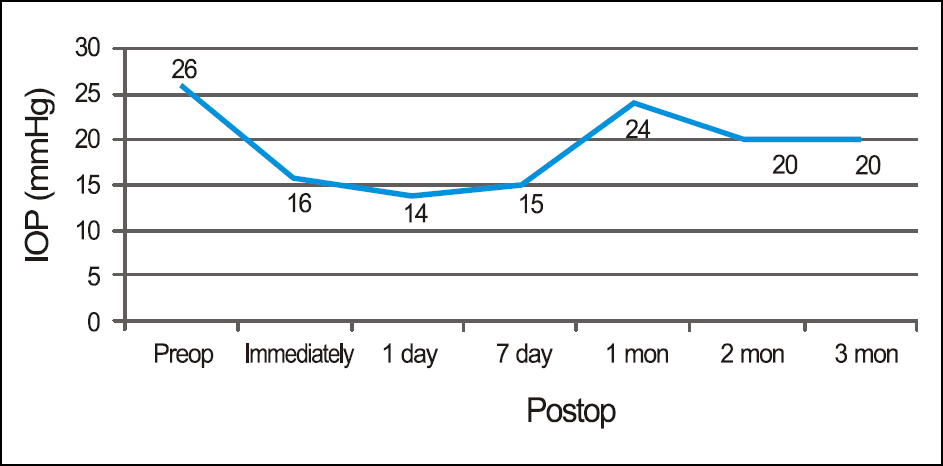
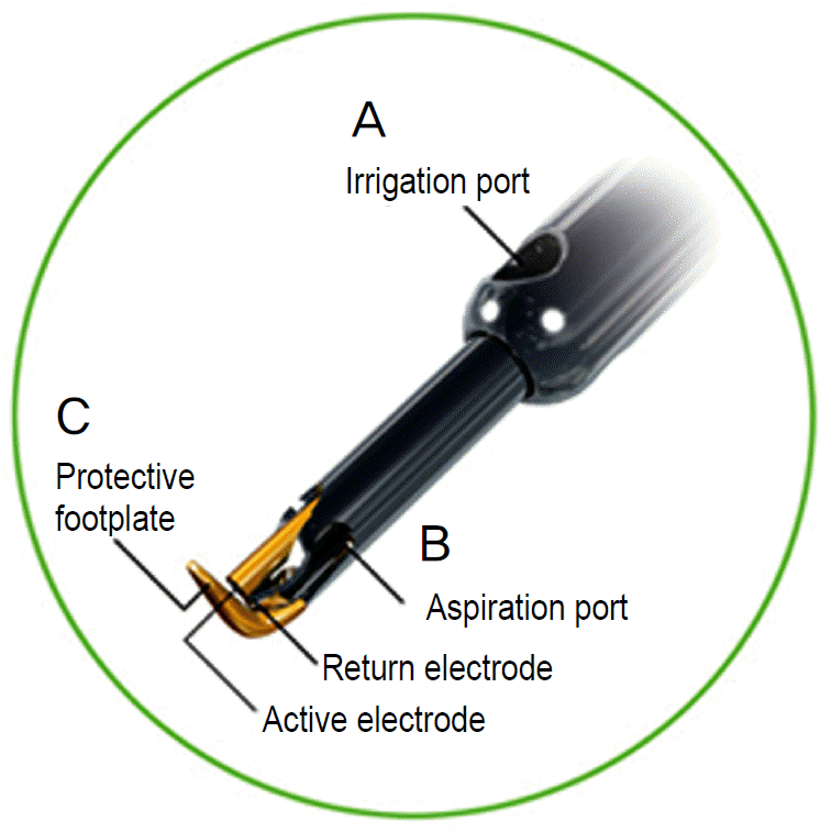
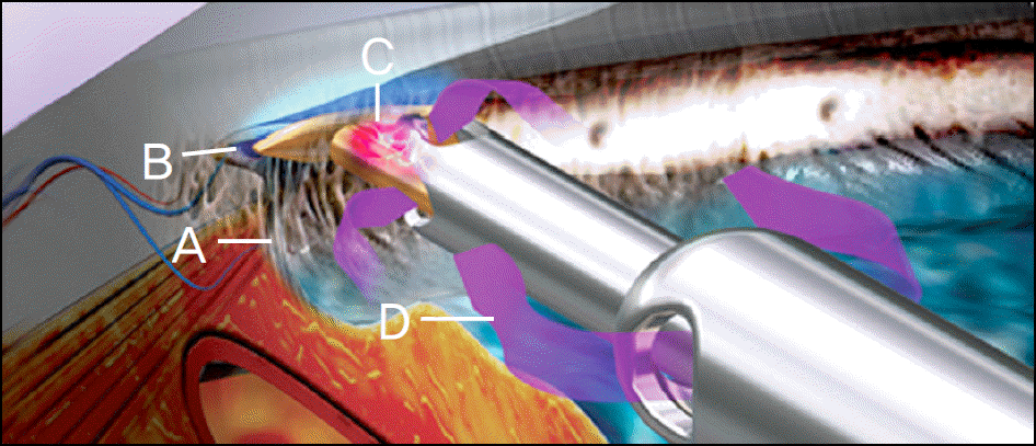
Trabectome® has microelectrocautery with simultaneous infusion and aspiration of debris and ablates a segment of trabecular meshwork and the inner wall of Schlemm’s canal. The patient, a 54-year-old man had uncontrolled intraocular pressure (IOP) with topical anti-glaucoma medications after trabeculectomy and Ahmed valve implantation for POAG. For the patient, ab interno trabeculotomy with Trabectome® was performed. There were no other postoperative complications except for microhyphema immediately after surgery. The IOP was controlled between 14 to 24 mm Hg up to 3 months postoperatively with topical anti-glaucoma medications (Cosopt®, Alphagan-P®, Lumigan®).
References
1. deLuise VP, Anderson DR. Primary infantile glaucoma (congenital glaucoma). Surv Ophthalmol. 1983; 28:1–19.

2. Luntz MH, Livingston DG. Trabeculotome ab externo and trabeculectomy in congenital and adult-onset glaucoma. Am J Ophthalmol. 1977; 83:174–9.
3. Kobayashi H, Kobayashi K, Okinami S. A comparison of the intraocular pressure-lowering effect and safety of viscocanalostomy and trabeculotomy with mitomycin C in bilateral open-angle glaucoma. Arch Clin Exp Ophthalmol. 2003; 241:359–66.
4. Nguyen QH. Trabectome: A novel approach to angle surgery in the treatment of glaucoma. Int Ophthalmol Clin. 2008; 48:65–72.

5. Minckler DS, Hill RA. Use of novel devices for control of intraocular pressure. Exp Eye Res. 2009; 88:792–8.

6. Minckler DS, Mosaed S, Dustin L, et al. Trabectome (trabeculectomy- internal approach): additional experience and extended follow-up. Trans Am Ophthalmol Soc. 2008; 106:149–60.
7. Mosaed S. Ab interno trabeculotomy with the trabectome surgical device. Techn Opthalmol. 2007; 5:63–6.

8. Godfrey DG, Fellman RL, Neelakantan A. Canal surgery in adult glaucoma. Curr Opin Ophthalmol. 2009; 20:116–21.
9. Francis BA, Minckler D, Dustin L, et al. Combined cataract extraction and trabeculotomy by internal approach for coexisting cataract and open-angle glaucoma. J Cataract Refract Surg. 2008; 34:1096–103.
10. Fontana H, Nouri-Mahdavi K, Caprioli J. Trabeculectomy with mitomycin C in pseudophakic patients with open angle glaucoma: outcomes and risk factors for failure. Am J Ophthalmol. 2006; 141:652–9.
11. Filippopoulos T, Rhee DJ. Novel surgical procedures in glaucoma: advances in penetrating glaucoma surgery. Curr Opin Ophthalmol. 2008; 19:149–54.

12. Vold SD, Dustin L. Impact of laser trabeculoplasty on Trabectome® outcomes. Ophthalmic Surg Lasers Imaging. 2010; 41:443–51.
Figure 1.
Trabectome surgical steps. (A) Create a 1.6-mm clear corneal incision. (B) Inject a small amount of viscoelastic at the incision site. (C) Place Goniolens on the cornea and verify the angle view. (D) Ablate the trabecular meshwork for approximately in counter-clockwise direction. (E) Rotate the tip. (F) Re-insert the tip in the clockwise direction and remove a similar arc. (G) Irrigate and aspirate viscoelastics from the anterior segment. (H) Place one suture across the incision. Arrow: direction of Trabectome® tip.

Figure 2.
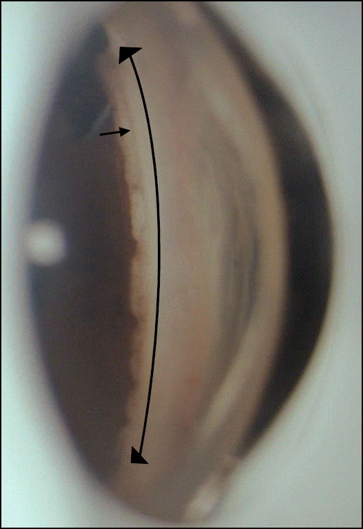
Gonioscopic findings of the patient at 3 months after surgery. Posterior wall of Schlemm’s canal is visible in the area where the trabecular meshwork is removed by Trabectome®. Long arrow, ablation range of the trabecular meshwork; small arrow, white shimmering of the posterior wall of Schlemm’s canal.


 PDF
PDF ePub
ePub Citation
 Print
Print


 XML Download