

 PDF
PDF ePub
ePub Citation
 Print
Print


Abstract
Purpose
To report a case of acute zonal occult outer retinopathy (AZOOR), initially misdiagnosed as retrobulbar optic neuritis, which was responsive to an immunosuppressive agent.
Case summary
A 28-year-old female with photopsia and a visual field defect in the left eye was referred to a hospital. There were no fundus abnormalities to explain her left visual field defect. Neurologic examination and brain MRI were normal. The patient was diagnosed with retrobulbar optic neuritis and treated with high-dose steroids. Four months after the onset of symptoms, she visited our hospital. Visual acuity was hand motion in the left eye. No relative afferent pupillary defect in the left eye and no anterior segment or fundus abnormalities were observed. A visual field examination of the right eye was normal and revealed field defect in the left eye. No abnormality was noted in the visual evoked potential test or fluorescein angiography. All examinations of the right eye were normal. In the left eye, fundus autofluorescence showed a hyperautofluorescent spot at the posterior pole, there was a decreased response in electroretinography and spectral domain optic coherence tomography showed that the junction between the photoreceptor inner and outer segments (IS/OS) was faintly visible only in the fovea. With the presumptive diagnosis of AZOOR, the patient was treated with an immunosuppressive agent. Visual acuity improved to 20/80 in the left eye at 10 months after the onset of symptoms.
Go to : 
References
1. Gass JDM. Acute zonal occult outer retinopathy. Donders Lecture: The Netherlands Ophthalmological Society, Maastricht, Holland, June 19, 1992. J Clin Neuroophthalmol. 1993; 13:79–97.
2. Gass JDM, Agarwal A, Scott IU. Acute zonal occult outer retinopathy: A long-term follow-up study. Am J Ophthalmol. 2002; 134:329–39.

3. Ogawa M, Mori H, Nemoto N, et al. Case of acute zonal occult outer retinopathy resembling retrobulbar optic neuropathy. Neuroophthalmology. 2001; 26:217–22.
4. Jacobson SG, Morales DS, Sun XK, et al. Pattern of retinal dys-function in acute zonal occult outer retinopathy. Ophthalmology. 1995; 102:1187–98.
5. Choi HJ, Hwang JM. A case report of acute zonal occult outer retinopathy. J Korean Ophthalmol Soc. 2003; 44:1384–91.
6. Mahajan VB, Stone EM. Patients with an acute zonal occult outer retinopathy-like illness rapidly improve with valacyclovir treatment. Am J Ophthalmol. 2010; 150:511–8.
7. Gass JDM. Are acute zonal occult outer retinopathy and the white spot syndromes (AZOOR Complex) specific autoimmune dis-eases? Am J Ophthalmol. 2003; 135:380–1.
8. Jampol LM, Wiredu A. MEWDS, MFC, PIC, AMN, AIBSE and AZOOR: one disease or many? Retina. 1995; 15:373–8.
9. Francis PJ, Marinescu A, Fitzke FW, et al. Acute zonal occult outer retinopathy: towards a set of diagnostic criteria. Br J Ophthalmol. 2005; 89:70–3.
10. Arai M, Nao-i N, Sawada A, Hayashida T. Multifocal electro-retinogram indicates visual field loss in acute zonal occult outer retinopathy. Am J Ophthalmol. 1998; 126:466–9.
11. Spaide RF. Collateral damage in acute zonal occult outer retinopathy. Am J Ophthalmol. 2004; 138:887–9.
12. Fine HF, Spaide RF, Ryan EH Jr, et al. Acute zonal occult outer retinopathy in patients with multiple evanescent white dot syndrome. Arch Ophthalmol. 2009; 127:66–70.
13. Fujiwara T, Imamura Y, Giovinazzo VH, Spaide RF. Fundus autofluorescence and optical coherence tomographic findings in acute zonal occult outer retinopathy. Retina. 2010; 30:1206–16.
14. Takai Y, Ishiko S, Kagokawa H, et al. Morphologic study of acute zonal occult outer retinopathy (AZOOR) by multiplanar optical coherence tomography. Acta Ophthalmol. 2009; 87:408–18.
15. Li D, Kishi S. Loss of photoreceptor outer segment in acute zonal occult outer retinopathy. Arch Ophthalmol. 2007; 125:1194–200.
16. Ohta K, Sato A, Fukui E. Spectral domain optical coherence tomographic findings at convalescent stage of acute zonal occult outer retinopathy. Clin Ophthalmol. 2009; 3:423–8.
17. Gass JDM. The acute zonal outer retinopathies. Am J Ophthalmol. 2000; 130:655–7.
18. Song ZM, Sheng YJ, Chen QS, et al. Clinical characteristics of Acute zonal occult outer retinopathy in Chinese patients. Ophthalmologica. 2008; 222:149–56.
19. Spaide RF, Koizumi H, Freund KB. Photoreceptor outer segment abnormalities as a cause of blind spot enlargement in acute zonal occult outer retinopathy-complex disease. Am J Ophthalmol. 2008; 146:111–20.
Go to :
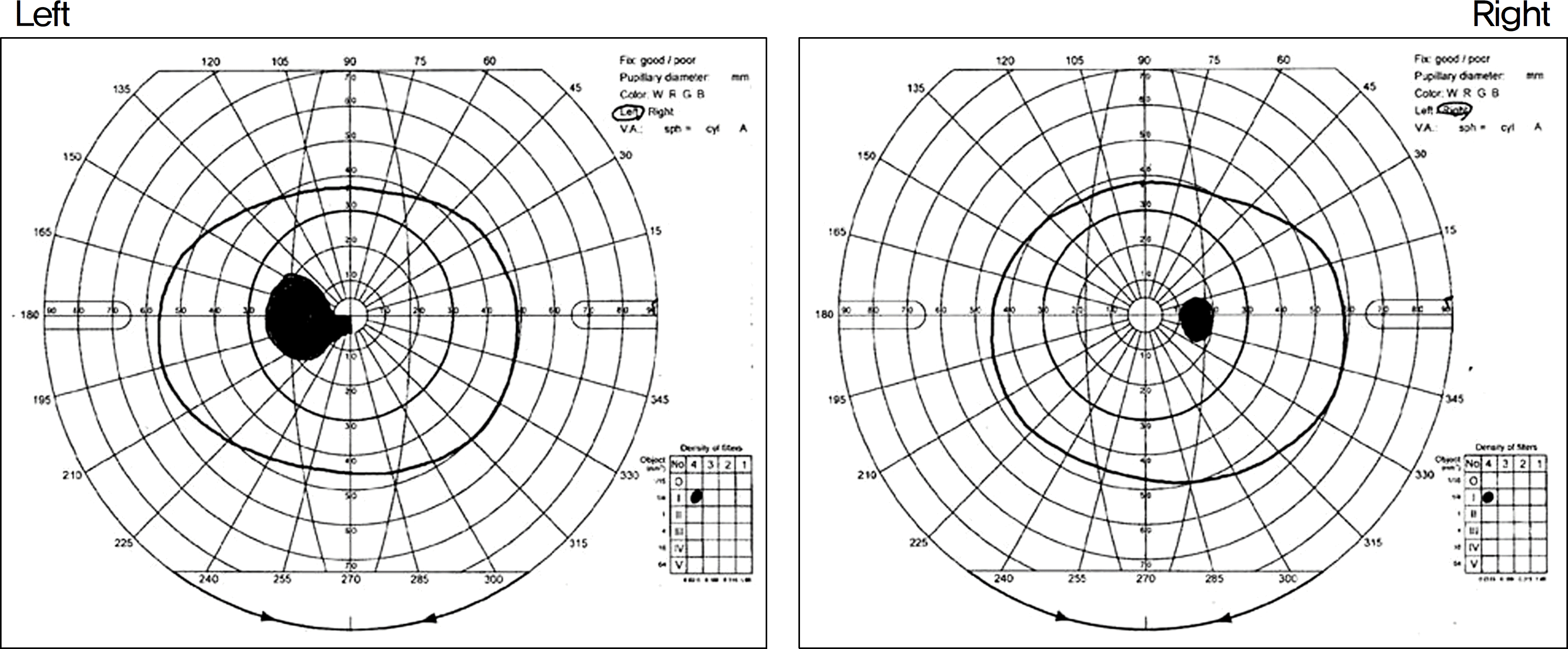 | Figure 1.Goldmann perimetry reveals enlarged blind spot and cecocentral scotoma in the left eye, and normal in the right eye. |
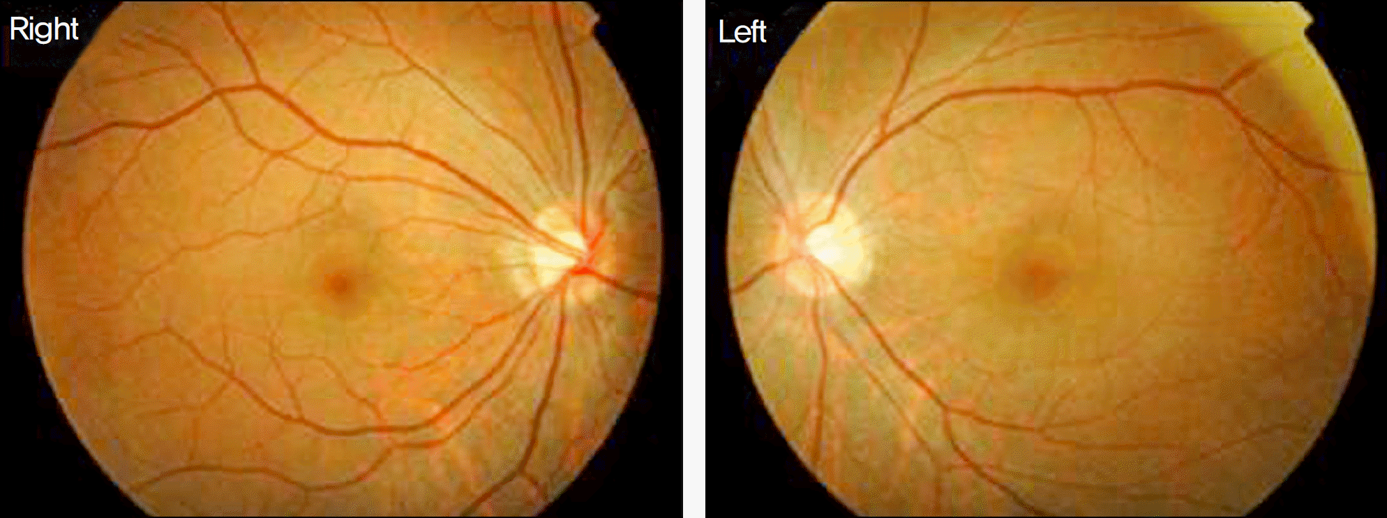 | Figure 2.Fundus photographs of both eyes show normal appearance of the optic disc and retina. |
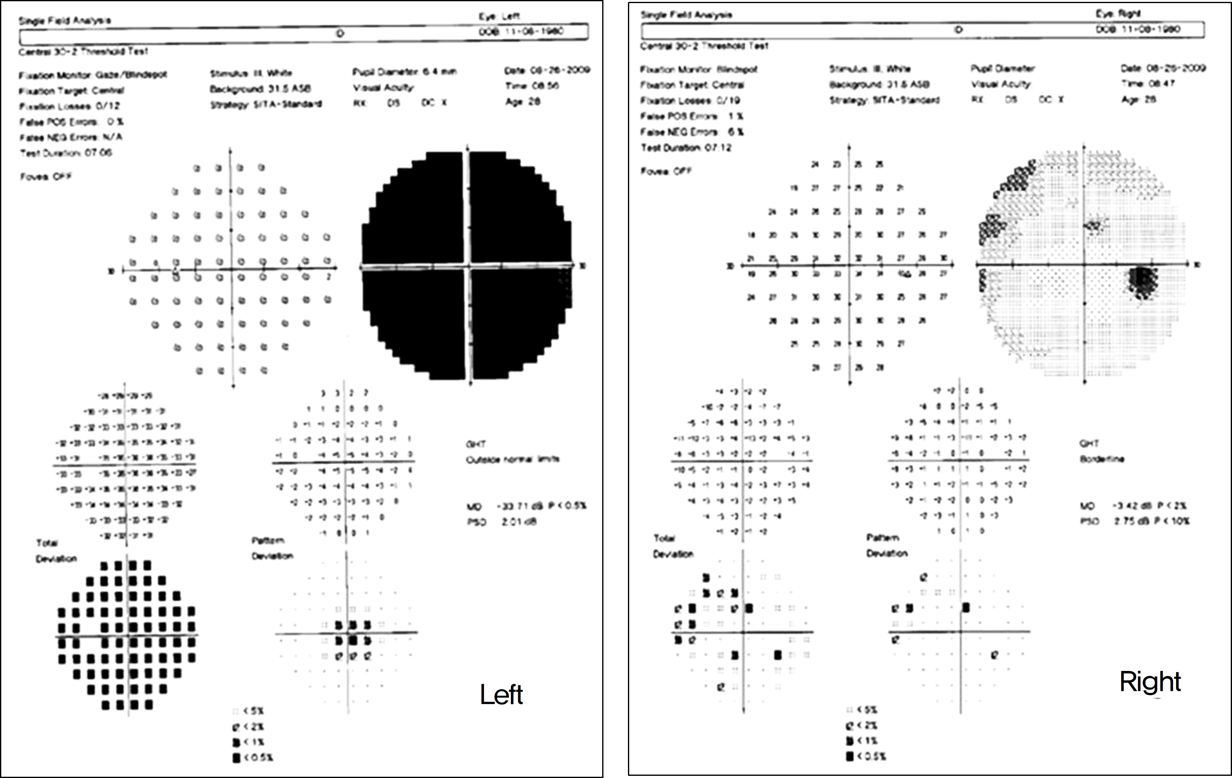 | Figure 3.Humphrey static perimetry reveals marked visual field defect in the left eye, and normal in the right eye. |
 | Figure 4.(A) Fundus autofluorescence show ill-defined hyperautofluorescent area at the posterior pole in the left eye (arrows) and normal in the right eye at the initial visit. (B) Fluorescein angiographs showed normal in both eyes. |
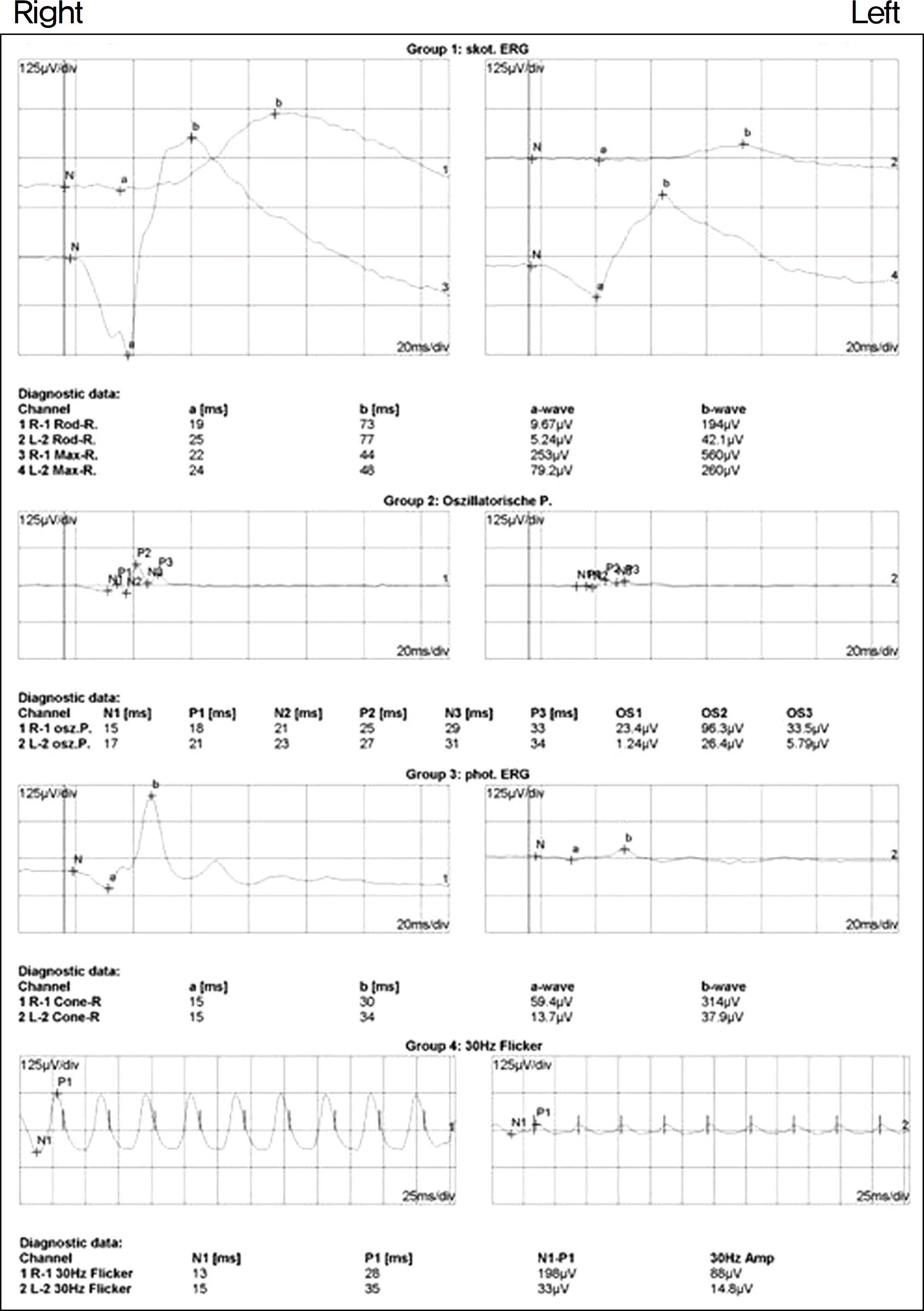 | Figure 5.Electroretinographic findings show normal in the right eye, abnormal in the left eye; subnormal rod specific ERG, both a-wave and b-wave amplitude reduction in maximal response, subnormal photopic single flash b-wave amplitude, and markedly delayed and subnormal 30-Hz flicker ERG. |
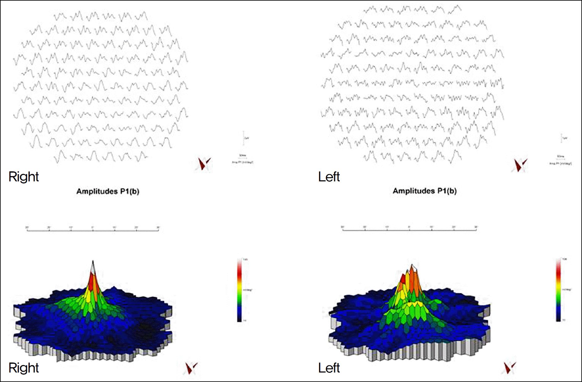 | Figure 6.Trace array and topographic maps of multifocal ERG show markedly reduced responses in the left eye and normal in the right eye. |
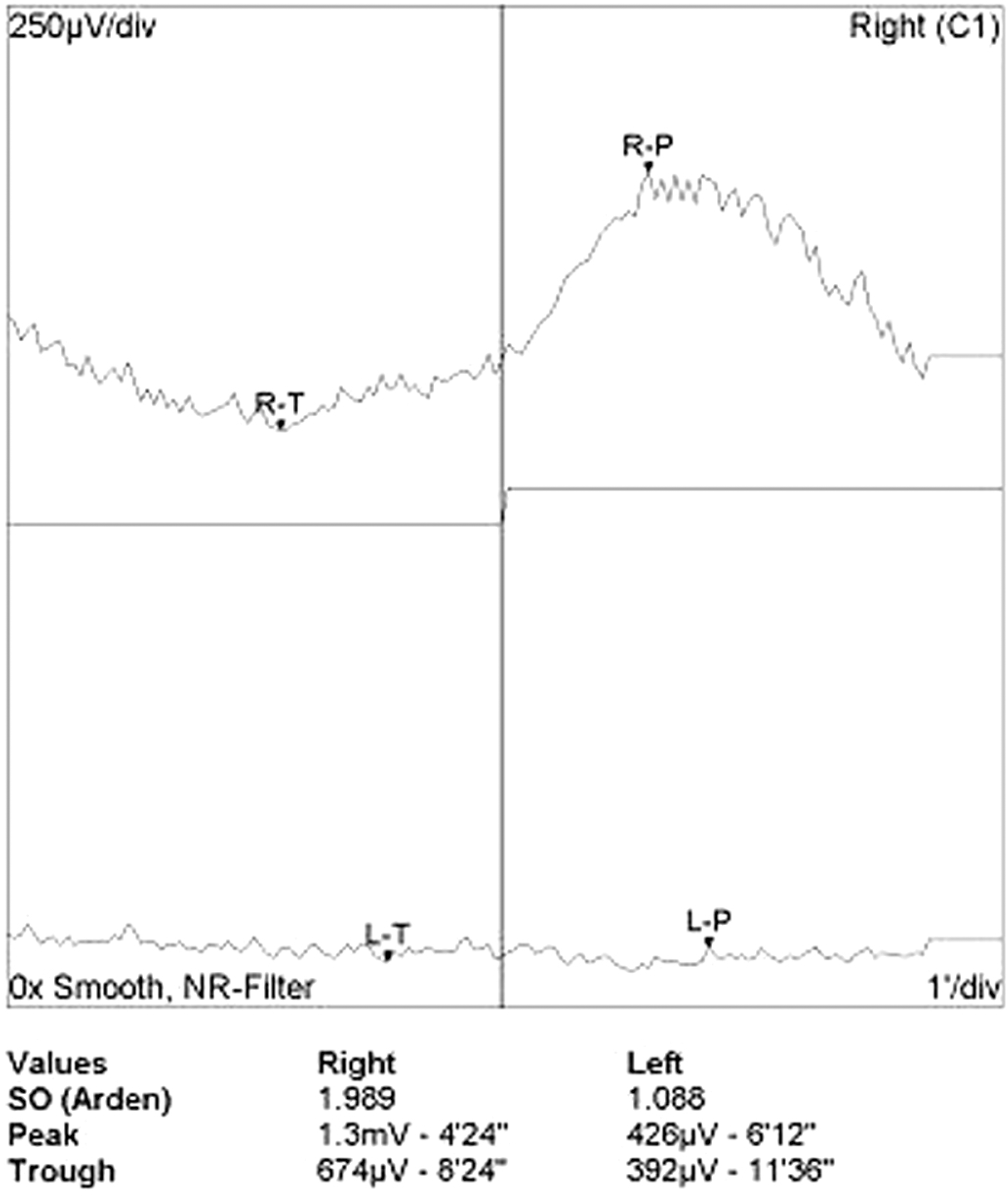 | Figure 7.Electro-oculogram of both eyes. The EOG light rise of the left eye is markedly reduced compared with that of the right eye. Accordingly, arden ratio of the left eye is markedly reduced. |
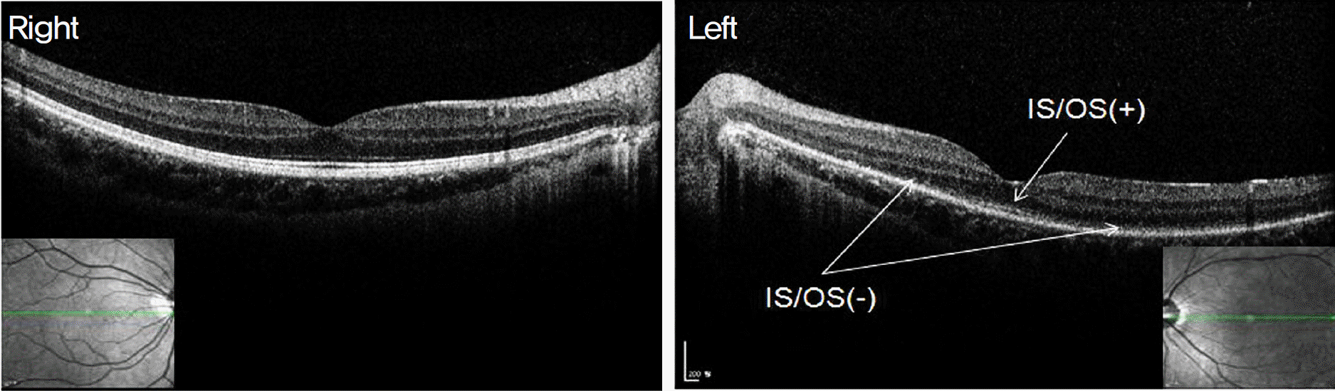 | Figure 8.Spectral domain optic coherence tomographic images at the first visit show that the junction between the photoreceptor inner and outer segments (IS/OS) is faintly visible only in the fovea and not visible outside the fovea in the left eye, and normal in the right eye. |
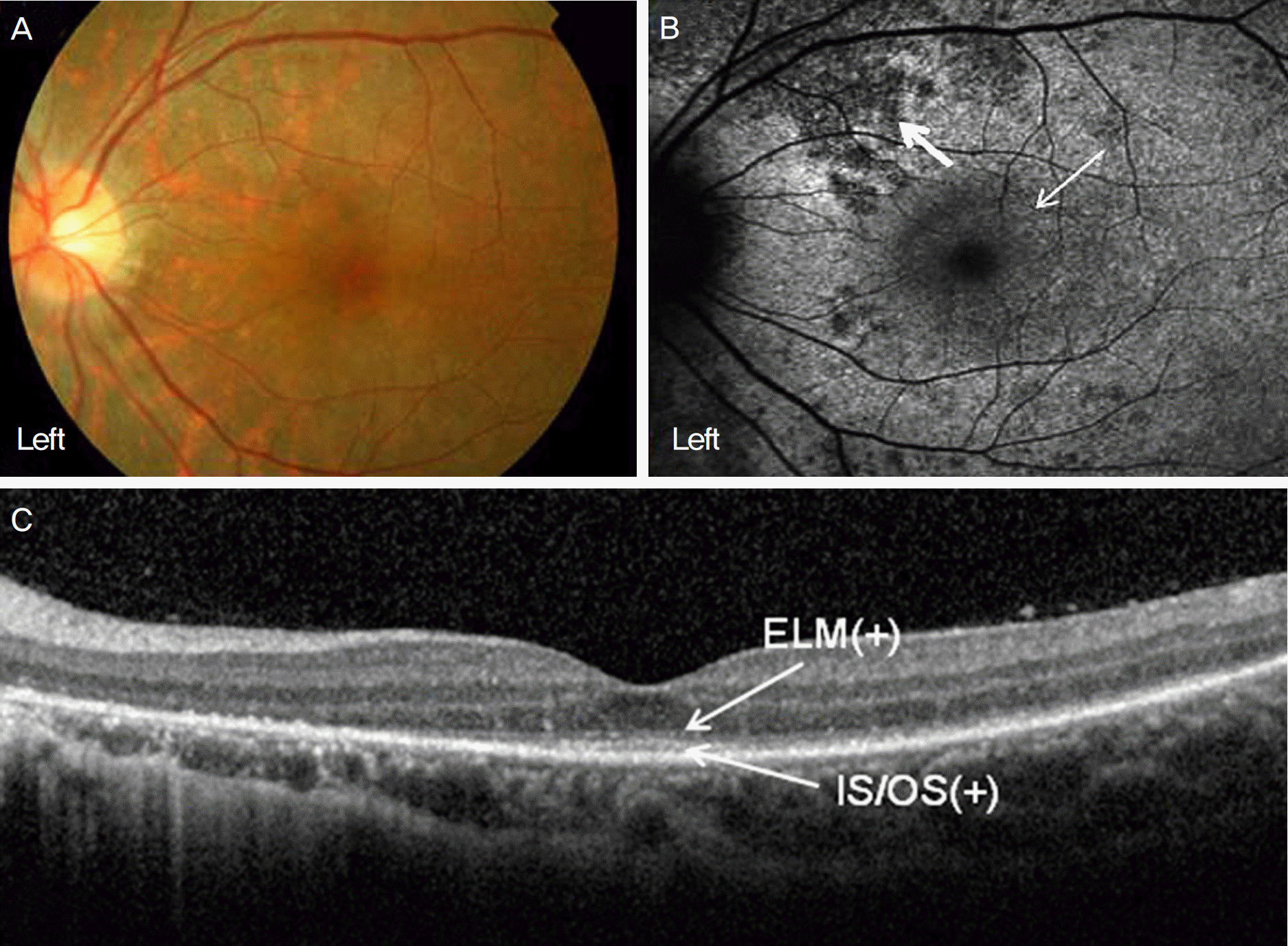 | Figure 9.Findings at 10 months after the initial visit. (A) Fundus photography shows normal in the left eye. (B) Fundus autofluorescence shows that the size of ill-defined hyperautofluorescent area (thin arrow) is decreased compared with that of the initial visit, but the number of multiple hypoautofluorescent dots is increased compared with that of the initial visit (thick ar-row). (C) The junction between the photoreceptor inner and outer segments (IS/OS) and ELM were clearly visible only in the fovea of the left eye. |
 XML Download