

 PDF
PDF ePub
ePub Citation
 Print
Print


Abstract
Purpose
To identify the risk factors associated with false negative findings of optical coherence tomography (Stratus OCT) in patients with photographic localized retinal nerve fiber layer (RNFL) defects.
Methods
Twenty-four patients with preperimetric glaucoma and 173 patients with perimetric glaucoma, all with localized RNFL defects were included in the present study. The patients were divided into 2 groups according to the presence or absence of detection of photographic defects by OCT. Gender, age, refractive error, diabetes, hypertension, central corneal thickness, type of glaucoma, mean deviation, pattern standard deviation, average RNFL thickness, disc area, and photographic RNFL defect related variables (location, number, and angular width) were compared between the 2 groups. Each variable was initially evaluated by univariate analysis and significant variables (p < 0.1) were included in the logistic regression analysis.
Results
Photographic RNFL defects were not detected by OCT in 51 (25.9%) of the 197 eyes. The angular locations and widths of RNFL defects by OCT were significantly correlated with those of RNFL defects by red-free RNFL photographs (Pearson correlation coefficient R = 0.98 and 0.64, respectively). Logistic regression analysis revealed the risk factors for false negative findings of OCT included average RNFL thickness (odds ratio = 1.106, 95% confidence interval [CI] =1.057-1.156, p < 0.001) and angular width of defect (odds ratio = 0.929, 95% CI = 0.884-0.977, p = 0.004).
References
1. Quigley HA, Addicks EM, Green WR. Optic nerve damage in human glaucoma. III. Quantitative correlation of nerve fiber loss and visual field defect in glaucoma, ischemic neuropathy, papilledema, and toxic neuropathy. Arch Ophthalmol. 1982; 100:135–46.
2. Sommer A, Katz J, Quigley HA, et al. Clinically detectable nerve fiber atrophy precedes the onset of glaucomatous field loss. Arch Ophthalmol. 1991; 109:77–83.

3. Airaksinen PJ, Alanko HI. Effect of retinal never fiber loss on the optic nerve head configuration in early glaucoma. Graefes Arch Clin Exp Ophthalmol. 1983; 220:193–6.
4. Tuulonen A, Lehtola J, Airaksinen PJ. Nerve fiber layer defects with normal visual fields. Do normal optic disc and normal visual field indicate absence of glaucomatous abnormality? Ophthalmology. 1993; 100:587–97.
5. Tuulonen A, Airaksinen PJ. Initial glaucomatous optic disk and retinal nerve fiber layer abnormalities and their progression. Am J Ophthalmol. 1991; 111:485–90.
6. Quigley HA, Reacher M, Katz J, et al. Quantitative grading of nerve fiber layer photographs. Ophthalmology. 1993; 100:1800–7.
7. Chen HY, Huang ML. Discrimination between normal and glaucomatous eyes using Stratus optical coherence tomography in Taiwan Chinese subjects. Graefes Arch Clin Exp Ophthalmol. 2005; 243:894–902.
8. Medeiros FA, Zangwill LM, Bowd C, et al. Evaluation of retinal nerve fiber layer, optic nerve head, and macular thickness measurements for glaucoma detection using optical coherence tomography. Am J Ophthalmol. 2005; 139:44–55.
9. Wollstein G, Ishikawa H, Wang J, et al. Comparison of three optical coherence tomography scanning areas for detection of glaucomatous damage. Am J Ophthalmol. 2005; 139:39–43.
10. Manassakorn A, Nouri-Mahdavi K, Caprioli J. Comparison of retinal nerve fiber layer thickness and optic disk algorithms with optical coherence tomography to detect glaucoma. Am J Ophthalmol. 2006; 141:105–15.
11. Song YM, Uhm KB. Discrimination between normal and early stage of glaucomatous eyes using the stratus optical coherence tomography. J Korean Ophthalmol Soc. 2007; 48:1675–85.
12. Paunescu LA, Schuman JS, Price LL, et al. Reproducibility of nerve fiber thickness, macular thickness, and optic nerve head measurements using StratusOCT. Invest Ophthalmol Vis Sci. 2004; 45:1716–24.
13. Budenz DL, Michael A, Chang RT, et al. Sensitivity and specificity of the StratusOCT for perimetric glaucoma. Ophthalmology. 2005; 112:3–9.
14. Kim TW, Park UC, Park KH, Kim DM. Ability of Stratus OCT to identify localized retinal nerve fiber layer defects in patients with normal standard automated perimetry results. Invest Ophthalmol Vis Sci. 2007; 48:1635–41.
15. Jonas JB, Schiro D. Localised wedge shaped defects of the retinal nerve fibre layer in glaucoma. Br J Ophthalmol. 1994; 78:285–90.
16. Airaksinen PJ, Mustonen E, Alanko HI. Optic disc haemorrhages precede retinal nerve fibre layer defects in ocular hypertension. Acta Ophthalmol (Copenh). 1981; 59:627–41.
17. Jeoung JW, Park KH, Kim TW, et al. Diagnostic ability of optical coherence tomography with a normative database to detect localized retinal nerve fiber layer defects. Ophthalmology. 2005; 112:2157–63.
18. Hwang JM, Kim TW, Park KH, et al. Correlation between topographic profiles of localized retinal nerve fiber layer defects as determined by optical coherence tomography and red-free fundus photography. J Glaucoma. 2006; 15:223–8.
19. Kanamori A, Escano MF, Eno A, et al. Evaluation of the effect of aging on retinal nerve fiber layer thickness measured by optical coherence tomography. Ophthalmologica. 2003; 217:273–8.
20. Budenz DL, Anderson DR, Varma R, et al. Determinants of normal retinal nerve fiber layer thickness measured by Stratus OCT. Ophthalmology. 2007; 114:1046–52.
21. Nagai-Kusuhara A, Nakamura M, Fujioka M, et al. Association of retinal nerve fibre layer thickness measured by confocal scanning laser ophthalmoscopy and optical coherence tomography with disc size and axial length. Br J Ophthalmol. 2008; 92:186–90.
22. Han JI, Lim HW, Song YM, Uhm KB. Factors influencing optic disc and retinal nerve fiber layer parameters measured by optical coherence tomography. J Korean Ophthalmol Soc. 2007; 48:1073–81.
23. Da Pozzo S, Iacono P, Marchesan R, et al. The effect of ageing on retinal nerve fibre layer thickness: an evaluation by scanning laser polarimetry with variable corneal compensation. Acta Ophthalmol Scand. 2006; 84:375–9.
24. Kang SM, Lee SB, Uhm KB. Diagnostic ability of stratus OCT using Korean normative database for early detection of normal-tension glaucoma. J Korean Ophthalmol Soc. 2008; 49:798–810.
Figure 1.
Example of false negative finding. Inferior localized retinal nerve fiber layer defect was observed on the red-free photograph, but optical coherence tomography result was normal at the corresponding location on the sector average analysis.
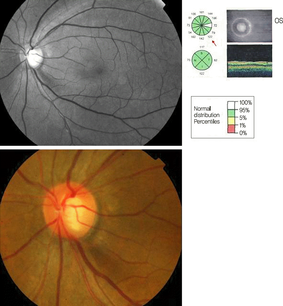
Figure 2.
The angular locations of localized retinal nerve fiber layer (RNFL) defects on red-free fundus photography were measured to evaluate the angular width and location of localized RNFL defects. Reference line was from the optic disc center to the temporal margin of the optic disc (0°). The angles between reference line and the line from the center of the disc to the points at which the upper and lower margins of the localized RNFL defect met with the optic disc margin were measured. The directional angle was assessed in a clockwise direction in right eyes and a counter-clockwise direction in left eyes.

Figure 3.
The number of eyes (total = 197) and retinal nerve fiber layer (RNFL) defects (total = 273) by location on red-free RNFL photographs.
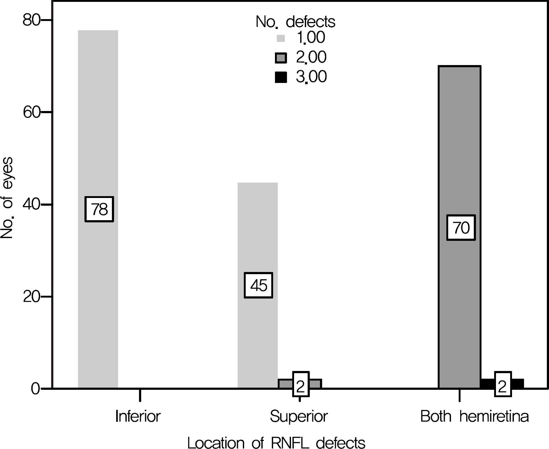
Figure 4.
Frequency distribution of photographic retinal nerve fiber layer (RNFL) defects by clock-hour location on red-free RNFL photographs. Defects were most commonly observed at 7 o’clock and 11 o’clock.
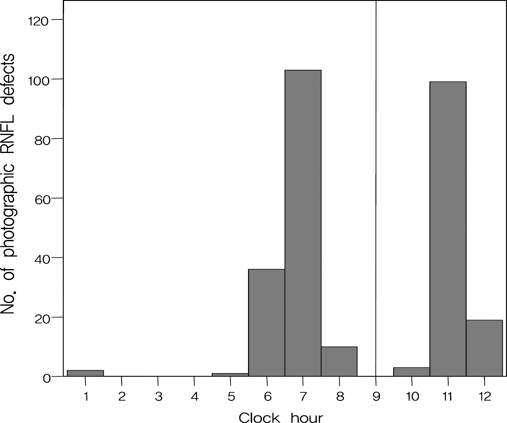
Figure 5.
Scatterplot of the angular locations of the retinal nerve fiber layer (RNFL) defects detected by red-free RNFL photograph and those of RNFL defects detected by optical coherence tomography (OCT) (R = 0.980).
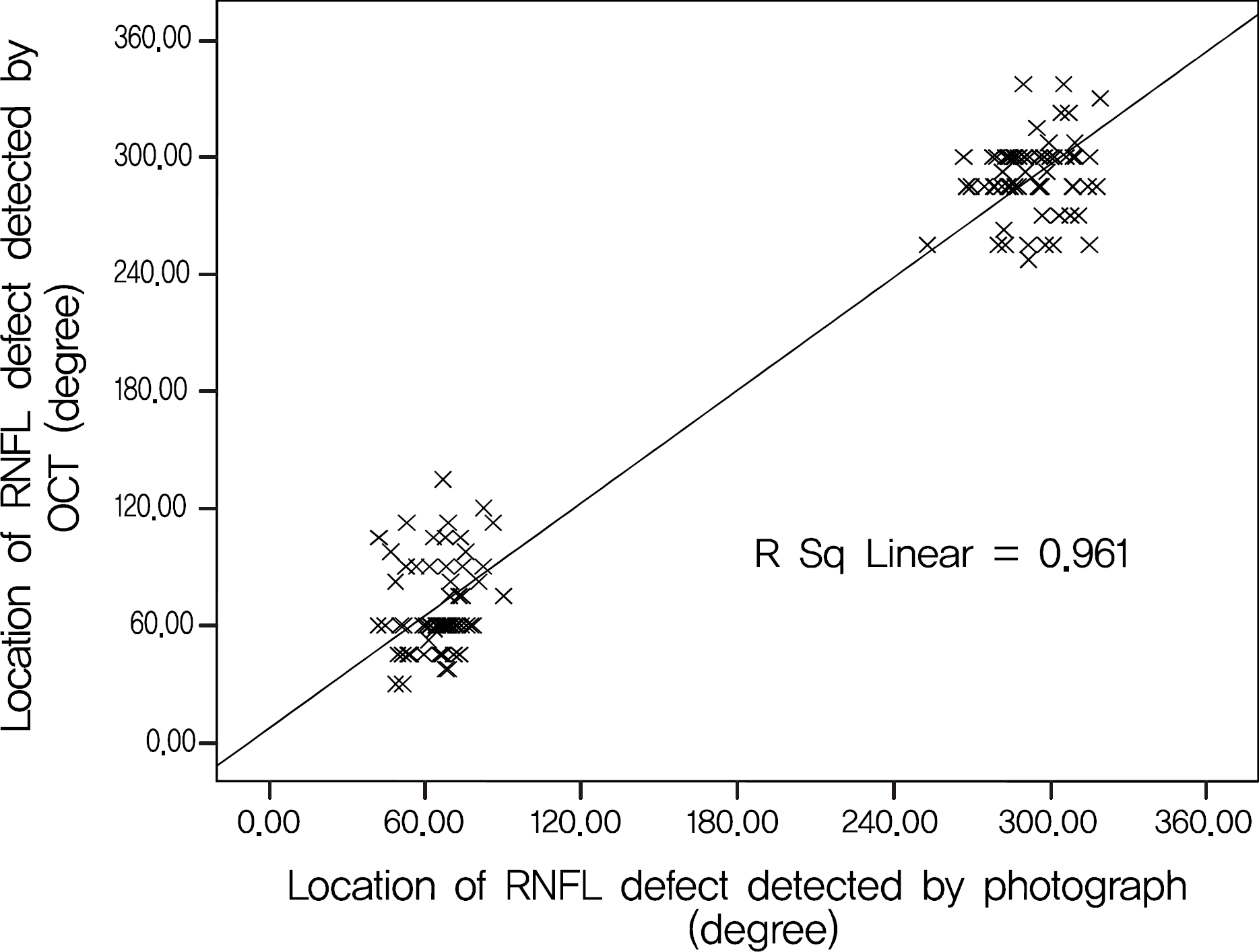
Figure 6.
Scatterplot of the angular widths of the retinal nerve fiber layer (RNFL) defects detected by red-free RNFL photograph and those of RNFL defects detected by optical coherence tomography (OCT) (R = 0.642).
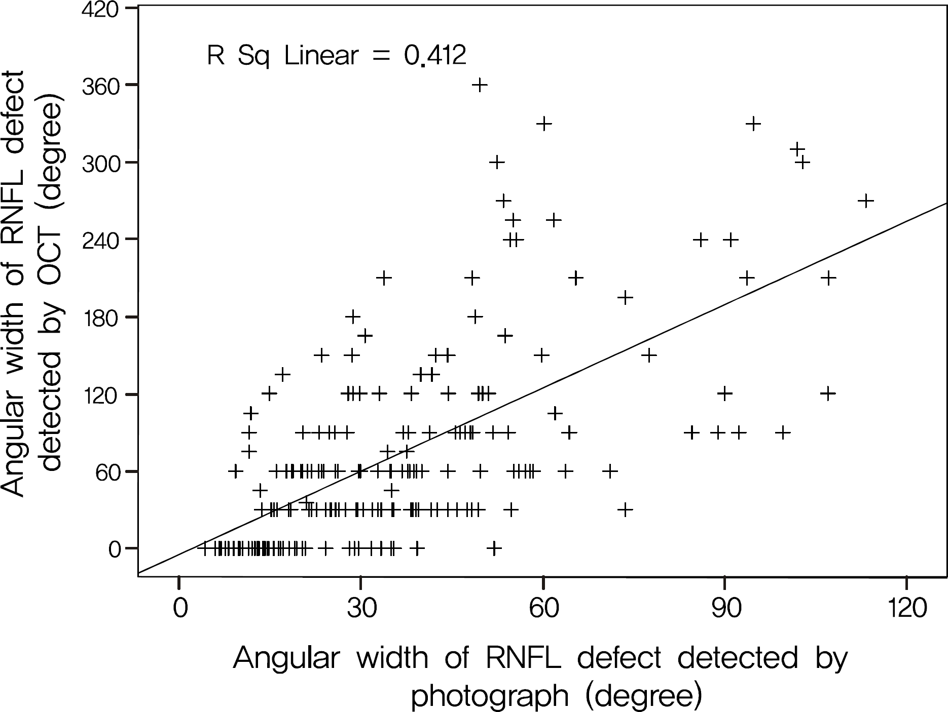
Table 1.
Comparison between photographic retinal nerve fiber layer (RNFL) defects not detected by optical coherence tomography (OCT) group and detected by OCT group
| Variable |
Not detected by OCT |
Detected by OCT |
p-value |
|---|---|---|---|
| (n = 51) | (n = 146) | ||
| Gender | 1.00* | ||
| Male | 29 (25.7%) | 84 (74.3%) | |
| Female | 22 (26.2%) | 62 (73.8%) | |
| Age (mean ± SD, yr) | 56.9 ± 12.3 | 56.5 ± 12.5 | 0.84† |
| Spherical equivalent (mean ± SD, diopter) | -0.83 ± 2.09 | -0.87 ± 2.65 | 0.91† |
| Hypertension | 0.08* | ||
| Yes | 21 (34.4%) | 40 (65.6%) | |
| No | 30 (22.1%) | 106 (77.9%) | |
| Diabetes | 0.22* | ||
| Yes | 13 (34.2%) | 25 (65.8%) | |
| No | 38 (23.9%) | 121 (76.1%) | |
| Central corneal thickness (mean ± SD, μ m) | 532.6 ± 35.0 | 538.6 ± 35.0 | 0.30† |
| Type of glaucoma | <0.001* | ||
| Preperimetric | 17 (70.8%) | 7 (29.2%) | |
| Perimetric | 34 (19.7%) | 139 (80.3%) | |
| SAP mean deviation (mean ± SD, dB) | -3.71 ± 2.93 | -7.95 ± 6.46 | <0.001† |
| SAP pattern standard deviation (mean ± SD, dB) | 3.66 ± 2.85 | 7.08 ± 4.13 | <0.001† |
| Photographic RNFL defect | |||
| Location | 0.01* | ||
| Inferior hemiretina | 21 (26.9%) | 57 (73.1%) | |
| Superior hemiretina | 19 (40.4%) | 28 (59.6%) | |
| Both hemiretina | 11 (15.3%) | 61 (84.7%) | |
| Number (mean ± SD) | 1.25 ± 0.48 | 1.43 ± 0.51 | 0.02‡ |
| Sum of angular width (mean ± SD, degrees)§ | 18.9 ± 11.1 | 42.3 ± 23.2 | <0.001† |
| OCT average RNFL thickness (mean ± SD, μ m) | 95.7 ± 10.7 | 74.9 ± 15.9 | <0.001† |
| HRT disc area (mean ± SD, mm2) | 2.09 ± 0.40 | 2.25 ± 0.54 | 0.05† |
Table 2.
Factors associated with false negative results of optical coherence tomography in eyes with localized retinal nerve fiber layer (RNFL) defects
| Factors |
Logistic regression |
p-value | |
|---|---|---|---|
| Odds ratio | 95% confidence interval | ||
| Average RNFL thickness (μ m) | 1.106 | 1.057-1.156 | <0.001 |
| Angular width of defect (degree) | 0.929 | 0.884-0.977 | 0.004 |
Table 3.
Retinal nerve fiber layer (RNFL) thickness and detection of photographic defects by optical coherence tomography (OCT)
| RNFL thickness (μ m) | No. of eyes | Mean ± SD (μ m) | Not detected by OCT | Detected by OCT | p-value* |
|---|---|---|---|---|---|
| Average† | <0.001 | ||||
| 31.0-74.8 | 66 | 60.6 ± 10.3 | 2 (3.0%) | 64 (97.0%) | |
| 74.9-82.8 | 66 | 81.7 ± 4.5 | 12 (18.2%) | 54 (81.8%) | |
| 97.2-100.4 | 65 | 98.8 ± 6.4 | 37 (56.9%) | 28 (43.1%) | |
| Inferior quadrant† | <0.001 | ||||
| 19.0-77.0 | 66 | 60.6 ± 11.8 | 1 (1.5%) | 65 (98.5%) | |
| 77.3-108.0 | 66 | 91.8 ± 8.7 | 10 (15.2%) | 56 (84.8%) | |
| 109.0-162.0 | 65 | 126.4 ± 12.4 | 40 (61.5%) | 25 (38.5%) | |
| Superior quadrant† | <0.001 | ||||
| 22.0-91.0 | 66 | 70.2 ± 16.9 | 3 (4.5%) | 63 (95.5%) | |
| 91.0-114.0 | 66 | 102.5 ± 6.7 | 12 (18.2%) | 54 (81.8%) | |
| 114.0-159.0 | 65 | 127.0 ± 9.8 | 36 (55.4%) | 29 (44.6%) |
Table 4.
Sum of angular width of photographic retinal nerve fiber layer (RNFL) defect (the summated width of the angle of RNFL defects around the disc) and detection of photographic defects by optical coherence tomography (OCT)
| Sum of angular width of photographic RNFL defect (degree) | Not detected by OCT | Detected by OCT | p-value* |
|---|---|---|---|
| <0.001 | |||
| ≤10 | 11 (91.7%) | 1 (8.3%) | |
| 11-20 | 24 (58.5%) | 17 (41.5%) | |
| 21-30 | 6 (13.3%) | 39 (86.7%) | |
| 31-40 | 8 (24.2%) | 25 (75.8%) | |
| >40 | 2 (3.0%) | 64 (97.0%) | |
| Total | 51 (25.9%) | 146 (74.1%) |
 XML Download