

 PDF
PDF ePub
ePub Citation
 Print
Print


Abstract
Purpose
To analyze retinal tears and to compare the clinical outcomes between retinal tear and rhegmatogenous retinal detachment (RRD) as the cause of dense non-diabetic vitreous hemorrhage in patients who underwent vitreoretinal surgery.
Methods
In a retrospective case series, the medical records of patients who presented dense non-diabetic vitreous hemorrhage and who underwent vitreoretinal surgery between January 2005 and June 2009 were reviewed. Among the 134 patients, 27 patients had dense vitreous hemorrhage caused by retinal tears. The first group had retinal tears only and the second group had accompanying RRD. A comparison of clinical features and postoperative prognoses between the two groups was performed.
Results
Among the 27 eyes with non-traumatic retinal tear and RRD, 18 were categorized into the retinal tear group and 9 to the RRD group. The demographic findings between the two studied groups exhibited no significant differences except for time between onset of symptoms and diagnosis. However, the time to diagnosis was significantly delayed in the group with RRD (22.67 ± 37.47 days) compared to the retinal tear group (5.00 ± 3.41 days) (p = 0.035). The amount of visual improvement was also greater in the retinal tear group than the RRD group (p = 0.002).
Conclusions
Retinal tears are a major cause of non-diabetic vitreous hemorrhage. Vitreous hemorrhage caused by retinal detachment may result in delayed diagnosis and poor visual recovery. Therefore, early examinations in suspicion of RRD and appropriate treatments are needed in non-diabetic vitreous hemorrhage.
Go to : 
References
1. Lindgren G, Lindblom B. Causes of vitreous hemorrhage. Curr Opin Ophthalmol. 1996; 7:13–9.

2. Spraul CW, Grossniklaus HE. Vitreous Hemorrhage. Surv Ophthalmol. 1997; 42:3–39.
3. Isernhagen RD, Smiddy WE, Michels RG, et al. Vitrectomy for nondiabetic vitreous hemorrhage. Not associated with vascular disease. Retina. 1988; 8:81–7.
4. Verbraeken H, Van Egmond J. Non-diabetic and non-oculotraumatic vitreous haemorrhage treated by pars plana vitrectomy. Bull Soc Belge Ophtalmol. 1999; 272:83–9.
5. Smiddy WE, Isernhagen RD, Michels RG, et al. Vitrectomy for nondiabetic vitreous hemorrhage. Retinal and choroidal vascular disorders. Retina. 1988; 8:88–95.
6. Oyakawa RT, Michels RG, Blase WP. Vitrectomy for nondiabetic vitreous hemorrhage. Am J Ophthalmol. 1983; 96:517–25.
7. Colyear BH Jr, Pischel DK. Preventive treatment of retinal detachment by means of light coagulation. Trans Pac Coast Otoophthalmol Soc Annu Meet. 1960; 41:193–217.
8. Davis MD. Natural history of retinal breaks without detachment. Arch Ophthalmol. 1974; 92:183–94.
9. Katsumi O, Hirose T, Kruger-Leite E, et al. Diagnosis and management of massive vitreous hemorrhage caused by retinal tear. Jpn J Ophthalmol. 1989; 33:177–84.
10. Jalkh AE, Jabbour N, Avila MP, et al. Ultrasonographic findings in eyes with giant retinal tears and opaque media. Retina. 1983; 3:154–8.
11. Rabinowitz R, Yagev R, Shoham A, Lifshitz T. Comparison between clinical and ultrasound findings in patients with vitreous hemorrhage. Eye. 2004; 18:253–6.
12. American Academy of Ophthalmology. Management of posterior vitreous detachment, retinal breaks, and lattice degeneration. Preferred Practice Pattern. San Francisco, CA: American Academy of Opthalmology;1998.
13. Wilkinson CP, Rice TA. Michels Retinal Detachment. 2nd ed.St. Louis: Mosby;1997. p. 1082–117.
14. Sarrafizadeh R, Hassan TS, Ruby AJ, et al. Incidence of retinal detachment and visual outcome in eyes presenting with posterior vitreous separation and dense fundus-obscuring vitreous hemorrhage. Ophthalmology. 2001; 108:2273–8.
15. DiBernardo C, Blodi B, Byrne SF. Echographic evaluation of retinal tears in patients with spontaneous vitreous hemorrhage. Arch Ophthalmol. 1992; 110:511–4.
16. Kelley LM, Walker JP, Wing GL, et al. Ultrasound-guided cry-otherapy for retinal tears in patients with vitreous hemorrhage. Ophthalmic Surg Lasers. 1997; 28:565–9.
17. Seelenfreund MH, Sternberg I, Hirsch I, Silverstone BZ. Retinal tears with total vitreous hemorrhage. Am J Ophthalmol. 1983; 95:659–62.
18. Peyman GA, Schulman JA. Intravitreal Surgery. 2nd ed.New Olrleans: Practice-Hall International Inc;1994. p. 813–50.
19. Dhingra N, Pearce I, Wong D. Early vitrectomy for fundus-obscur-ing dense vitreous haemorrhage from presumptive retinal tears. Graefes Arch Clin Exp Ophthalmol. 2007; 245:301–4.
Go to :
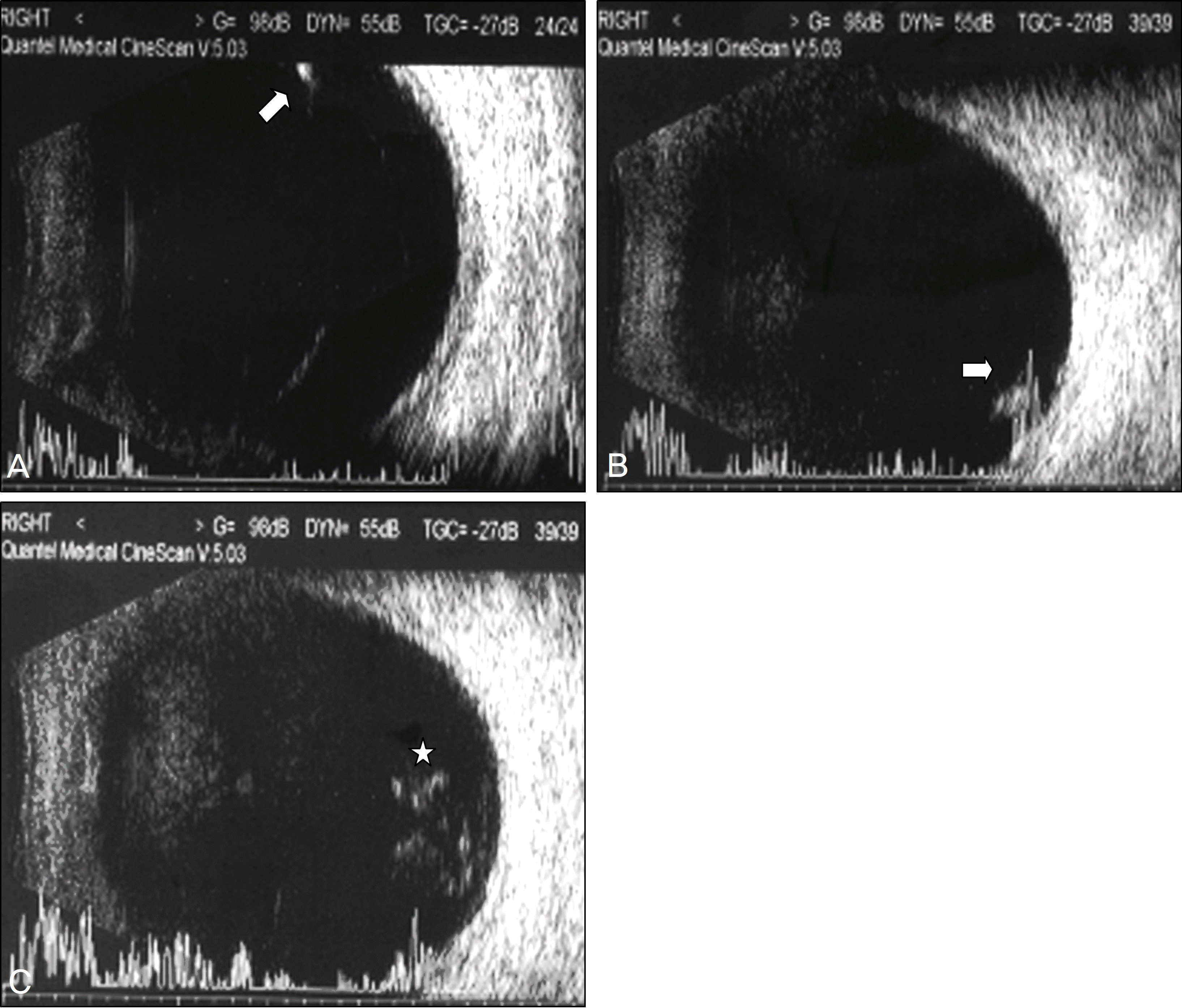 | Figure 1.(A) Transverse echogram shows echo-genic retinal ridges adherent to the posterior vitreous cortex that were interpreted as suspicious retinal tears, but no retinal tears could be confirmed by indirect ophthalmoscopy. (B) Vertical echograms. (C) Vertical echograms shows dense vitreous hemorrhage. |
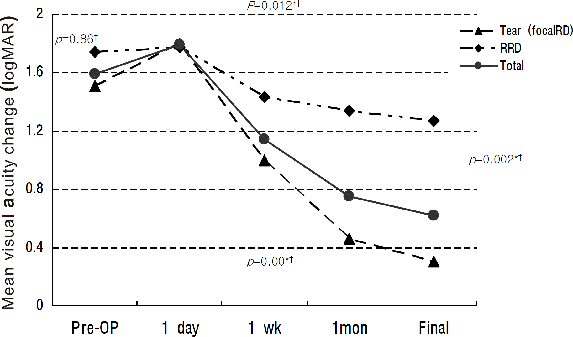 | Figure 2.Change of preoperative and postoperative mean visual acuity. * Statistically significant; † Wilcoxon matched-pairs signed-ranks test; ‡ Mann-whitney U test. |
Table 1.
Distribution of patients
| Retinal tear group | Rhegmatogenous retinal detachment group | p-value | Total patients | |
|---|---|---|---|---|
| Number of patients | 18 | 9 | 27 | |
| Age (mean ± SD, yr) | 53.89 ± 11.23 | 48.56 ± 14.58 | 0.253† | 52.11 ± 12.43 |
| Sex (male:female) | 6:12 | 6:3 | 0.127‡ | 12:15 |
| Laterality (right:left) | 10:8 | 4:5 | 0.695‡ | 14:13 |
| Preoperative V/A (mean ± SD, logMAR) | 1.51 ± 1.03 | 1.74 ± 1.08 | 0.860† | 1.59 ± 1.03 |
| Preoperative IOP (mean ± SD, mmHg) | 12.61 ± 1.88 | 10.44 ± 2.79 | 0.067† | 11.89 ± 2.41 |
| Symptoms → Visit (mean ± SD, day) | 5.00 ± 3.41 | 22.67 ± 37.47 | 0.035†* | 10.89 ± 22.62 |
| Visit → Surgery (mean ± SD, day) | 3.89 ± 4.14 | 2.22 ± 0.67 | 0.668† | 3.33 ± 3.46 |
| Follow up (mean ± SD, mon) | 10.37 ± 8.87 | 10.92 ± 10.54 | 1.000† | 10.56 ± 9.26 |
 XML Download