

 PDF
PDF ePub
ePub Citation
 Print
Print


Abstract
Purpose
The purpose of the present study is to compare Nd:YAG capsulotomy rates between spherical and aspheric intraocular lenses.
Methods
The present retrospective study enrolled patients who received cataract surgery by a single surgeon between March 1, 2006 and October 31, 2009. Patients included in the study were implanted with SA60AT spherical intraocular lenses (Alcon, Fort Worth, TX, USA, 66 eyes), SN60AT spherical intraocular lenses (Alcon, 48 eyes; a total of 114 eyes), or SN60WF aspheric intraocular lenses (Alcon, 187 eyes). The Nd:YAG capsulotomy rates were compared between the two groups 6 months after the operation. Ten patients who were implanted with a spherical intraocular lens in one eye and an aspheric intraocular lens in the contralateral eye were analyzed separately.
Results
Nd:YAG capsulotomy was performed in 2 of 114 eyes (1.8%) in the spherical intraocular lens group and 7 of 187 eyes (3.2%) in the aspheric intraocular lens group; no significant difference was found (p = 0.359). Among the 10 patients who were implanted with 2 different intraocular lenses, Nd:YAG capsulotomy was performed in only 1 eye in the aspheric intraocular lens group; no significant difference was found (p = 0.500).
Go to : 
References
1. Schaumberg DA, Dana MR, Christen WG, Glynn RJ. A systematic overview of the incidence of posterior capsule opacification. Ophthalmology. 1998; 105:1213–21.

2. Vock L, Menapace R, Stifter E, et al. Posterior capsule opacification and neodymium:YAG laser capsulotomy rates with a round-edged silicone and a sharp-edged hydrophobic acrylic intraocular lens 10 years after surgery. J Cataract Refract Surg. 2009; 35:459–65.
3. Awasthi N, Guo S, Wagner BJ. Posterior capsular opacification: a problem reduced but not yet eradicated. Arch Ophthalmol. 2009; 127:555–62.
4. Meacock WR, Spalton DJ, Stanford MR. Role of cytokines in the pathogenesis of posterior capsule opacification. Br J Ophthalmol. 2000; 84:332–6.
5. Nishi O. Posterior capsule opacification. Part 1: Experimental investigations. J Cataract Refract Surg. 1999; 25:106–17.
6. Jung HW, Kim IC. Posterior capsular opacification and Nd:YAG laser capsulotomy in 811B, SI40NB, MA60BM intraocular lens. J Korean Ophthalmol Soc. 2003; 44:1072–8.
7. Aykan U, Bilge AH, Karadayi K, Akin T. The effect of capsulorhexis size on development of posterior capsule opacification: small (4.5 to 5.0 mm) versus large (6.0 to 7.0 mm). Eur J Ophthalmol. 2003; 13:541–5.
8. Bolz M, Menapace R, Findl O, et al. Effect of anterior capsule polishing on the posterior capsule opacification-inhibiting properties of a sharp-edged, 3-piece, silicone intraocular lens: three- and 5-year results of a randomized trial. J Cataract Refract Surg. 2006; 32:1513–20.
9. Hollick EJ, Spalton DJ, Meacock WR. The effect of capsulorhexis size on posterior capsular opacification: one-year results of a randomized prospective trial. Am J Ophthalmol. 1999; 128:271–9.
10. Sacu S, Menapace R, Wirtitsch M, et al. Effect of anterior capsule polishing on fibrotic capsule opacification: three-year results. J Cataract Refract Surg. 2004; 30:2322–7.
11. Lee MJ, Lee JH. The factors affecting early development of posterior capsular opacification after cataract surgery. J Korean Ophthalmol Soc. 2007; 48:493–8.
12. Suh SW, Kim MS. A study of factors influencing after cataract development. J Korean Ophthalmol Soc. 2001; 42:1685–90.
13. Biber JM, Sandoval HP, Trivedi RH, et al. Comparison of the incidence and visual significance of posterior capsule opacification between multifocal spherical, monofocal spherical, and monofocal aspheric intraocular lenses. J Cataract Refract Surg. 2009; 35:1234–8.
14. Buehl W, Findl O. Effect of intraocular lens design on posterior capsule opacification. J Cataract Refract Surg. 2008; 34:1976–85.
15. Buehl W, Menapace R, Sacu S, et al. Effect of a silicone intraocular lens with a sharp posterior optic edge on posterior capsule opacification. J Cataract Refract Surg. 2004; 30:1661–7.
16. Li N, Chen X, Zhang J, et al. Effect of AcrySof versus silicone or polymethyl methacrylate intraocular lens on posterior capsule opacification. Ophthalmology. 2008; 115:830–8.
17. Sacu S, Menapace R, Buehl W, et al. Effect of intraocular lens optic edge design and material on fibrotic capsule opacification and capsulorhexis contraction. J Cataract Refract Surg. 2004; 30:1875–82.
18. Rönbeck M, Zetterström C, Wejde G, Kugelberg M. Comparison of posterior capsule opacification development with 3 intraocular lens types: five-year prospective study. J Cataract Refract Surg. 2009; 35:1935–40.
19. Dewey S. Posterior capsule opacification. Curr Opin Ophthalmol. 2006; 17:45–53.
20. Burq MA, Taqui AM. Frequency of retinal detachment and other complications after neodymium:Yag laser capsulotomy. J Pak Med Assoc. 2008; 58:550–2.
21. Holweger RR, Marefat B. Intraocular pressure change after neo-dymium:YAG capsulotomy. J Cataract Refract Surg. 1997; 23:115–21.
22. Steinert RF, Puliafito CA, Kumar SR, et al. Cystoid macular edema, retinal detachment, and glaucoma after Nd:YAG laser posterior capsulotomy. Am J Ophthalmol. 1991; 112:373–80.
23. Trinavarat A, Atchaneeyasakul L, Udompunturak S. Neodymium: YAG laser damage threshold of foldable intraocular lenses. J Cataract Refract Surg. 2001; 27:775–80.
24. Trueb PR, Albach C, Montés-Micó R, Ferrer-Blasco T. Visual acuity and contrast sensitivity in eyes implanted with aspheric and spherical intraocular lenses. Ophthalmology. 2009; 116:890–5.
25. Rocha KM, Soriano ES, Chamon W, et al. Spherical aberration and depth of focus in eyes implanted with aspheric and spherical intraocular lenses: a prospective randomized study. Ophthalmology. 2007; 114:2050–4.
26. Nanavaty MA, Spalton DJ, Boyce J, et al. Wavefront aberrations, depth of focus, and contrast sensitivity with aspheric and spherical intraocular lenses: fellow-eye study. J Cataract Refract Surg. 2009; 35:663–71.
27. Ohtani S, Miyata K, Samejima T, et al. Intraindividual comparison of aspherical and spherical intraocular lenses of same material and platform. Ophthalmology. 2009; 116:896–901.
28. Cadarso L, Iglesias A, Ollero A, et al. Postoperative optical aberrations in eyes implanted with AcrySof spherical and aspheric intraocular lenses. J Refract Surg. 2008; 24:811–6.
29. Marshall J, Cionni RJ, Davison J, et al. Clinical results of the blue-light filtering AcrySof Natural foldable acrylic intraocular lens. J Cataract Refract Surg. 2005; 31:2319–23.
30. Monnet D, Tépenier L, Brézin AP. Objective assessment of inflammation after cataract surgery: comparison of 3 similar intraocular lens models. J Cataract Refract Surg. 2009; 35:677–81.
31. Beauchamp CL, Stager DR Jr, Weakley DR Jr, et al. Surgical findings with the tinted AcrySof intraocular lens in children. J AAPOS. 2007; 11:166–9.
32. Leibovitch I, Lai T, Porter N, et al. Visual outcomes with the yel-low intraocular lens. Acta Ophthalmol Scand. 2006; 84:95–9.
33. Nixon DR, Woodcock MG. Pattern of posterior capsule opacification models 2 years postoperatively with 2 single-piece acrylic intraocular lenses. J Cataract Refract Surg. 2010; 36:929–34.
34. Shah VC, Russo C, Cannon R, et al. Incidence of Nd:YAG Capsulotomy After Implantation of AcrySof Multifocal and Monofocal Intraocular Lenses: A Case Controlled Study. J Refract Surg. 2010; 26:565–8.
35. Kugelberg M, Wejde G, Jayaram H, Zetterström C. Posterior capsule opacification after implantation of a hydrophilic or a hydrophobic acrylic intraocular lens: one-year follow-up. J Cataract Refract Surg. 2006; 32:1627–31.
36. Kugelberg M, Wejde G, Jayaram H, Zetterström C. Two-year fol-low-up of posterior capsule opacification after implantation of a hydrophilic or hydrophobic acrylic intraocular lens. Acta Ophthalmol. 2008; 86:533–6.
37. Bertelmann E, Kojetinsky C. Posterior capsule opacification and anterior capsule opacification. Curr Opin Ophthalmol. 2001; 12:35–40.
Go to :
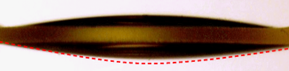 | Figure 1.Horizontal shape of SN60WF lens: a 9% reduction of central thickness than SN60AT lens. Red dotted line: an imaginary line of spherical SN60AT margin (provided from Alcon, Fort Worth, TX, USA). |
Table 1.
Preoperative patient characteristics
 XML Download