Abstract
Purpose
To report a case of a macular hole resulting from accidental exposure to tattoo removal by the Q-Switched Nd:YAG laser, which was treated successfully by vitrectomy and silicone oil infusion.
Case summary
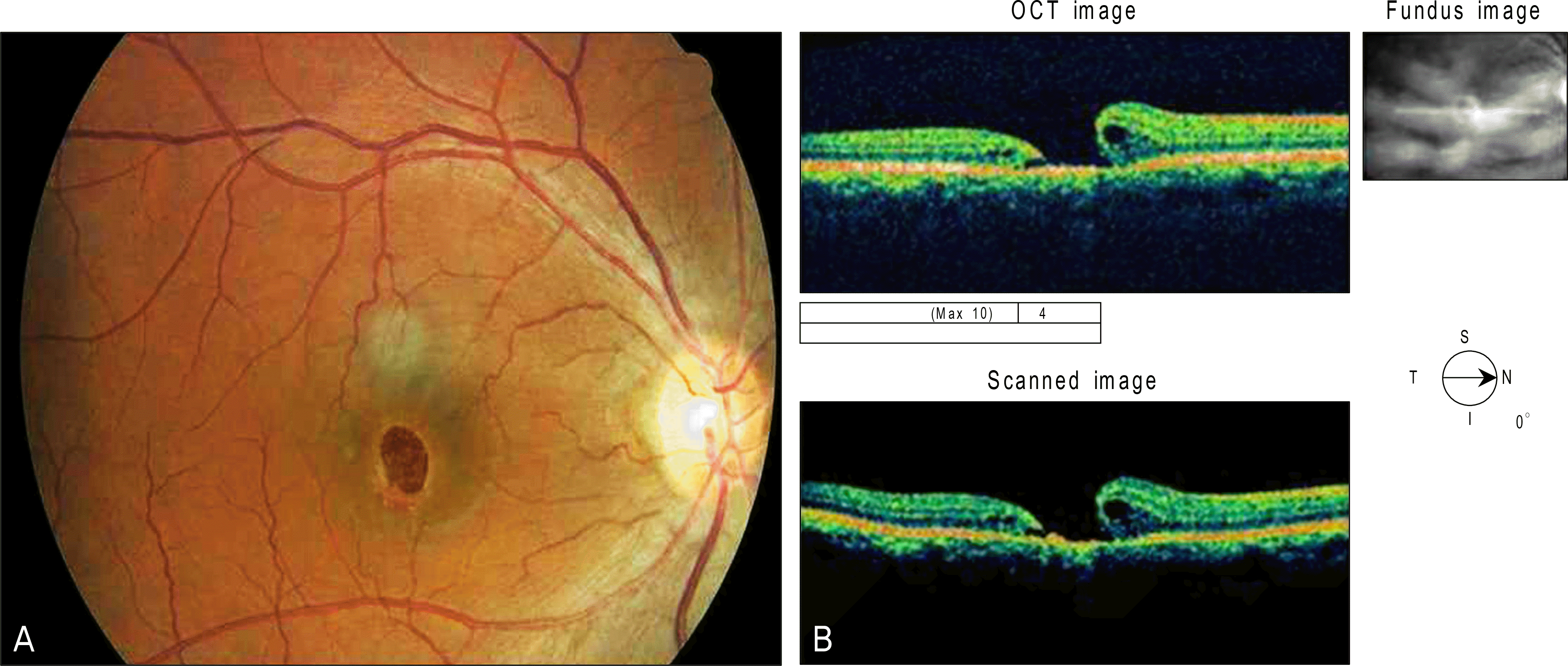
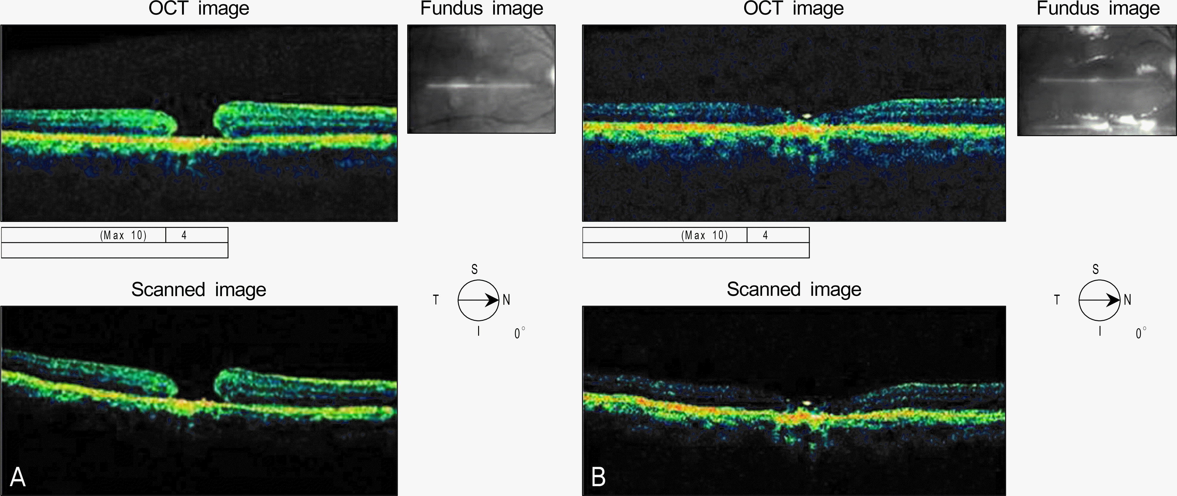
A 33-year-old man presented with decreased visual acuity after accidental exposure to a Q-Switched Nd:YAG laser. According to fundus examination, vitreous hemorrhage and macular edema were observed. After 21 days, a macular hole had developed which was treated by standard pars plana vitrectomy and gas tamponade. Unfortunately, closure was not obtained on the first attempt. Therefore, a second attempt using silicone oil infusion was performed. Four months after the initial visit, BCVA had increased to 20/50, and anatomical occlusion was achieved.
References
2. NamGung M, Park JS, Choi YI. A case of Nd: YAG laser injury to the macula. J Korean Ophthalmol Soc. 2004; 45:1756–60.
3. Liu HF, Gao GH, Wu DC, et al. Ocular injuries from accidental laser exposure. Health Phys. 1989; 56:711–6.
4. Kearney JJ, Cohen HB, Stuck BE, et al. Laser injury to multiple retinal foci. Lasers Surg Med. 1987; 7:499–502.

5. Krauss JM, Puliafito CA, Steinert RF. Laser interactions with the cornea. Surv Ophthalmol. 1986; 31:37–53.

6. Fine S, Feigen L, MacKeen D. Corneal injury threshold to carbon dioxide laser irradiation. Am J Ophthalmol. 1968; 66:1–15.

7. Wu J, Seregard S, Algvere PV. Photochemical damage of the retina. Surv Ophthalmol. 2006; 51:461–81.

8. Alhalel A, Glovinsky Y, Treister G, et al. Long-term follow up of accidental parafoveal laser burns. Retina. 1993; 13:152–4.

9. Thach AB, Lopez PF, Snady-McCoy LC, et al. Accidental Nd:YAG laser injuries to the macula. Am J Ophthalmol. 1995; 119:767–73.

10. Chuang LH, Lai CC, Yang KJ, et al. A traumatic macular hole secondary to a high-energy Nd:YAG laser. Ophthalmic Surg Lasers. 2001; 32:73–6.

11. Marshall J. Structural aspects of laser-induced damage and their functional implications. Health Phys. 1989; 56:617–24.

12. Marshall J. Thermal and mechanical mechanisms in laser damage to the retina. Invest Ophthalmol. 1970; 9:97–115.
13. Mainster MA. Wavelength selection in macular photocoagulation. Tissue optics, thermal effects, and laser systems. Ophthalmology. 1986; 93:952–8.
14. Manning JR, Davidorf FH, Keates RH, et al. Neodymium:YAG laser lesion in the human retina: accidental/experimenta l. Contemp Ophthalmic Forum. 1986; 4:86–91.
15. Potthöfer S, Foerster MH. Vitrectomy and autologous thrombocyte adhesion of an accidental macular hole caused by Nd:YAG laser. Br J Ophthalmol. 1997; 81:803–4.
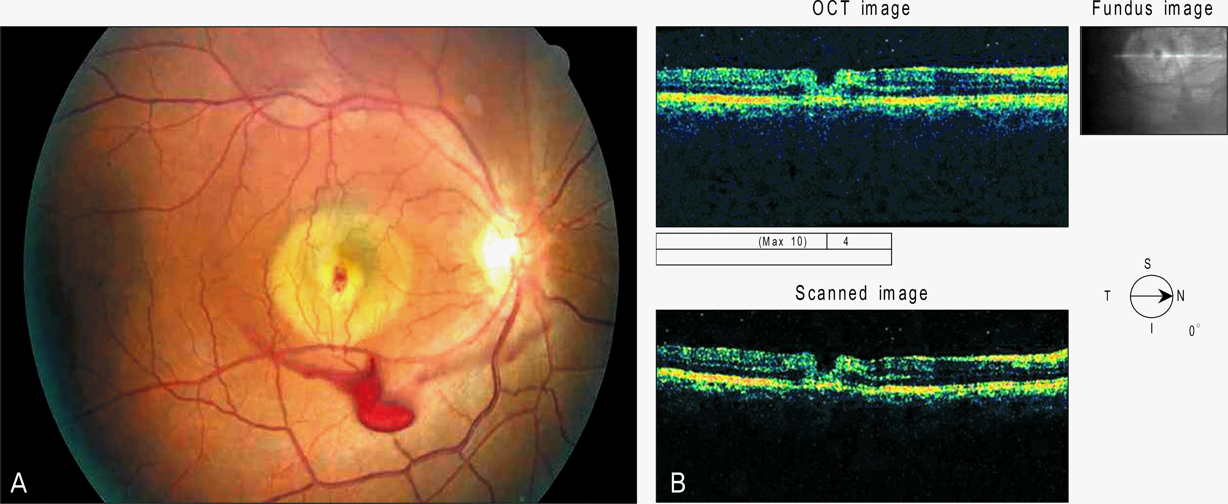
Figure 1.
Fundus photograph (A) at presentation shows dot hemorrhage at the center of the fovea and halo of subretinal fluid. Vitreous hemorrhage observed also. Optical coherence tomography (B) confirms partial defect of the neurosensory retina.


 PDF
PDF ePub
ePub Citation
 Print
Print


 XML Download