Abstract
Purpose
To present a rare case of Muir-Torre syndrome characterized by the association of sebaceous skin tumors and systemic malignancies.
Case summary
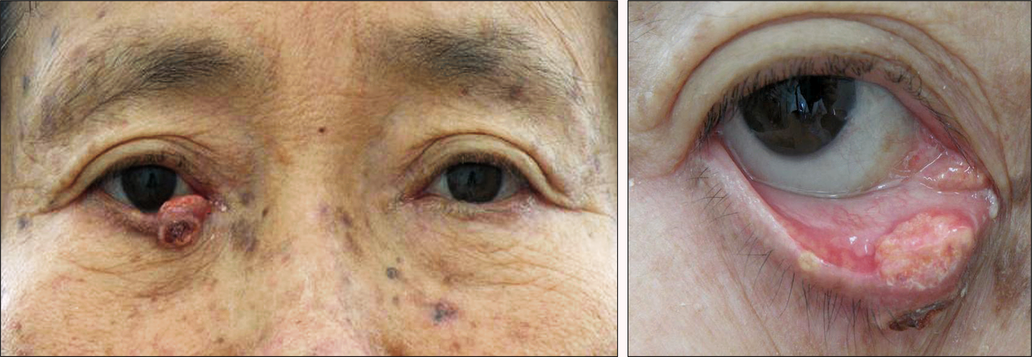
A 65-year-old female visited our clinic with an irregular nodular mass of the right lower eyelid, which developed 1 year earlier. An excisional biopsy and lower lid reconstruction with Tenzel's semicircular rotational flap was performed under local anesthesia. Histopathologic examination showed well-differentiated sebaceous cells, consistent with sebaceous adenoma. The patient had undergone total abdominal hysterectomy and lower anterior resection due to endo-metrial cancer and sigmoid colon cancer 5 years before, and nephroureterectomy due to papillary urothelial carcinoma 3 years before. Based on the history of systemic malignancy and sebaceous skin cancer, a diagnosis of Muir-Torre syndrome was made.
References
1. Muir EG, Bell AJ, Barlow KA. Multiple primary carcinomata of the colon, duodenum, and larynx associated with kerato-acantho-mata of the face. Br J Surg. 1967; 54:191–5.

3. Rishi K, Font RL. Sebaceous gland tumors of the eyelids and conjunctiva in the Muir-Torre syndrome: a clinicopathologic study of five cases and literature review. Ophthal Plast Reconstr Surg. 2004; 20:31–6.
4. Font RL, Rishi K. Sebaceous gland adenoma of the tarsal conjunctiva in a patient with Muir-Torre syndrome. Ophthalmology. 2003; 110:1833–6.

5. Cohen PR, Kohn SR, Kurzrock R. Association of sebaceous gland tumors and internal malignancy: the Muir-Torre syndrome. Am J Med. 1991; 90:606–13.

6. Stockl FA, Dolmetsch AM, Codère F, Burnier MN Jr. Sebaceous carcinoma of the eyelid in an immunocompromised patient with Muir-Torre syndrome. Can J Ophthalmol. 1995; 30:324–6.
7. Tay E, Schofield JB, Rowell NP, Jones CA. Ophthalmic pre-sentation of the Muir Torre syndrome. Ophthal Plast Reconstr Surg. 2003; 19:402–4.

8. Akhtar S, Oza KK, Khan SA, Wright J. Muir-Torre syndrome: case report of a patient with concurrent jejunal and ureteral cancer and a review of the literature. J Am Acad Dermatol. 1999; 41:681–6.

9. Schwartz RA, Torre DP. The Muir-Torre syndrome: a 25-year retrospect. J Am Acad Dermatol. 1995; 33:90–104.

10. Tillawi I, Katz R, Pellettiere EV. Solitary tumors of meibomian gland origin and Torre's syndrome. Am J Ophthalmol. 1987; 104:179–82.

11. Kruse R, Rütten A, Lamberti C, et al. Muir-Torre phenotype has a frequency of DNA mismatch-repair-gene mutations similar to that in hereditary nonpolyposis colorectal cancer families defined by the Amsterdam criteria. Am J Hum Genet. 1998; 63:63–70.

12. Lynch HT, Leibowitz R, Smyrk T, et al. Colorectal cancer and the Muir-Torre syndrome in a Gypsy family: a review. Am J Gastroenterol. 1999; 94:575–80.

13. Fusaro RM, Lemon SJ, Lynch HT. Muir-Torre syndrome and de-fective DNA mismatch repair genes. J Am Acad Dermatol. 1996; 35:493–4.

Figure 1.
Clinical photograph showing an irregular nodular mass in the lower tarsal conjunctiva of the right eye.


 PDF
PDF ePub
ePub Citation
 Print
Print


 XML Download