Abstract
Purpose
To determine if a difference exists in surgically-induced astigmatism (SIA) and the mean change in keratometric astigmatism in patients who underwent microcoaxial cataract surgery (MCCS).
Methods
A prospective study including 193 eyes with astigmatism of greater than 0.5 diopters was performed. The eyes were randomized into two groups: (1) 95 eyes with steep axis incision, and (2) 98 eyes with temporal incision. A 2.2-mm microcoaxial phacoemulsification was performed. The UCVA, BCVA and corneal topography (Orbscan II, Bausch & Lomb) were measured preoperatively and three months postoperatively. Surgically induced astigmatism was calculated via vector analysis, and the mean change in keratometric astigmatism was also calculated.
Results
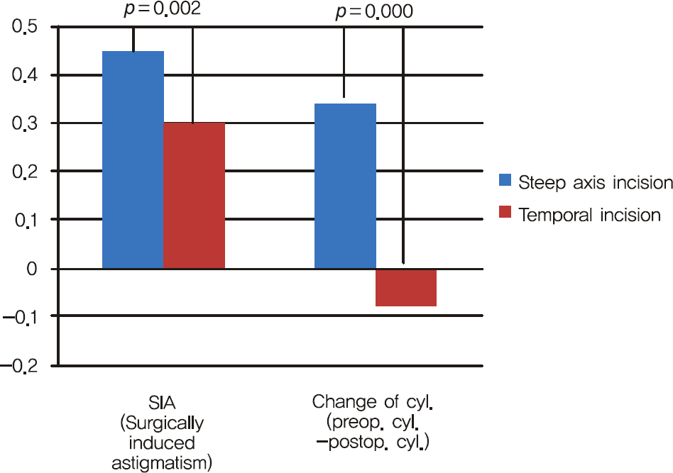
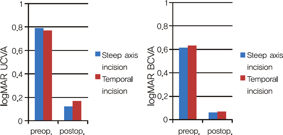
There were no significant differences in UCVA or BCVA between the two groups three months postoperative. The mean SIA was 0.45 ± 0.27 diopters in the steep axis incision group and 0.30 ± 0.17 diopters in the temporal incision group. In the steep axis incision group, the mean keratometric astigmatism showed a mean reduction of 0.31 ± 0.37 diopter (WTR: 0.37 D; oblique: 0.35D; ATR: 0.16 D), while the mean keratometric astigmatism showed a mean increase of 0.06 ± 0.29 diopters (WTR: 0.15 D increased; oblique: 0.11 D increased; ATR: 0.13 D reduced) in the temporal incision group. There were statistically significant differences in SIA and change in astigmatism between the two groups (p = 0.002, p = 0.000).
Go to : 
References
1. Kershner RM. Clear corneal cataract surgery and the correction of myopia, hyperopia, and astigmatism. Ophthalmology. 1997; 104:381–9.

3. Amesbury EC, Miller KM. Correction of astigmatism at the time of cataract surgery. Curr Opin Ophthalmol. 2009; 20:19–24.

4. Osher RH, Injev VP. Microcoaxial phacoemulsification Part 1: laboratory studies. J Cataract Refract Surg. 2007; 33:401–7.
5. Osher RH. Microcoaxial phacoemulsification Part 2: clinical study. J Cataract Refract Surg. 2007; 33:408–12.
6. Praveen MR, Vasavada AR, Gajjar D, et al. Comparative quantification of ingress of trypan blue into the anterior chamber after microcoaxial, standard coaxial, and bimanual phacoemulsification: randomized clinical trial. J Cataract Refract Surg. 2008; 34:1007–12.
7. Wirbelauer C, Pham DT. Surgical experience with microcoaxial phacoemulsification. Klin Monbl Augenheilkd. 2008; 225:212–6.
8. Long DA, Monica ML. A prospective evaluation of corneal curvature changes with 3.0- to 3.5-mm corneal tunnel phacoemulsification. Ophthalmology. 1996; 103:226–32.

9. Alió JL, Rodríguez-Prats JL, Galal A, Ramzy M. Outcomes of microincision cataract surgery versus coaxial phacoemulsification. Ophthalmology. 2005; 112:1997–2003.

10. Alió JL, Rodríguez-Prats JL, Vianello A, Galal A. Visual outcome of microincision cataract surgery with implantation of an Acri. Smart lens. J Cataract Refract Surg. 2005; 31:1549–56.
11. Chernyak DA. Cyclotorsional eye motion occurring between wavefront measurement and refractive surgery. J Cataract Refract Surg. 2004; 30:633–8.

12. Olsen T, Dam-Johansen M, Bek T, et al. Corneal versus scleral tunnel incision in cataract surgery: a randomized study. J Cataract Refract Surg. 1997; 23:337–41.

13. Borasio E, Mehta JS, Maurino V. Surgically induced astigmatism after phacoemulsification in eyes with mild to moderate corneal astigmatism: temporal versus on-axis clear corneal incisions. J Cataract Refract Surg. 2006; 32:565–72.
14. Masket S, Wang L, Belani S. Induced astigmatism with 2.2- and 3.0-mm coaxial phacoemulsification incisions. J Refract Surg. 2009; 25:21–4.

15. Rainer G, Kiss B, Dallinger S, et al. Effect of small incision cataract surgery on ocular blood flow in cataract patients. J Cataract Refract Surg. 1999; 25:964–8.

16. Kohnen T, Dick B, Jacobi KW. Comparison of the induced astigmatism after temporal clear corneal tunnel incisions of different sizes. J Cataract Refract Surg. 1995; 21:417–24.

17. Borasio E, Mehta JS, Maurino V. Torque and flattening effects of clear corneal temporal and on-axis incisions for phacoemulsification. J Cataract Refract Surg. 2006; 32:2030–8.

18. Cravy TV. Calculation of the change in corneal astigmatism following cataract extraction. Ophthalmic Surg. 1979; 10:38–49.
19. Boote C, Dennis S, Meek K. Spatial mapping of collagen fibril organisation in primate cornea-an X-ray diffraction investigation. J Struct Biol. 2004; 146:359–67.

20. Boote C, Dennis S, Newton RH, et al. Collagen fibrils appear more closely packed in the prepupillary cornea: optical and biomechanical implications. Invest Ophthalmol Vis Sci. 2003; 44:2941–8.

Go to : 
 | Figure 2.SIA (surgically induced astigmatism) and change in cylinder (absolute value of preoperative cylinder– absolute value of postoperative cylinder) shows statistically sigsigicant difference between 2 groups). In change in cylinder, positive result indicates decrease in astigmatism and negative result indicates increase in astigmatism.
*Mann-Whitney U test.
|
Table 1.
Demographics and preoperative data in each group
| | (1) Steep axis incision | (2) Temporal incision | * p value |
|---|---|---|---|
| Eyes | 95 | 98 | |
| With-the-rule | 50 (52.6%) | 48 (49.0%) | |
| Oblique | 22 (23.3%) | 30 (30.6%) | |
| Against-the-rule | 23 (24.2%) | 20 (20.4%) | |
| Ages (mean year ± SD) | 70.77 ± 9.52 | 67.52 ± 10.28 | 0.750 |
| Preoperative visual acuity (logMAR) | | | |
| UCVA | 0.79 ± 0.43 | 0.77 ± 0.50 | 0.309 |
| BCVA | 0.61 ± 0.30 | 0.63 ± 0.35 | 0.665 |
| Mean tophographic cylinder (mean power ± SD) | 1.02 ± 0.47 | 0.96 ± 0.69 | 0.125 |
Table 2.
Surgically indeced astigmatism according to preoperative astigmatism in both (1) steep axis incision group and (2) temporal incision group
| | With-the-rule | Against-the-rule | Oblique | † p value |
|---|---|---|---|---|
| (1) Steep axis incision | 0.52 ± 0.28 | 0.26 ± 0.12 | 0.47 ± 0.25 | p<0.05 |
| (2) Temporal incision | 0.31 ± 0.25 | 0.25 ± 0.15 | 0.34 ± 0.27 | p = 0.11 |
| * p value | p<0.05 | p = 0.77 | p<0.05 | |
Table 3.
Change in cylinder according to preoperative astigmatism in both (1) steep axis incision group and (2) temporal incision group. Change in cylinder: absolute value of preoperative cylinder– absolute value of postoperative cylinder. Positive result indicates decrease in astigmatism and negative result indicates increase in astigmatism
| | With-the-rule | Against-the-rule | oblique | † p value |
|---|---|---|---|---|
| (1) Steep axis incision | 0.37 ± 0.19 | 0.16 ± 0.15 | 0.35 ± 0.18 | p<0.05 |
| (2) Temporal incision | −0.15 ± 0.26 | 0.13 ± 0.18 | −0.11 ± 0.24 | p<0.05 |
| * p value | p<0.05 | p = 0.83 | p<0.05 | |


 PDF
PDF ePub
ePub Citation
 Print
Print


 XML Download