

 PDF
PDF ePub
ePub Citation
 Print
Print


Abstract
Purpose
To present a case of leukemic infiltration of the optic nerve head as the initial manifestation of leukemic relapse.
Case summary
A 65-year-old woman was diagnosed with acute myeloid leukemia. Complete remission was achieved after 4 complete courses of chemotherapy. She complained of a sudden decrease in visual acuity in her left eye. Fundus examination showed severe optic disc edema with peripapillary hemorrhage and serous retinal detachment. Visual acuity and fundus continued to aggravate and high-dose intravenous steroid therapy was instituted. Visual acuity and fundus deteriorated more after treatment. Brain magnetic resonance imaging and CSF study were normal but intrathecal chemotherapy and focal irradiation were performed on account of the suspected CNS involvement of leukemia. Morphologic improvement in the retinal structure was achieved, however, optic atrophy remained and her vision did not recover.
Conclusions
The present case shows the involvement of the optic nerve head as the initial isolated manifestation for the relapse in a patient with complete remission. CNS involvement is rare in acute myeloid leukemia and in particular, the optic nerve is rarely reported as the initial isolated presentation for the relapse. Moreover, the disease progression relatively aggravated after treatment. In the atypical aspects of leukemic relapse, the present case was noticeable.
References
1. Schachat AP, Markowitz JA, Guyer DR, et al. Ophthalmic manifestations of leukemia. Arch Ophthalmol. 1989; 107:697–700.

2. Kim JW, Baek CM, Kim KS. A case of chronic myelogenous leukemia involving retina and optic nerve. J Korean Ophthalmol Soc. 2003; 44:2687–93.
3. Sharma T, Grewal J, Gupta S, Murray PI. Ophthalmic manifestations of acute leukaemias: the ophthalmologist's role. Eye. 2004; 18:663–72.
4. Lin YC, Wang AG, Yen MY, Hsu WM. Leukaemic infiltration of the optic nerve as the initial manifestation of leukaemic relapse. Eye. 2004; 18:546–50.
5. Mayo GL, Carter JE, McKinnon SJ. Bilateral optic disk edema and blindness as initial presentation of acute lymphocytic leukemia. Am J Ophthalmol. 2002; 134:141–2.
6. Castagnola C, Nozza A, Corso A, Bernasconi C. The value of combination therapy in adult acute myeloid leukemia with central nervous system involvement. Haematologica. 1997; 82:577–80.
7. Rosenthal AR. Ocular manifestations of leukemia. A review. Ophthalmology. 1983; 90:899–905.
8. Russo V, Scott IU, Querques G, et al. Orbital and ocular manifestations of acute childhood leukemia: clinical and statistical analysis of 180 patients. Eur J Ophthalmol. 2008; 18:619–23.
9. Costagliola C, Rinaldi M, Cotticelli L, et al. Isolated optic nerve involvement in chronic myeloid leukemia. Leuk Res. 1992; 16:411–3.
10. Nikaido H, Mishima H, Ono H, et al. Leukemic involvement of the optic nerve. Am J Ophthalmol. 1988; 105:294–8.
11. Wallace RT, Shields JA, Shields CL, et al. Leukemic infiltration of the optic nerve. Arch Ophthalmol. 1991; 109:1027.
Figure 1.
Fundus photograph and OCT finding on the first visit. Optic disc edema with peripapillary hemorrhages is observed (A). Associated serous retinal detachment is seen (B). Retinal nerve fiber layer (RNFL) thickness obtained with a circular scan around the optic disc is out of normal range, suggesting optic disc edema (C). Retinal thickness map also revealed increased thickness of macula and nasal side due to serous retinal detachment (D). INF=inferior; NAS=nasal; SUP=superior; TEMP=temporal.
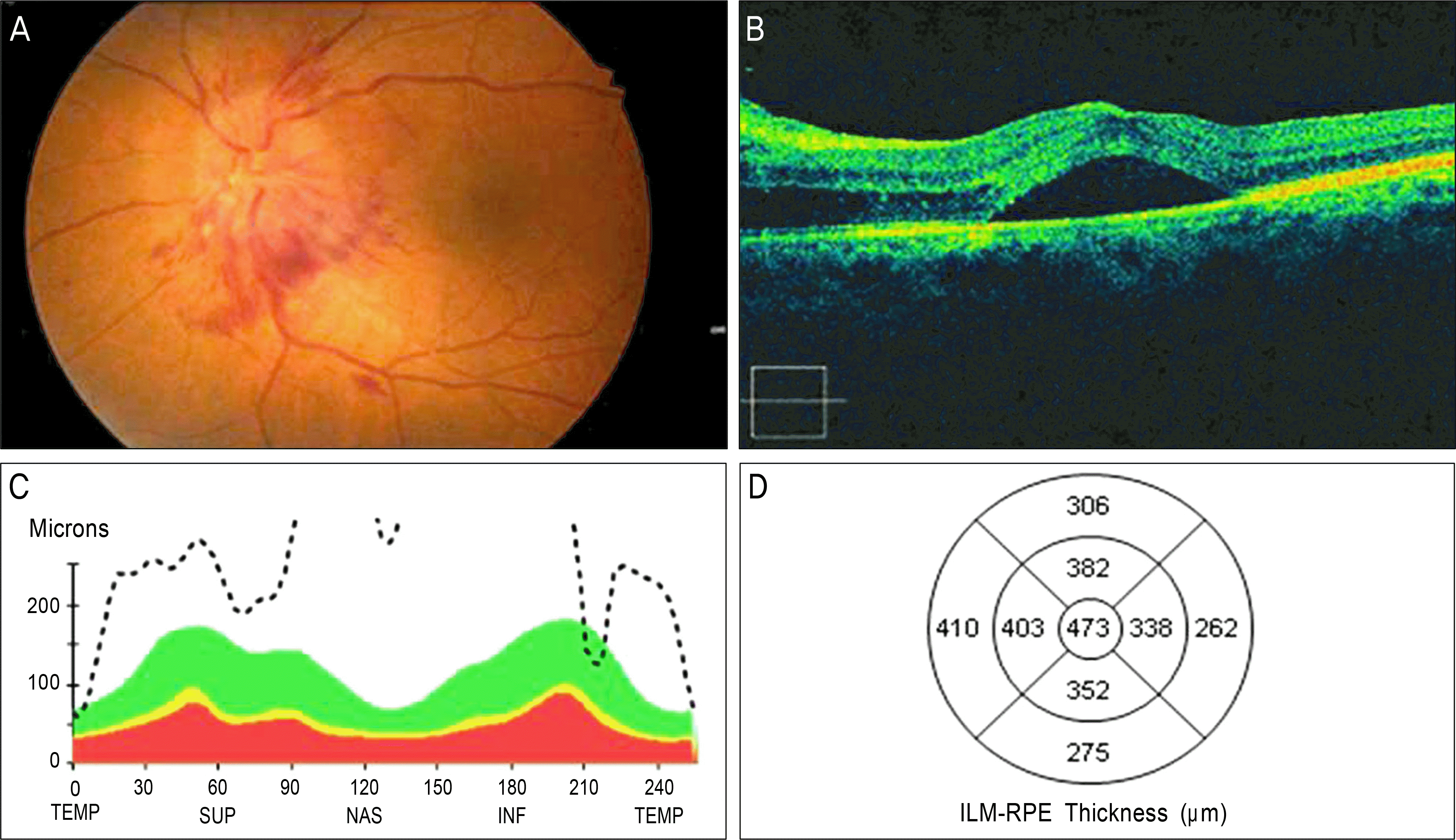
Figure 2.
Fluorescein angiogram on the 12 th day of admission. Severe leakage around the optic disc which started in the early phase and continued to the late phase was shown. Dilated vessels are also noticed (A-D).

Figure 3.
Fundus photographs and OCT findings since the 12 th day of admission. Compared with initial findings, optic disc edema and peripapillary hemorrhages are aggravated and multiple retinal hemorrhages were also noted (A). Moreover, serous retinal detachment in perimacular lesion was much progressed (B). After starting high-dose intravenous corticosteroids therapy on the 15 th day of admission, optic disc edema with peripapillary hemorrhages was rather more deteriorated (C), but on the contrary to disc edema, serous retinal detachment was much diminished (D). Two months later, previous optic disc edema and peripapillary hemorrhages were nearly disappeared but optic atrophy remains (E). Previous serous retinal detachment was also absorbed, and morphologic improvement in the retinal structure is noticed (F).

 XML Download