Abstract
Purpose
To compare postoperative recurrence rates between conjunctival autograft transplantation alone and conjunctival autograft transplantation with amniotic membrane transplantation in primary pterygium surgery.
Methods
The authors conducted a retrospective analysis of 66 eyes from 62 patients who underwent primary pterygium surgery from January 2001 to May 2009. Twenty three eyes underwent conjunctival autograft transplantation alone, 43 eyes underwent conjunctival autograft transplantation with amniotic membrane transplantation.
Results
Recurrence of pterygium was observed in 5 of 23 eyes that received conjunctival autograft transplantation alone. There were 2 cases of recurrence of 43 eyes that received conjunctival autograft transplantation and amniotic membrane transplantation. No major complications such as necrotizing scleritis, sclera ulcer, or corneal perforation were observed in either group after surgery.
Go to : 
References
1. Donnenfeld ED, Perry HD, Fromer S, et al. Subconjunctival mitomycin C as adjunctive therapy before pterygium excision. Ophthalmology. 2003; 110:1012–6.

2. Hong SB, Oh SJ, Oh JH. The effects and complications of mitomycin-C for prevention of recurrence after pterygium operation. J Korean Ophthalmol Soc. 1998; 39:2013–8.
3. Mastropasqua L, Carpineto P, Ciancaglini M, Enrico Gallenga P. Long-term results of intraoperative mitomycin C in the treatment of recurrent pterygium. Br J Ophthalmol. 1996; 80:288–91.
4. Amano S, Motojyma Y, Oshika T, et al. Comparative study of intraoperative mitomycin C and beta irradiation in pterygium surgery. Br J Ophthalmol. 2000; 84:618–21.

5. Lee SH, Jeong HJ. Immune reactions in pterygium. J Korean Ophthalmol Soc. 1987; 28:927–33.
7. Kim CH, Lee JK, Park DJ. Recurrence rates of amniotic membrane transplantation, conjunctival autograft and conjunctivolimbal autograft in primary pterygium. J Korean Ophthalmol Soc. 2009; 50:1780–8.

8. Manning CA, Kloess PM, Diaz MD, Yee RW. Intraoperative mitomycin in primary pterygium excision. A prospective, randomized trial. Ophthalmology. 1997; 104:844–8.
9. Allan BD, Short P, Crawford GJ, et al. Pterygium excision with conjunctival autografting: an effective and safe technique. Br J Ophthalmol. 1993; 77:698–701.

10. Cho JW, Chung SH, Seo KY, Kim EK. Conjunctival mini-flap technique and conjunctival autotransplantation in pterygium surgery. J Korean Ophthalmol Soc. 2005; 46:1471–7.
11. Prabhasawat P, Barton K, Burkett G, Tseng SC. Comparison of conjunctival autografts, amniotic membrane grafts, and primary closure for pterygium excision. Ophthalmology. 1997; 104:974–85.

12. Tan DT, Chee SP, Dear KB, Lim AS. Effect of pterygium morphology on pterygium recurrence in a controlled trial comparing conjunctival autografting with bare sclera excision. Arch Ophthalmol. 1997; 115:1235–40.

13. Mutlu FM, Sobaci G, Tatar T, Yildirim E. A comparative study of recurrent pterygium surgery: limbal conjunctival autograft transplantation versus mitomycin C with conjunctival flap. Ophthalmology. 1999; 106:817–21.

14. Barraquer JI, Binder PS, Buxton JN. Etiology and treatment of pterygium; Symposium on Medical and Surgical Diseases of the Cornea. Transactions of the New Orleans Academy of Ophthalmology. St. Louis: Mosby;1980. p. 167–78.
15. Ozer A, Yildirim N, Erol N, Yurdakul S. Long-term results of bare sclera, limbal-conjunctival autograft and amniotic membrane graft techniques in primary pterygium excisions. Ophthalmologica. 2009; 223:269–73.

16. Kenyon KR, Tseng SC. Limbal autograft transplantation for ocular surface disorders. Ophthalmology. 1989; 96:709–22.

17. van Herendael BJ, Oberti C, Brosens I. Microanatomy of the human amniotic membrane. A light microscopic, transmission, and scanning electron microscopic study. Am J Obstet Gynecol. 1978; 131:872–80.
18. Terranova VP, Lyall RM. Chemotaxis of human gingival epithelial cells to laminin. A mechanism for epithelial cell apical migration. J Periodontol. 1986; 57:311–7.
19. Sonnenberg A, Calafat J, Janssen H, et al. Integrin alpha 6/beta 4 complex is located in hemidesmosomes, suggesting a major role in epidermal cell-basement membrane adhesion. J Cell Biol. 1991; 113:907–17.

20. Streuli CH, Bailey N, Bissel MJ. Control of mammary epithelial differentiation: basement membrane induces tissue-specific gene expression in the absence of cell-cell interaction and morphological polarity. J Cell Biol. 1991; 115:1383–95.

Go to : 
 | Figure 1.Surgical technique of conjunctival autograft transplantation. (A) Removal of pterygium from the cornea with a Beaver blade. (B) Tenon's capsule and subconjunctival fibrovascular tissue are undermined and excised extensively with a pair of spring scissors. (C) Marked the donor conjunctiva with gentian violet made to measure the size of the conjunctival defect. (D) Conjunctival graft is attached to the conjunctival edges and episclera with interrupted 10-0 nylon sutures. |
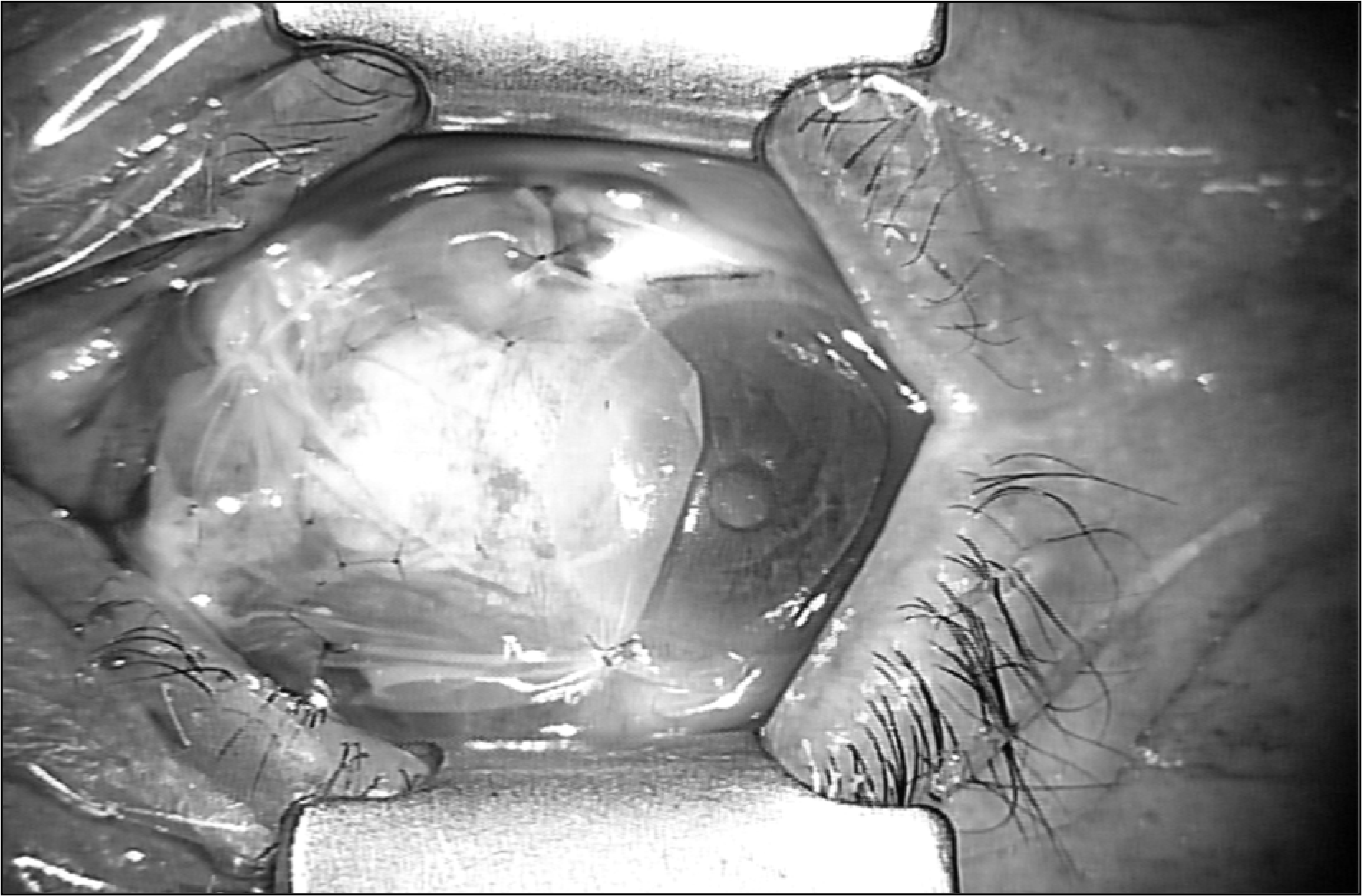
 | Figure 2.Surgical technique of amniotic membrane transplantation. Amniotic membrane is cut a proper-sized graft, which will cover the conjunctival transplantation area and corneal epithelial defect area. The membrane is placed over the epithelial side on top. Pinch together the membrane and conjunctiva, interrupted suture is done with 10-0 nylon. |
Table 1.
Characteristics of patients
| Control group† (23 eyes) | Study group‡ (43 eyes) | p-value | |
|---|---|---|---|
| Age (yr, Mean ± SD∗) | 46.3 ± 13.7 | 51.3 ± 10.8 | 0.345§ |
| Sex (Male:Female) | 9:14 | 19:24 | 0.796∏ |
| Followup period (months, Mean ± SD∗) | 11.7 ± 24.2 | 12.4 ± 16.8 | 0.891§ |
| Preoperative grading (T2:T3) | 8:12 (3 eyes were unrecorded) | 12:22 (9 eyes were unrecorded) | 0.735∏ |
Table 2.
Differences between recurred cases and non-recurred cases in control group†
| Recurred cases (5 eyes) | Non-recurred cases (18 eyes) | p-value | |
|---|---|---|---|
| Age (yr, Mean ± SD∗) | 48.8 ± 12.1 | 45.6 ± 14.5 | 0.658‡ |
| Sex (Male:Female) | 1:4 | 8:10 | 0.611§ |
| Preoperative grading (T2:T3) | 2:3 | 6:9 (3 eyes were unrecorded) | 0.693§ |
Table 3.
Differences between recurred cases and non-recurred cases in study group†
| Recurred cases (2 eyes) | Non-recurred cases (41 eyes) | p-value | |
|---|---|---|---|
| Age (yr, Mean ± SD∗) | 70.0 ± 1.4 | 50.4 ± 10.2 | 0.011‡ |
| Sex (Male:Female) | 0:2 | 19:22 | 0.495§ |
| Preoperative grading (T2:T3) | 0:2 | 12:20 (9 eyes were unrecorded) | 0.512§ |


 PDF
PDF ePub
ePub Citation
 Print
Print


 XML Download