

 PDF
PDF ePub
ePub Citation
 Print
Print


Abstract
Purpose
To report a rare case of a 62-year-old man who presented with bilateral eyelid swelling, chemosis, and hyperemia of the conjunctiva, which wax and waned, being the initial manifestation of relapsing polychondritis.
Case summary
A 62-year-old man presented with pain and erythematous swelling of the right eyelid for 2 days. There were no other symptoms except chemosis and hyperemia of the conjunctiva. After 1 week, the same symptoms occurred in the left eye, while the lesion of the right eye underwent improvement and aggravation repeatedly. Blood culture tests, Orbital CT, and MRI were performed, but could not confirm a diagnosis. During the followup period, erythematous swelling of the left auricle and laryngopharyngitis appeared and erythematous plaques were scattered on the extremities. Punch biopsies were performed; histopathologic examinations concluded to the diagnosis of chondritis. The diagnosis of relapsing polychondritis was confirmed through histologically diagnosed chondritis and repeated ocular symptoms with systemic features.
References
1. McAdam LP, O'Hanlan MA, Bluestone R, Pearson CM. Relapsing polychondritis: Prospective study of 23 patients and a review of the literature. Medicine (Baltimore). 1976; 55:193–215.
2. Jaksch-Wartenhorst R. Polychondropathia. Wien Arch Int Med. 1923; 6:93–100.
3. Zeuner M, Straub RH, Rauh G, et al. Relapsing polychondritis: clinical and immunogenetic analysis of 62 patients. J Rheumatol. 1997; 24:96–101.
4. Foidart JM, Shigeto A, Martin G, et al. Antibodies to typeII colla-gen in relapsing polychondritis. N Engl J Med. 1978; 299:1203–7.
5. Kent PD, Michet CJ Jr, Luthra HS. Relapsing polychondritis. Curr Opin Rheumatol. 2004; 16:56–61.

6. Gleicher N, Barad DH. Gender as risk factor for autoimmune diseases. J Autoimmun. 2007; 28:1–6.
7. Magargal LE, Donoso LA, Goldberg RE, et al. Ocular manifestations of relapsing polychondritis. Retina. 1981; 1:96–9.
8. Isaak BL, Liesegang TJ, Michet CJ Jr. Ocular and systemic findings in relapsing polychondritis. Ophthalmology. 1986; 93:681–9.
9. Rafailidis PI, Falagas ME. Fever and periorbital edema: A review. Surv Ophthalmol. 2007; 52:422–33.
10. Butterton JR, Collier DS, Romero JM, Zembowicz A. Case records of the massachusetts general hospital. case 14-2007. A 59-year-old man with fever and pain and swelling of both eyes and the right ear. N Engl J Med. 2007; 356:1980–8.
11. Park JJ, Lee JC, Kim JH, et al. Clinical analysis of relapsing polychondritis: 16 Cases in Korea. J Korean Rheum Assoc. 2005; 12:213–21.
12. Damiani JM, Levine HL. Relapsing polychondritis report of ten cases. Laryngoscope. 1979; 89:929–46.
13. Letko E, Zafirakis P, Baltatzis S, et al. Relapsing polychondritis: a clinical review. Semin Arthritis Rheum. 2002; 31:384–95.
14. Balsa A, Expinosa A, Cuesta M, et al. Joint symptoms in relapsing polychondritis. Clin Exp Rheumatol. 1995; 13:425–30.
15. Lahmer T, Treiber M, von Werder A, et al. Relapsing polychondritis: An autoimmune disease with many faces. Autoimmun Rev. 2010; 9:540–6.
16. Choi YS, Yim HB, Kim KB. A case of episcleritis with relapsing polychondritis. J Korean Ophthalmol Soc. 2002; 43:626–30.
17. Joo SH, Choe JK. A case of posterior scleritis associated with relapsing polychondritis. J Korean Ophthalmol Soc. 1989; 30:665–70.
18. Yim JS, Oum BS, Park D. A case of relapsing polychondritis complicated with chorioretinitis without scleritis. J Korean Ophthalmol Soc. 2007; 48:1716–22.
19. Lim TH, Han JI. A case of gypopyon uveitis associated with relapsing polychondritis. J Korean Ophthalmol Soc. 2009; 50:486–90.
20. Barranco VP, Minor DB, Soloman H. Treatment of relapsing polychondritis with dapsone. Arch Dermatol. 1976; 112:1286–8.
21. Martin J, Roenigk HH, Lynch W, Tingwald FR. Relapsing polychondritis treated with dapsone. Arch Dermatol. 1976; 112:1272–4.
22. Lee KH, Hong YS, Kang HJ, et al. A case of relapsing polychondritis associated with Sjögren's syndrome. J Korean Rheum Assoc. 2001; 8:198–202.
23. Choy EH, Chikanza IC, Kingsley GH, Panayi GS. Chimaeric an-ti-CD4 monoclonal antibody for relapsing polychondritis. Lancet. 1991; 338:450.
24. van der Lubbe PA, Miltenburg AM, Breedveld FC. Anti-CD4 monoclonal antibody for relapsing polychondritis. Lancet. 1991; 337:1349.
Figure 1.
Orbit CT reveals mild asymmetric swelling of the right eyelid with no evidence of mass-like lesions. Orbit CT of the left eye shows normal findings.
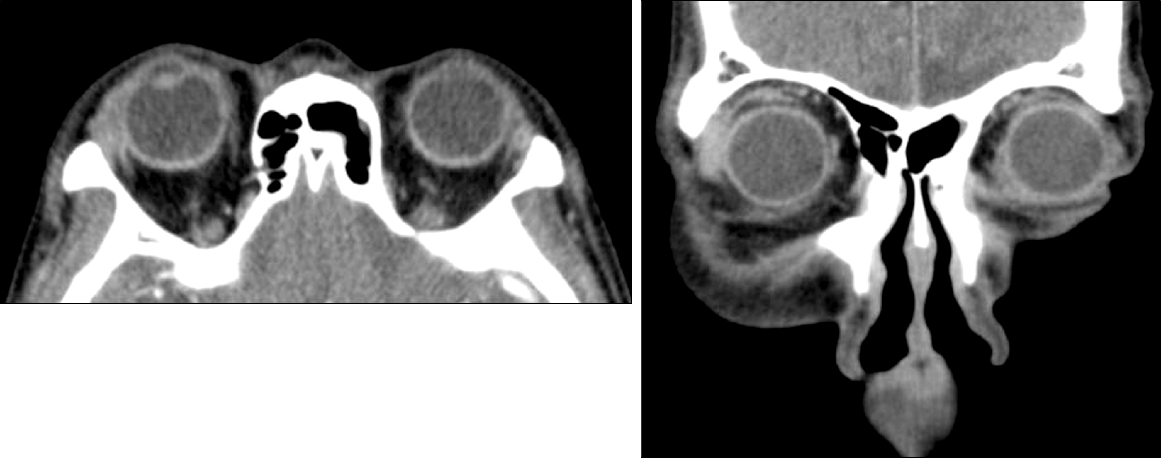
Figure 2.
One week after initial treatment, photograph shows normal appearance of the right eyelid and conjunctiva. However, severe erythematous swelling occurred previously on the right eyelid can be seen on the left eyelid.
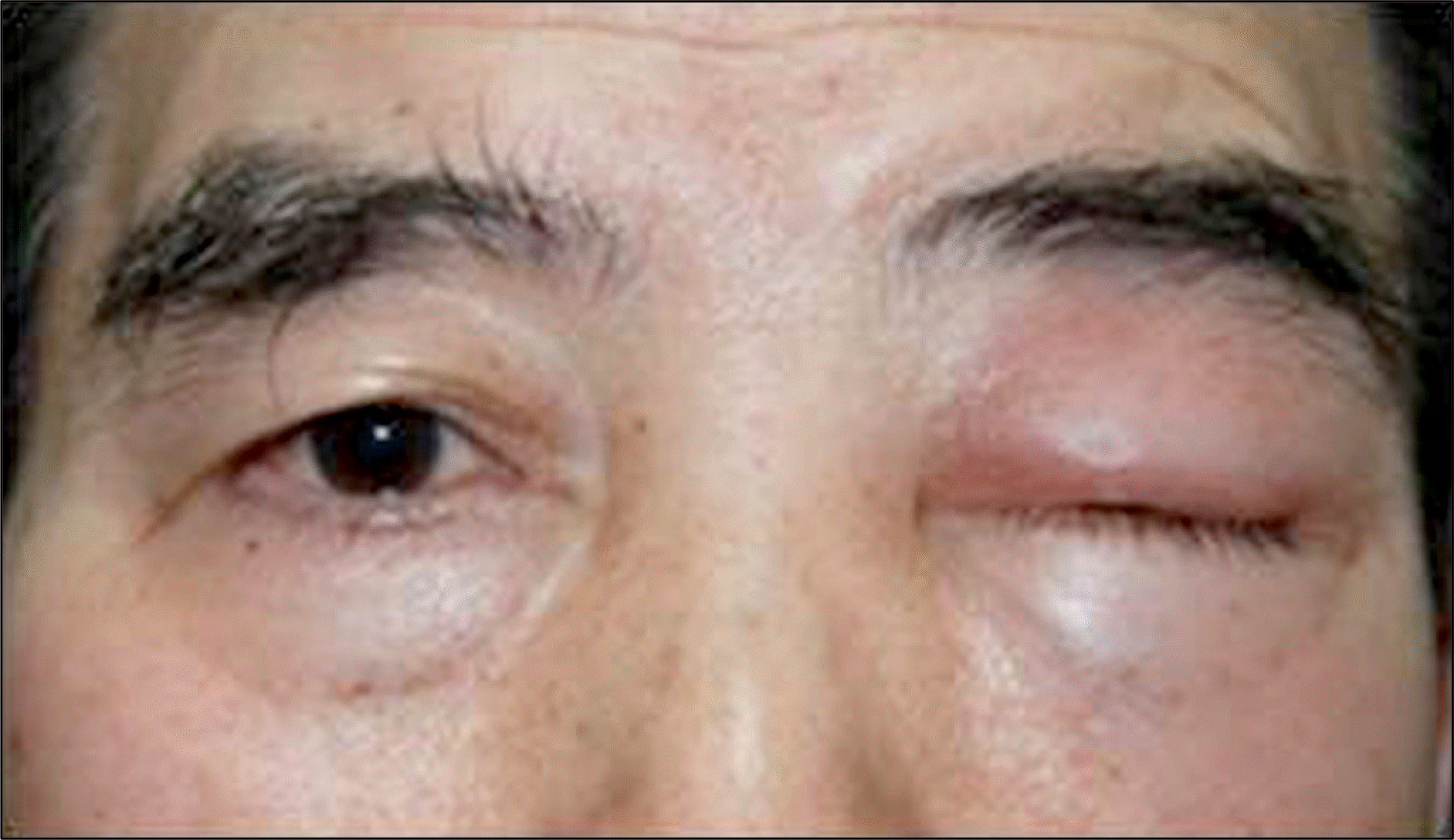
Figure 3.
(A, B) There is no evidence of mass-like lesions in T1 and T2 images. (C, D) Enhanced view shows strong enhancement of left lacrimal area and superior orbital tissue.

Figure 4.
Photograph shows recurred erythematous eyelid swelling and conjunctival hyperemia of the right eye. Left eyelid swelling improved from 1 week ago, but swelling and hyperemia are still present. The patient also experienced bilateral hearing loss, sore throat, and fever.
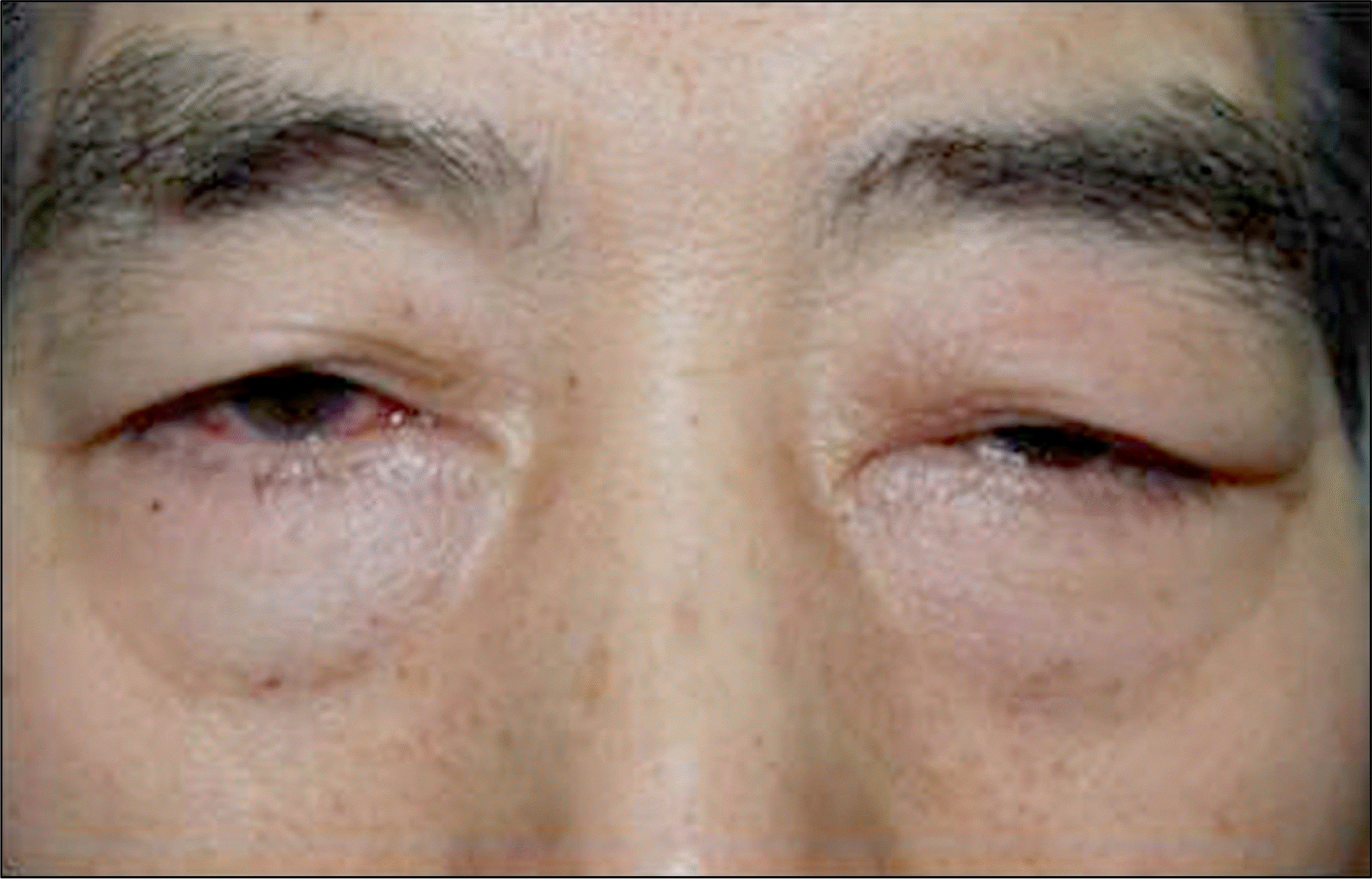
Figure 5.
(A) Photograph shows erythematous swelling of the left auricle. Erythematous swelling is localized to the cartilaginous portion of the helix sparing the ear lobe. (B) Photograph shows pea- to-walnut-sized erythematous plaques scattered on the extremities.
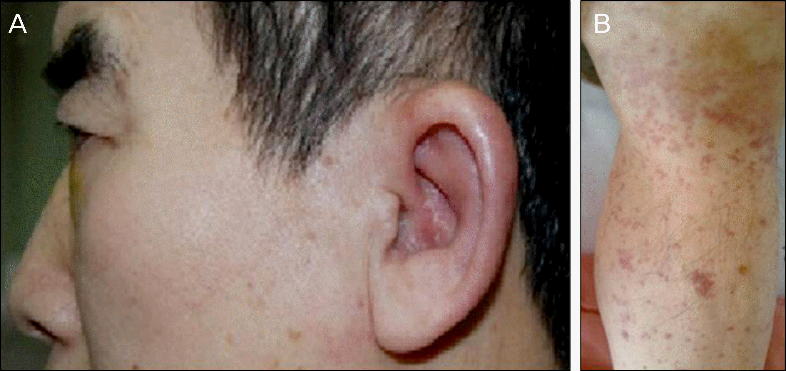
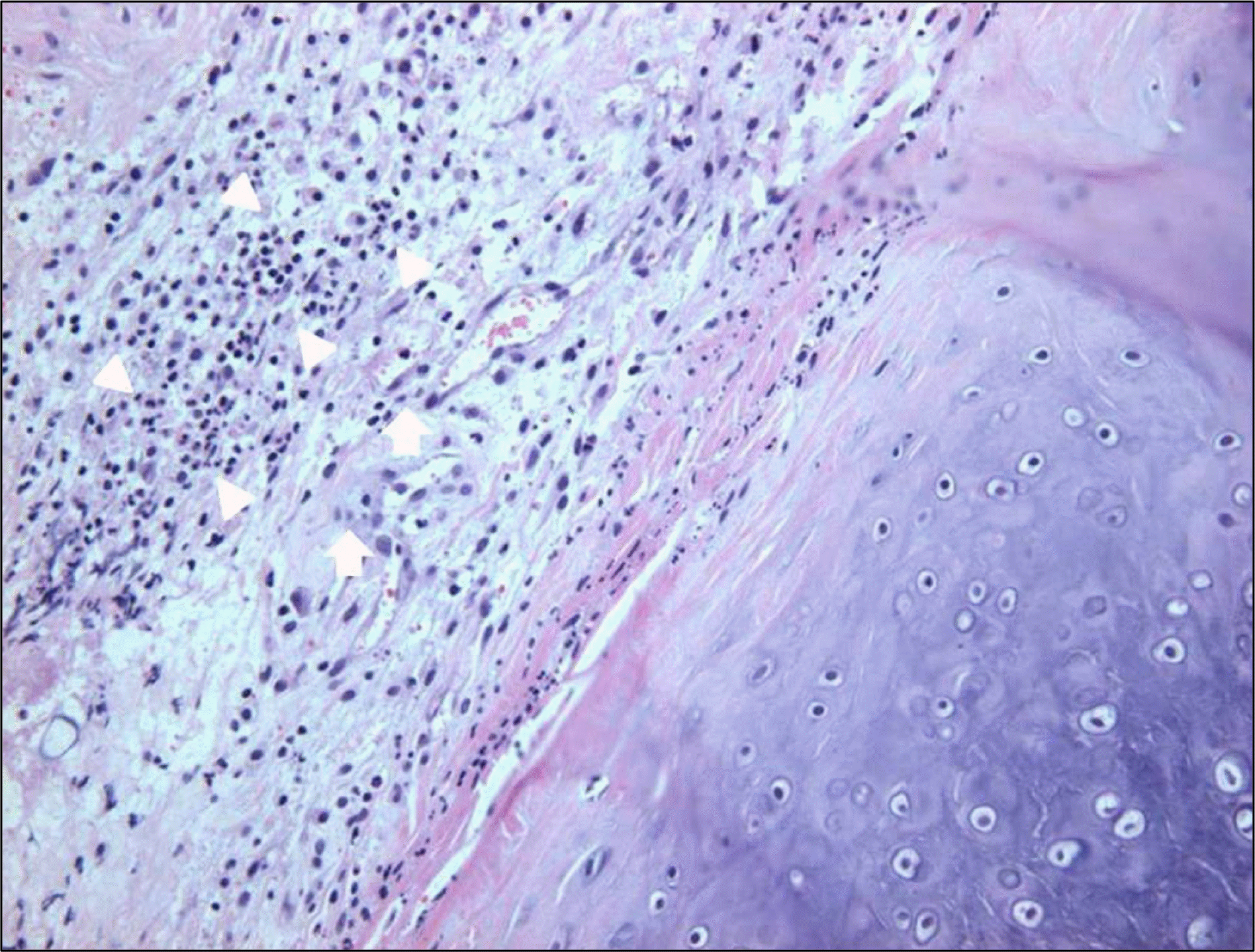
Figure 6.
Pathological findings of the auricular cartilage (HE staining, ×200). Infiltration of neutrophils and lymphocytes (white arrowhead) are noted in the perichondrium. Infiltration and congestion of dermal perivascular mixed cells (white arrow) are also seen in the soft tissue and perichondrium.
 XML Download