Abstract
Purpose
To report a rare case of basosquamous carcinoma of the eyelid, an aggressive tumor with a higher tendency for recurrence and metastasis.
Case summary
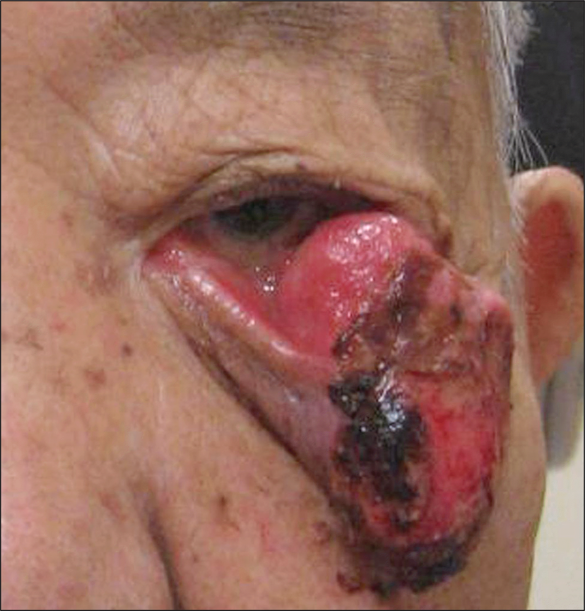
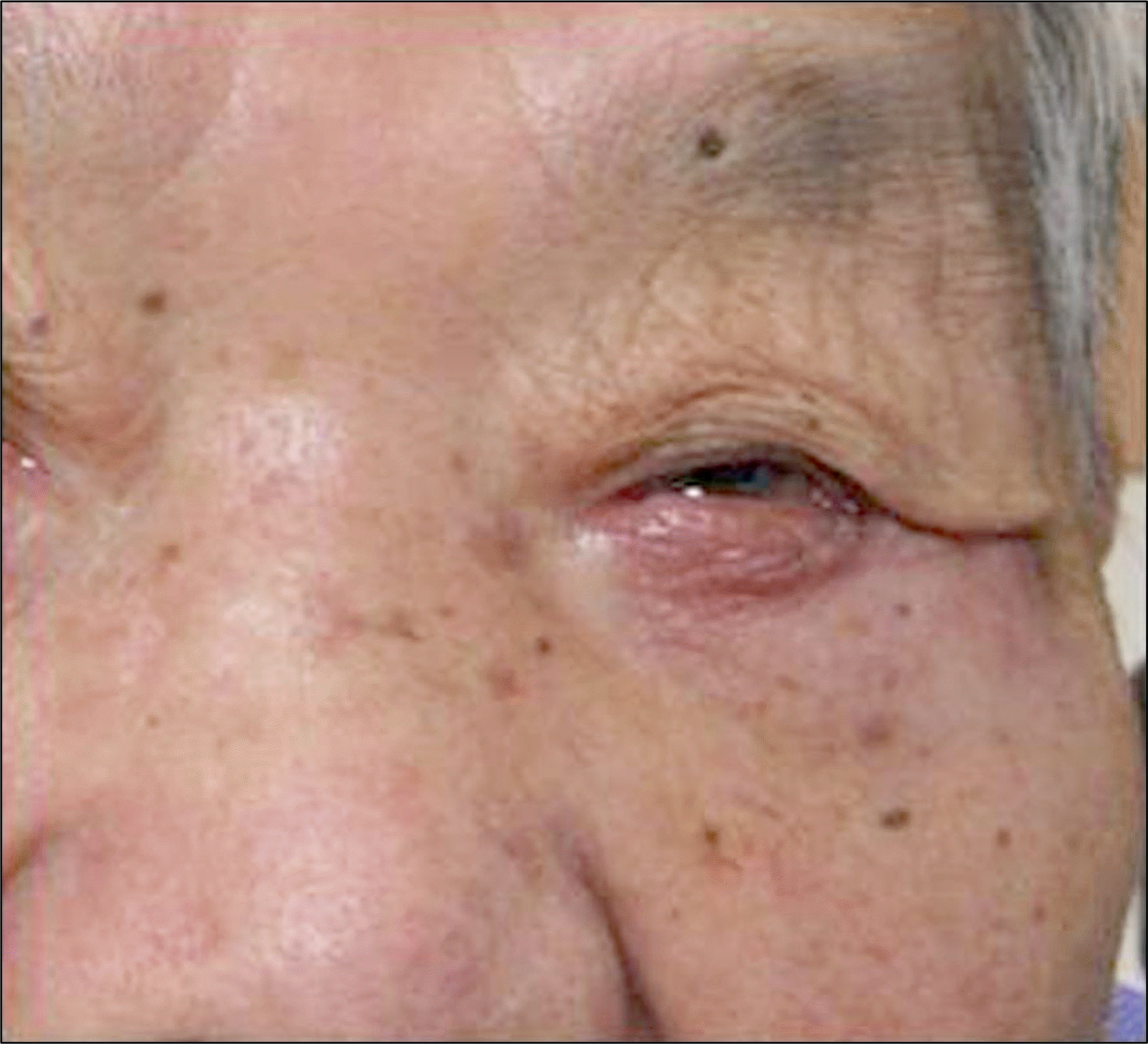
An 87-year-old woman presented with a painful mass and bloody exudates at the left lateral lower eyelid. Four years previous, the patient was diagnosed with basosquamous carcinoma of the left lower eyelid after biopsy at an-other hospital. At that time, she was unable to receive operation because she had suffered from a serious heart condition. With time, the left lower eyelid mass continued to grow and symptoms and signs of pain and bloody exudates appeared. The patient underwent surgery for complete tumor resection and repair and the biopsy of a specimen showed tumor-free margins and no metastasis to distal sites.
Conclusions
Basosquamous carcinoma is a subtype of basal cell carcinoma with aggressive behavior and a higher tendency for recurrence and metastasis. However, our case showed no recurrence with no metastasis to the nearby lymph nodes, vessels, or nerves. We report a case of basosquamous carcinoma of the eyelid at old age that was cured after operative resection.
References
3. De Faria J. Basal cell carcinoma of the skin with areas of squamous cell carcinoma: a basosquamous cell carcinoma? J Clin Pathol. 1985; 38:1273–7.

4. Kazantseva IA, Khlebnikova AN, Babaev VR. Immunohisto-chemical study of primary and recurrent basal cell and metatypical carcinomas of the skin. Am J Dermatopathol. 1996; 18:35–42.

5. Beer TW, Shepherd P, Theaker JM. Ber EP4 and epithelial membrane antigen aid distinction of basal cell, squamous cell and basosquamous carcinomas of the skin. Histopathology. 2000; 37:218–23.

6. Jones MS, Helm KF, Maloney ME. The immunohistochemical characteristics of the basosquamous cell carcinoma. Dermatol Surg. 1997; 23:181–4.

7. Crowson AN. Basal cell carcinoma: biology, morphology and clinical implications. Mod Pathol. 2006; 19(Suppl 2):S127–47.

9. Torosian MH, Daly JM. Nutritional support in the cancer-bearing host. Effects on host and tumor. Cancer. 1986; 58:1915–29.

10. Nixon DW, Lawson DH, Kutner M, et al. Hyperalimentation of the cancer patient with protein-calorie undernutrition. Cancer Res. 1981; 41:2038–45.
11. Borel DM. Cutaneous basosquamous carcinoma. Review of the literature and report of 35 cases. Arch Pathol. 1973; 95:293–7.
12. Martin RC 2nd, Edwards MJ, Cawte TG, et al. Basosquamous carcinoma: analysis of prognostic factors influencing recurrence. Cancer. 2000; 88:1365–9.
Figure 1.
Clinical appearance of the left eyelid lesion at initial presentation shows a 30 (horizontal) × 50 (vertical) × 15 (height) mm3 sized huge mass.

Figure 2.
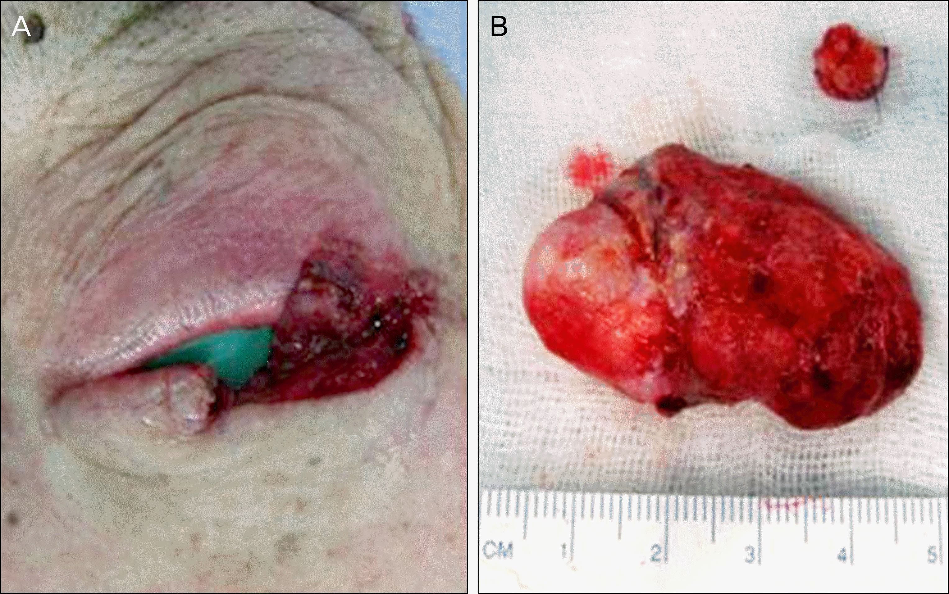
(A) Intraoperative photographs show a widely excised lesion. (B) Upper) 7 (horizontal) × 4 (vertical) × 1 (height) mm3 sized upper eyelid mass, Lower) 30 (horizontal) × 50 (vertical)× 15 (height) mm3 sized lower eyelid mass.

Figure 3.
Hematoxylin and eosin staining of the excised lesion. (A) Low magnification view exhibits squamous differentiation in center of some nodules (arrow). (H & E staining, magnification ×100). (B) Basosquamous carcinoma has an aggressive-growth in-filtrative pattern with tongues of tumor cells embedded in dense stroma containing proplastic fibroblasts (arrow). (H & E staining, magnification ×200).


 PDF
PDF ePub
ePub Citation
 Print
Print


 XML Download