

 PDF
PDF ePub
ePub Citation
 Print
Print


Abstract
Purpose
To determine whether the expression of mutant myocilin can lead to death of human trabecular meshwork (HTM) cells and to determine whether the mechanism by which this occurs is apoptosis.
Methods
HTM cells were transduced with a recombinant adenovirus expressing human mutant (Q368X) myocilin. The apoptotic death of HTM cells caused by expression of mutant myocilin was examined using a cell proliferation assay, flow cytometry, Western blot analysis, and immunocytochemistry.
Go to : 
References
1. Stone EM, Fingert JH, Alward WLM, et al. Identification of a gene that causes primary open angle glaucoma. Science. 1997; 275:668–70.

2. Polansky JR, Kurtz RM, Alvarado JA, et al. Eicosanoid production and glucocorticoid regulatory mechanisms in cultured human trabecular meshwork cells. Prog Clin Biol Res. 1989; 312:113–38.
3. Nguyen TD, Chen P, Huang WD, et al. Gene structure and proper-ties of TIGR, an olfactomedin-related glycoprotein cloned from glucocorticoid-induced trabecular meshwork cells. J Biol Chem. 1998; 273:6341–50.
4. Sohn S, Hur W, Joe MK, et al. Expression of wild-type and trun-cated myocilins in trabecular meshwork cells: their subcellular lo-calizations and cytotoxicities. Invest Ophthalmol Vis Sci. 2002; 43:3680–5.
5. Fingert JH, Stone EM, Sheffield VC, Alward WL. Myocilin glaucoma. Surv Ophthalmol. 2002; 47:547–61.
6. Alward WL, Fingert JH, Coote MA, et al. Clinical features associated with mutations in the chromosome 1 openangle glaucoma gene (GLC1A). N Engl J Med. 1998; 338:1022–7.
7. Fingert JH, Heon E, Liebmann JM, et al. Analysis of myocilin mutations in 1703 glaucoma patients from five different populations. Hum Mol Genet. 1999; 8:899–905.
8. Yoon SJ, Kim HS, Moon JI, et al. Mutations of the TIGR/MYOC gene in primary openangle glaucoma in Korea. Am J Hum Genet. 1999; 64:1775–8.
9. Wiggs JL, Vollrath D. Molecular and clinical evaluation of a patient hemizygous for TIGR/MYOC. Arch Ophthalmol. 2001; 119:1674–8.
10. Lam DS, Leung YF, Chua JK, et al. Truncations in the TIGR gene in individuals with and without primary openangle glaucoma. Invest Ophthalmol Vis Sci. 2000; 41:1386–91.
11. Kim BS, Savinova OV, Reedy MV, et al. Targeted disruption of the myocilin gene (Myoc) suggests that human glaucoma-causing mutations are gain of function. Mol Cel Biol. 2001; 21:7707–13.
12. Zhou ZH, Vollrath D. A cellular assay distinguishes normal and mutant TIGR/myocilin protein. Hum Mol Genet. 1999; 8:2221–8.
14. Jacobson N, Andrews M, Shepard AR, et al. Non-secretion of mutant proteins of the glaucoma gene myocilin in cultured trabecular meshwork cells and in aqueous humor. Hum Mol Genet. 2001; 10:117–25.
15. Joe MK, Sohn S, Hur W, et al. Accumulation of mutant myocilins in ER leads to ER stress and potential cytotoxicity in human trabecular meshwork cells. Biochem Biophys Res Commun. 2003; 312:592–600.
16. Liu Y, Vollrath D. Reversal of mutant myocilin non-secretion and cell killing: implications for glaucoma. Hum Mol Genet. 2004; 13:1193–204.
17. Yam GH, Gaplovska-Kysela K, Zuber C, Roth J. Aggregated myocilin induces Russell bodies and causes apoptosis - Implications for the pathogenesis of myocilin-caused primary openangle glaucoma. Am J Pathol. 2007; 170:100–9.
18. Karali A, Russell P, Stefani FH, Tamm ER. Localization of myocilin/trabecular meshwork–inducible glucocorticoid response protein in the human eye. Invest Ophthalmol Vis Sci. 2000; 41:729–40.
19. Rutkowski DT, Hegde RS. Regulation of basal cellular physiology by the homeostatic unfolded protein response. J Cell Biol. 2010; 189:783–94.
20. Kim R, Emi M, Tanabe K, Murakami S. Role of the unfolded protein response in cell death. Apoptosis. 2006; 11:5–13.
21. Moretti L, Cha YI, Niermann KJ, Lu B. Switch between apoptosis and autophagy: radiation-induced endoplasmic reticulum stress? Cell Cycle. 2007; 6:793–8.
22. Joe MK, Tomarev SI. Expression of myocilin mutants sensitizes cells to oxidative stress-induced apoptosis: implication for glaucoma pathogenesis. Am J Pathol. 2010; 176:2880–90.
Go to :
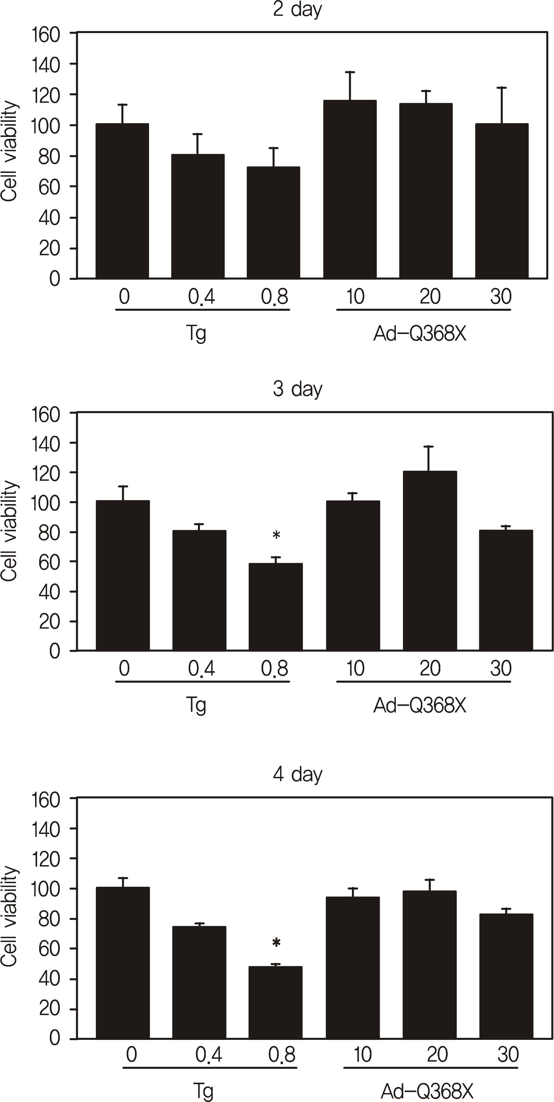 | Figure 1.Effect of mutant myocilin expression on cell viability. HTM cells were transduced at an MOI indicated with an adenovirus expressing mutant (Q368X) myocilin (Ad-Q368X), and 2, 3, and 4 days after transduction, subjected to cell proliferation assay. Cells treated for 48 hours with Tg were included as positive controls. Each column rep-resents the means from three different wells of three independent experiments with the bars denoting the standard deviation. ∗ p < 0.01 vs. control (Dunnet's test). |
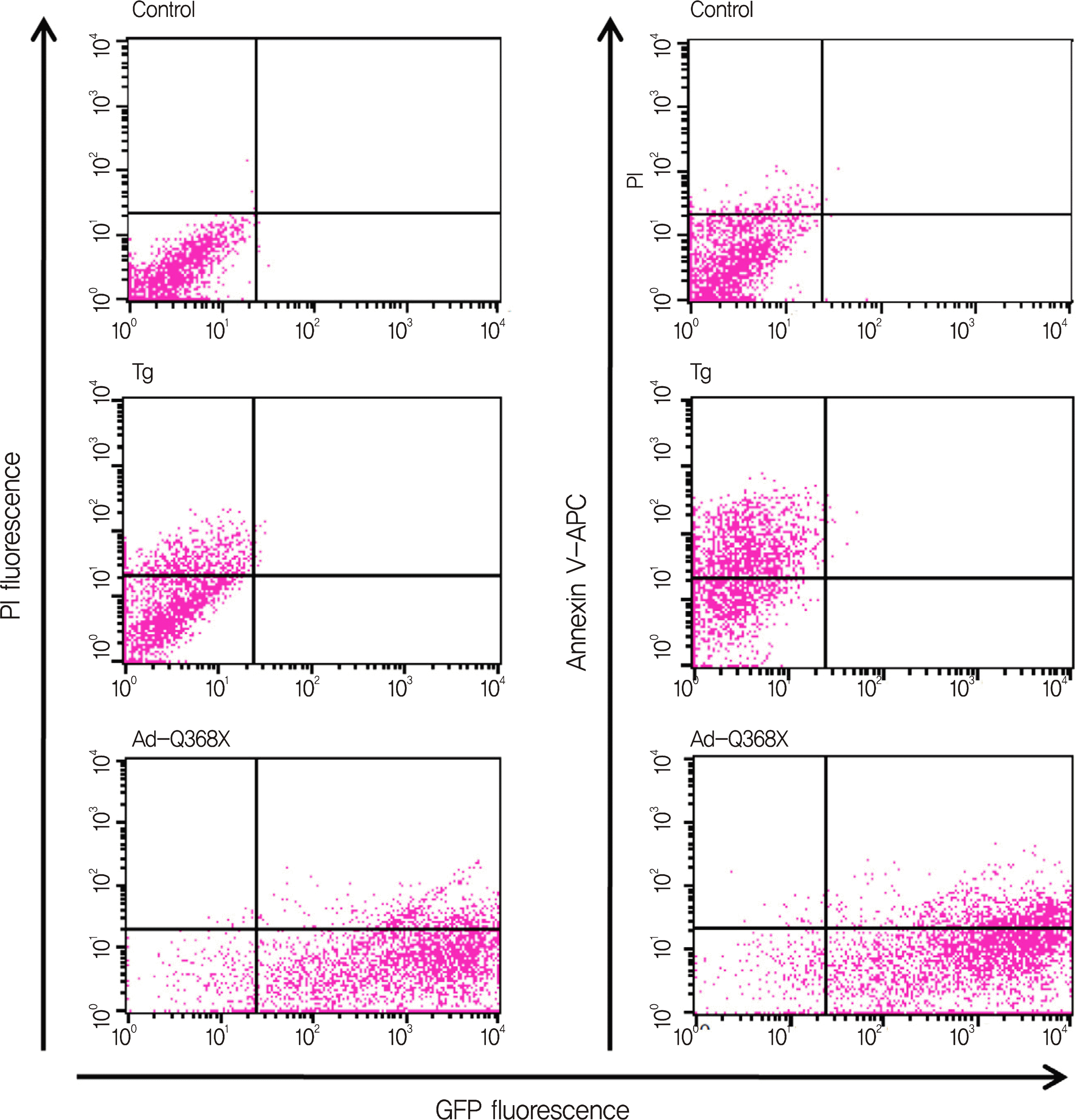 | Figure 2.Quantitative analysis of apoptotic cell death caused by mutant myocilin expression. HTM cells were transduced at an MOI of 50 pfu per cell with AdQ368X, and 48 hours later, stained with Annexin V-APC or propi-dium iodide (PI) and then analyzed by flow cytometry. Cells treated for 48 hours with 400 uM Tg served as positive controls. Shown are representative of three independent experiments with similar results. |
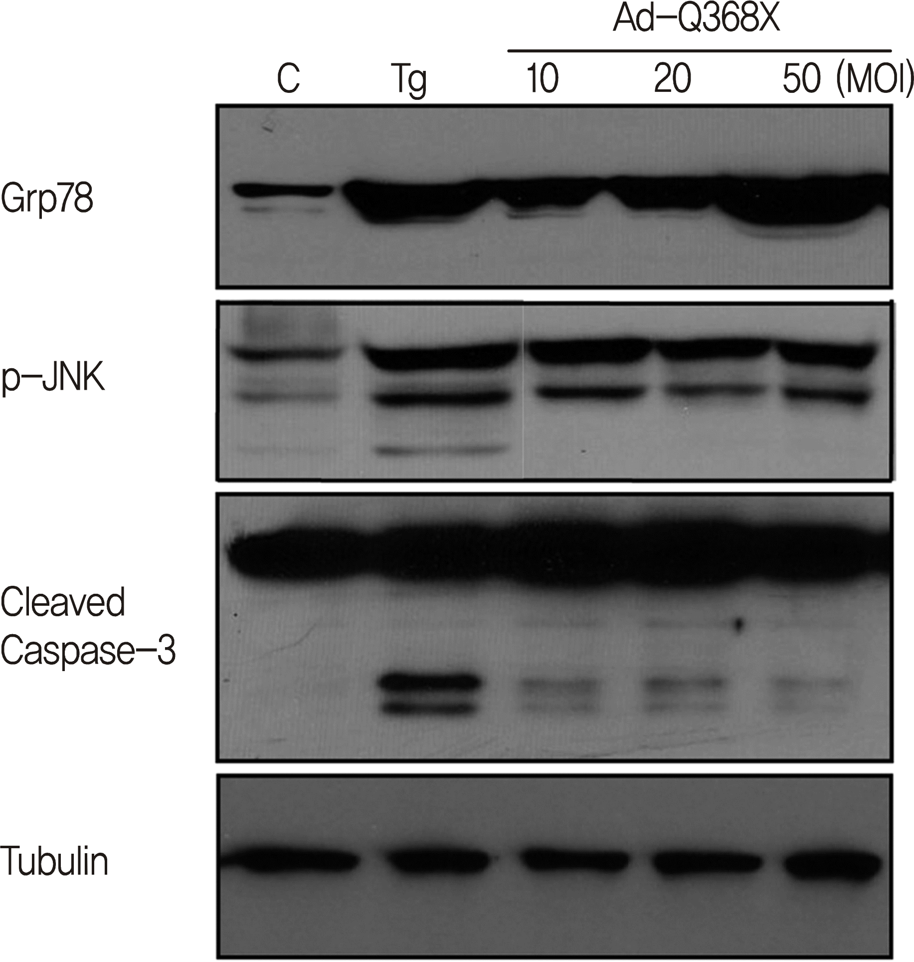 | Figure 3.Western blots showing the effect of mutant myocilin expression on the level of Grp78, p-JNK, and cleaved cas-pase-3. HTM cells transduced at an MOI indicated with Ad-Q368X. After 48 hours, cell lysates were assayed for Grp78, p-JNK and cleavaged caspase 3 by Western blot analysis. Data were normalized with α-tubulin expression. |
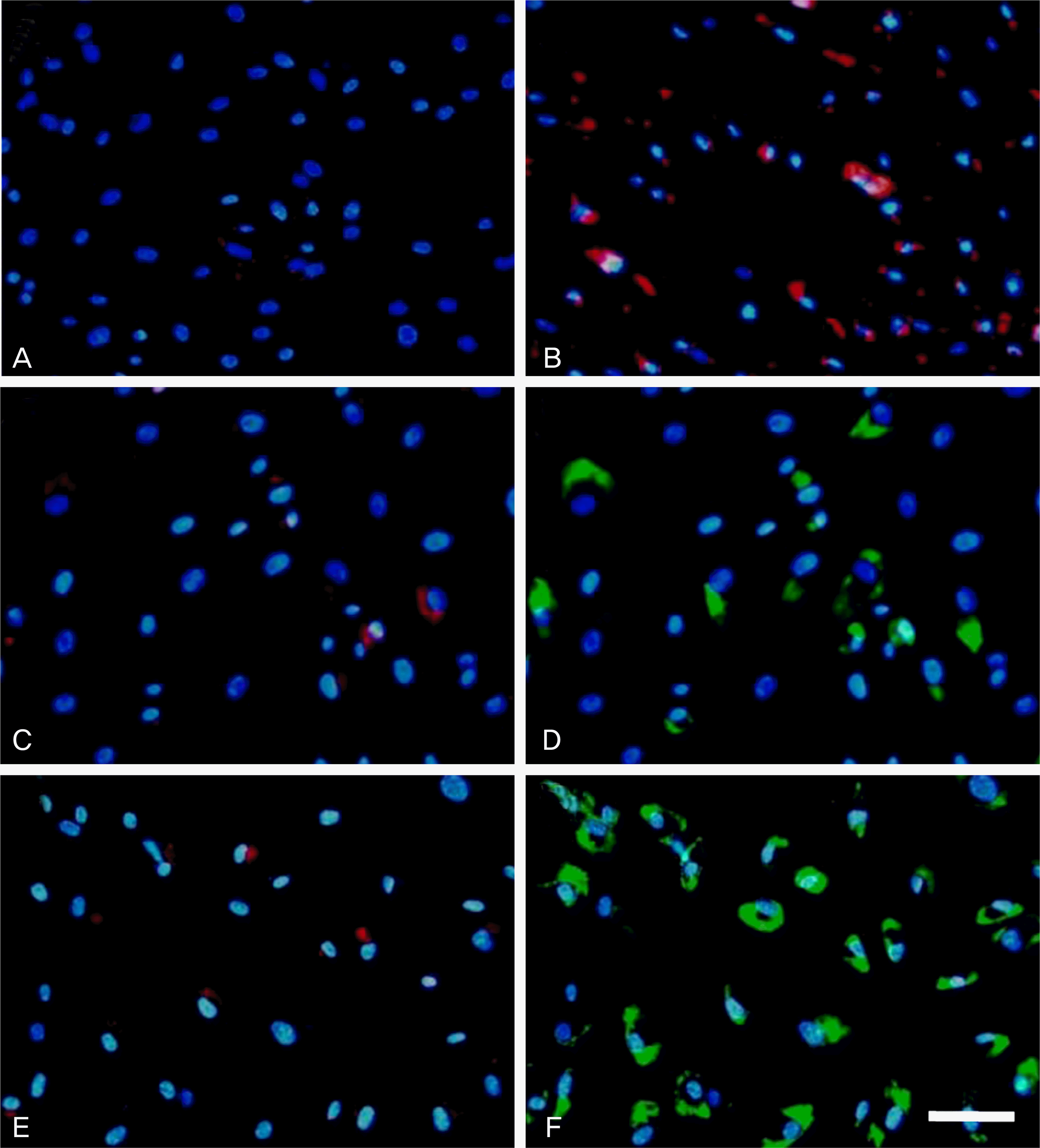 | Figure 4.Effect of mutant myocilin expression on the activation of caspsae-3. HTM cells were transduced at an MOI of 50 (C, D) or 100 pfu (E, F) per cell with Ad-Q368X, and after 48 hours, stained with aspartylglutamy-lalanylaspartic acid (DEVD) coupled to cresyl violet, MR-(DEVD)2 (A, B, C, and E). Cells untreated (A) or treated (B) with 400 uM of Tg served as controls. Red fluorescence and green fluorescence represent caspase-3 activity and GFP-tagged myocilin expression, respectively. Blue fluorescence corresponds to nuclei stained with Hoechst 33342. Bar: 100 um. |
 XML Download