Abstract
Purpose
To report the visual outcome and to determine the stability of WIOL-CF® (GELMED, Praha, Czech Republic) hydrogel full-optics accommodative intraocular lens (IOL).
Methods
The present study evaluated 20 eyes of 10 patients who underwent routine cataract surgery and WIOL-CF® accommodative IOL implantation. Measurement included uncorrected/best corrected visual acuities (VA) at near and distant, degree of IOL decentration, degree of IOL tilting and anterior chamber depth on postoperative day 1, 1 month, 2 months, 6 months and 12 months.
Results
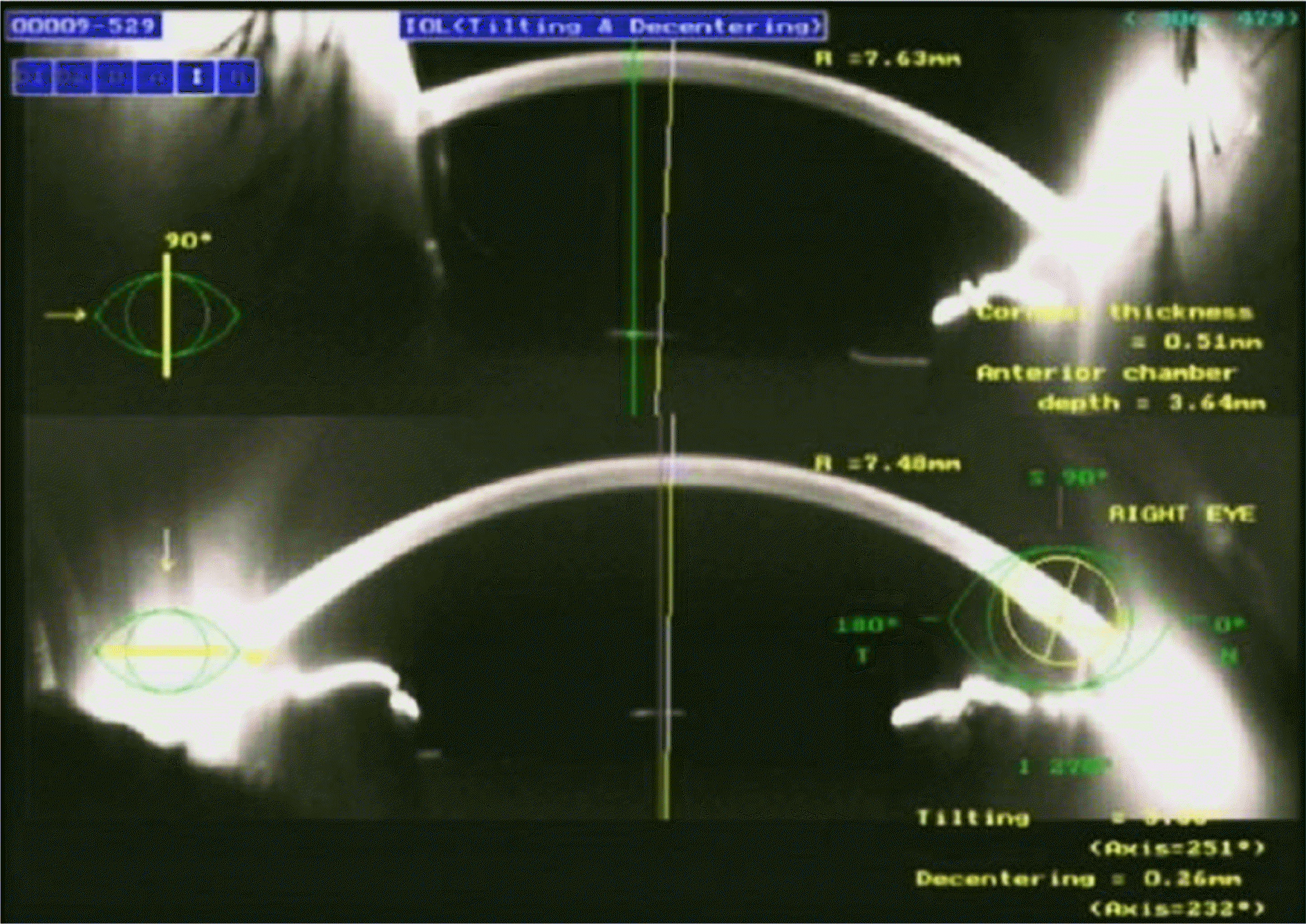
Result analysis was performed with 19 eyes of 10 patients, except for 1 complicated eye. At 12 months, the uncorrected distance VA of 11 eyes (57.9%) and 17 eyes (89.5%) were 20/25 and 20/40 or better, respectively. At 12 months, the uncorrected near VA of 12 eyes (63.2%) and 18 eyes (94.7%) were 20/25 and 20/40 or better, respectively. There was no statistically significant difference in anterior chamber depth (p > 0.05), IOL decentration (p > 0.05) and IOL tilting (p > 0.05) on postoperative day 1, 1 month, 2 months, 6 months and 12 months.
Go to : 
References
1. Becker KA, Martin M, Rabsilber TM, et al. Prospective, non-rand-omised, long term clinical evaluation of a foldable hydrophilic single piece intraocular lens: results of the Centerflex FDA study. Br J Ophthalmol. 2006; 90:971–4.

2. Olson RJ, Werner L, Mamalis N, Cionni R. New intraocular lens technology. Am J Ophthalmol. 2005; 140:709–16.

4. Leyland M, Zinicola E. Multifocal versus monofocal intraocular lenses in cataract surgery; a systematic review. Ophthalmology. 2003; 110:1789–98.
5. Findl O, Leydolt C. Meta-analysis of accommodating intraocular lenses. J Cataract Refract Surg. 2007; 33:522–7.

6. Lee JM, Seo KY, Kim EK. Comparison of optical aberrations and contrast sensitivity between monofocal and multifocal intraocular lens. J Korean Ophthalmol Soc. 2002; 43:1882–6.
7. Song MJ, Lee MK, Park BI. A Clinical study of 3M multifocal intraocular lens implant. J Korean Ophthalmol Soc. 1991; 32:234–40.
8. Montés-Micó R, España E, Bueno I, et al. Visual performance with multifocal intraocular lenses: mesopic contrast sensitivity under distance and near conditions. Ophthalmology. 2004; 111:85–96.
9. Sen HN, Sarikkola AU, Uusitalo RJ, Laatikainen L. Quality of vision after AMO Array multifocal intraocular lens implantation. J Cataract Refract Surg. 2004; 30:2483–93.

10. Schmitz S, Dick HB, Krummenauer F, et al. Contrast sensitivity and glare disability by halogen light after monofocal and multifocal lens implantation. Br J Ophthalmol. 2000; 84:1109–12.

11. Allen ED, Burton RL, Webber SK, et al. Comparison of a diffractive bifocal and a monofocal intraocular lens. J Cataract Refract Surg. 1996; 22:446–51.

12. Rossetti L, Carraro F, Rovati M, Orzalesi N. Performance of diffractive multifocal intraocular lenses in extracapsular cataract surgery. J Cataract Refract Surg. 1994; 20:124–8.

13. Kamlesh , Dadeya S, Kaushik S. Contrast sensitivity and depth of focus with aspheric multifocal versus conventional monofocal intraocular lens. Can J Ophthalmol. 2001; 36:197–201.

14. Davison JA, Simpson MJ. History and development of the apo-dized diffractive intraocular lens. J Cataract Refract Surg. 2006; 32:849–58.

15. Slagsvold JE. 3M diffractive multifocal intraocular lens: eight year followup. J Cataract Refract Surg. 2000; 26:402–7.

16. Cumming JS, Ritter JA. The measurement of vitreous cavity length and its comparison pre- and postoperatively. Eur J Implant Refract Surg. 1994; 6:261–72.

17. Cumming JS, Slade SG, Chayet A. Clinical evaluation of the mod-el AT-45 silicone accommodating intraocular lens: results of feasi-bility and the initial phase of a Food and Drug Administration clinical trial. Ophthalmology. 2001; 108:2005–9.

18. Cumming JS, Colvard DM, Dell SJ, et al. Clinical evaluation of the Crystalens AT-45 accommodating intraocular lens: results of the U.S. Food and Drug Administration clinical trial. J Cataract Refract Surg. 2006; 32:812–25.
19. Kwon JW, Kang S, Chung SK, Baek NH. Clinical results of Crystalens® (AT-45) accommodating intraocular Lens. J Korean Ophthalmol Soc. 2009; 50:1179–83.
20. Kim C, Kwon JW, Wee WR, et al. Factors affecting the visual outcome of cataract surgery in the very elderly. J Korean Ophthalmol Soc. 2007; 48:905–10.
21. Morrison JD, Jay JL. Changes in visual function with normal ageing, cataract and intraocular lenses. Eye. 1993; 7:20–5.

22. Jay JL, Mammo RB, Allan D. Effect of age on visual acuity after cataract extraction. Br J Ophthalmol. 1987; 71:112–5.

23. Dogru M, Honda R, Omoto M, et al. Early visual results with the 1CU accommodating intraocular lens. J Cataract Refract Surg. 2005; 31:895–902.

Go to : 
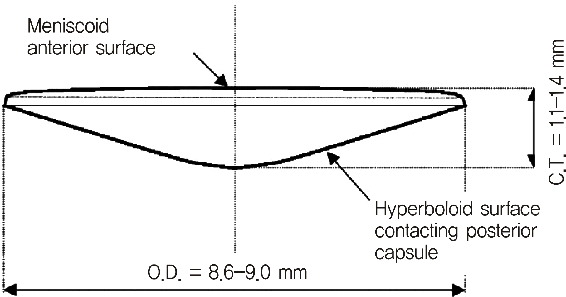 | Figure 1.The design of WIOL-CF® polyfocal full-optics accommodative intraocular lens. O.D. = optic diameter; C.T.= central thickness. |
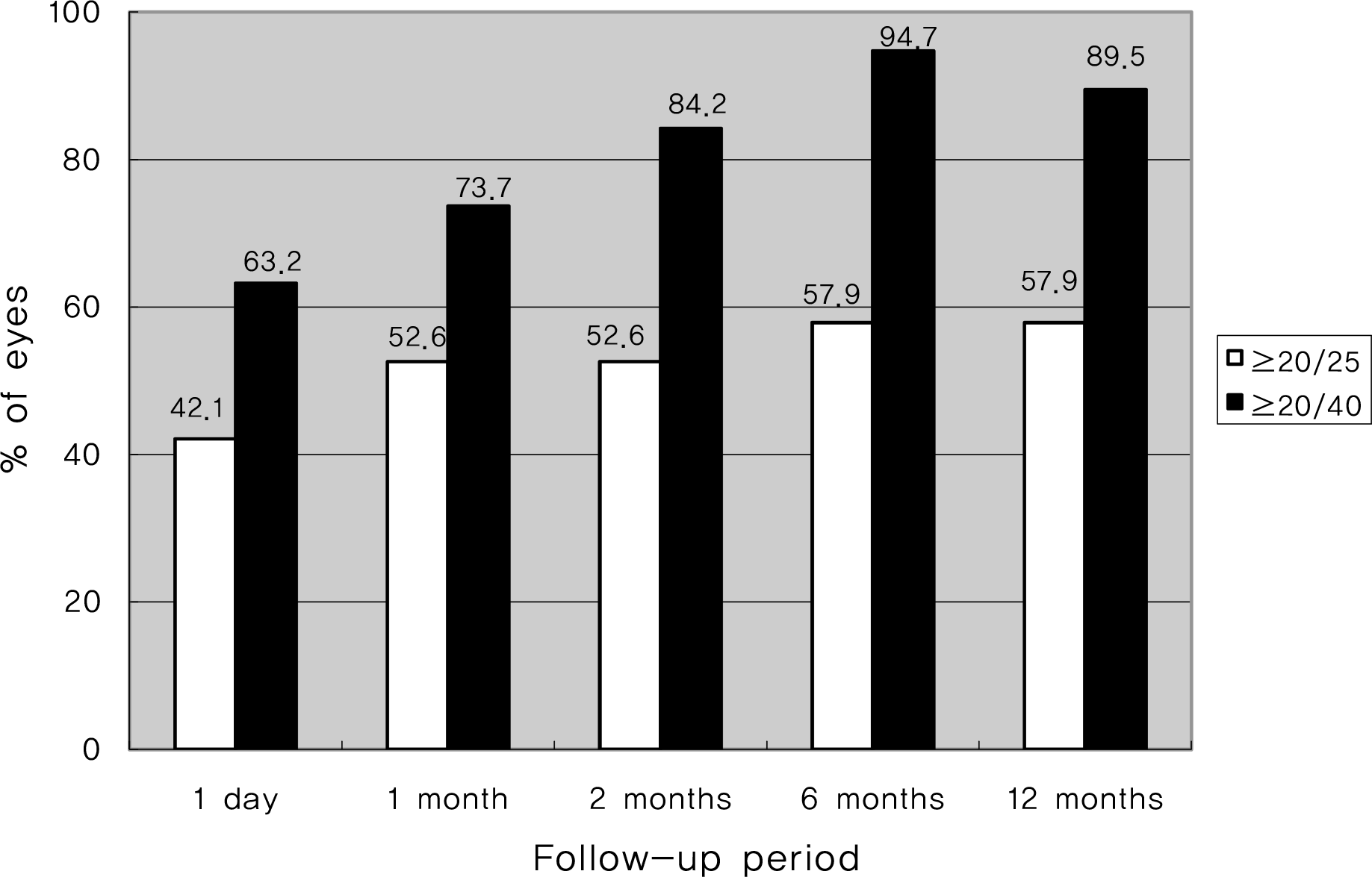 | Figure 3.Uncorrected distant visual acuity after WIOL-CF® accommodative intraocular lens implantation, according to the followup period (n = 19). |
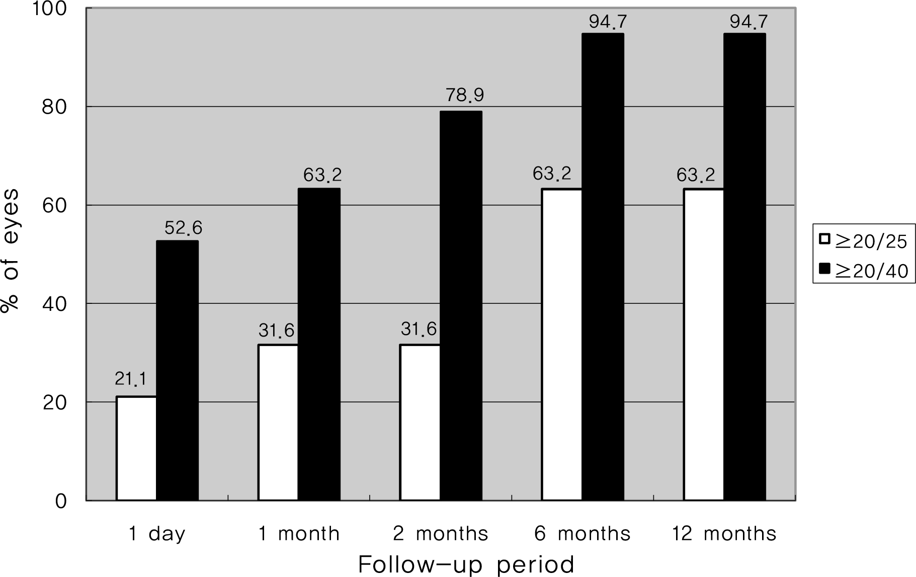 | Figure 4.Uncorrected near visual acuity after WIOL-CF® accommodative intraocular lens implantation, according to the followup period (n = 19). |
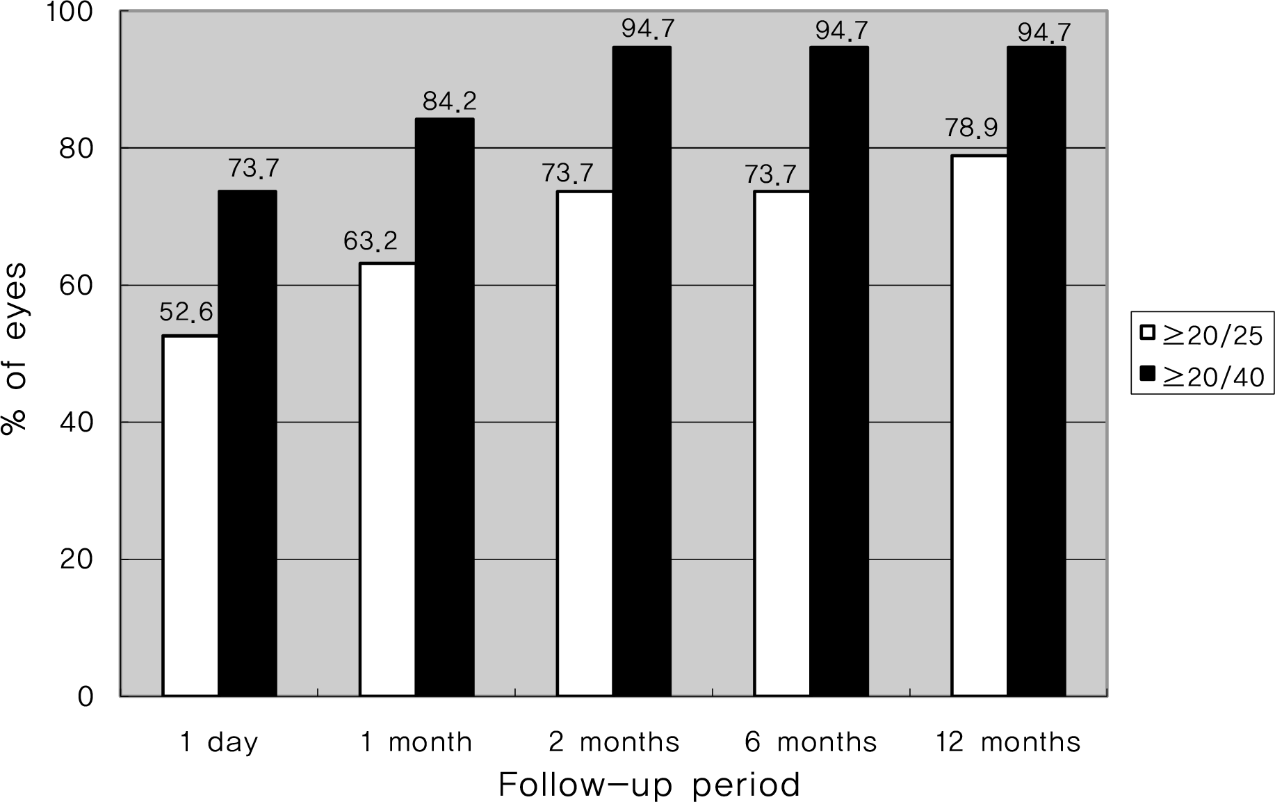 | Figure 5.Best corrected distant visual acuity after WIOL-CF® accommodative intraocular lens implantation, according to the followup period (n = 19). |
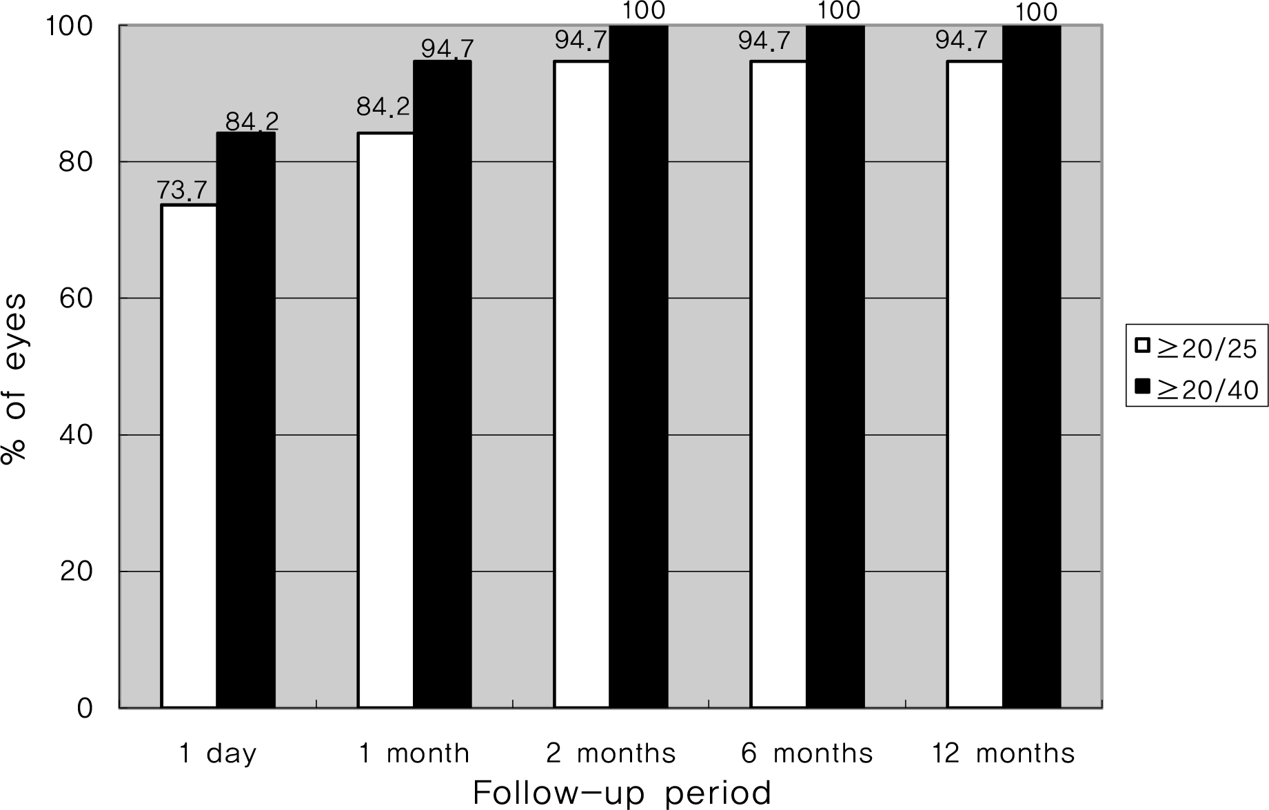 | Figure 6.Best corrected near visual acuity after WIOL-CF® accommodative intraocular lens implantation, according to the followup period (n = 19). |
Table 1.
Patient demographics
| Sex (M/F) | 3/7 |
| Age (yr) | 67 ± 9.7 |
| Axial length (mm) | 23.61 ± 0.86 |
| Predicted postoperative refraction (diopter) | -0.12 ± 0.09 |
Table 2.
Postoperative changes in the intraocular lens decentration (mm), tilting (°) and anterior chamber depth (mm)
|
Postoperative followup periods |
p-value∗ | |||||
|---|---|---|---|---|---|---|
| 1 day | 1 month | 2 months | 6 months | 12 months | ||
| Decentration (mm) | 0.19 ± 0.07 | 0.20 ± 0.07 | 0.21 ± 0.09 | 0.19 ± 0.07 | 0.19 ± 0.09 | 0.27 |
| Tilting (°) | 3.05 ± 0.47 | 3.11 ± 0.50 | 3.24 ± 0.48 | 3.14 ± 0.50 | 3.18 ± 0.46 | 0.24 |
| Anterior chamber depth (mm) | 3.60 ± 0.29 | 3.60 ± 0.32 | 3.59 ± 0.33 | 3.60 ± 0.34 | 3.60 ± 0.31 | 0.57 |


 PDF
PDF ePub
ePub Citation
 Print
Print


 XML Download