

 PDF
PDF ePub
ePub Citation
 Print
Print


Abstract
Purpose
To analyze the incidence and clinical course of epithelial ingrowth after laser in situ keratomileusis (LASIK) using a femtosecond laser.
Methods
We retrospectively evaluated the results of 1158 eyes of 581 patients who received LASIK with the flap created by a femtosecond laser from February 2006 to March 2009 at our institute. We investigated the incidence and clinical course of LASIK in which the flap was created by a femtosecond laser.
Results
Epithelial ingrowth was first detected one week after surgery in 12 (57%) of 21 eyes and within one month in 19 eyes (90%). Epithelial ingrowth was localized most commonly near the temporal and nasal flap edge in 12 eyes (57%). In two eyes (9.5%), an isolated ingrowth mass was located in the pupillary area. The development of more than 2.0 mm of epithelial ingrowth was observed in three eyes (0.25%). Among 28 eyes with loose epithelium or epithelial detachment during surgery, epithelial ingrowth was observed in six eyes (21.4%) compared with 15 (1.32%) of 1130 eyes without loose epithelium or epithelial detachment. Therefore, epithelial detachment during surgery was significantly associated with epithelial ingrowth (p = 0.00).
Go to : 
References
1. Ambrosio R Jr, Wilson SE. Complications of laser in situ keratomileusis: etiology, prevention, and treatment. J Refract Surg. 2001; 17:350–79.

2. Kim SW, Byun YJ, Kim EK, Kim TI. Treatment of epithelial ingrowth after laser in situ keratomileusis using amniotic membrane patch. J Korean Ophthalmol Soc. 2007; 48:230–7.
3. Jacobs JM, Taravella MJ. Incidence of intraoperative flap complications in laser in situ keratomileusis. J Cataract Refract Surg. 2002; 28:23–8.
4. Asano-Kato N, Toda I, Hori-Komai Y, et al. Risk factors for in-sufficient fixation of microkeratome during laser in situ keratomileusis. J Refract Surg. 2002; 18:47–50.
5. Domniz Y, Comaish IF, Lawless MA, et al. Epithelial ingrowth: causes, prevention, and treatment in 5 cases. J Cataract Refract Surg. 2001; 27:1803–11.
6. Naoumidi I, Papadaki T, Zacharopoulos I, et al. Epithelial ingrowth after laser in situ keratomileusis: a histopathologic study in human corneas. Arch Ophthalmol. 2003; 121:950–5.
7. Farah SG, Azar DT, Gurdal C, Wong J. Laser in situ keratomileusis: literature review of a developing technique. J Cataract Refract Surg. 1998; 24:989–1006.
8. Helena MC, Meisler D, Wilson SE. Epithelial growth within the lamellar interface after laser in situ keratomileusis (LASIK). Cornea. 1997; 16:300–5.
9. Walker MB, Wilson SE. Incidence and prevention of epithelial growth within the interface after laser in situ keratomileusis. Cornea. 2000; 19:170–3.
10. Wright JD Jr, Neubaur CC, Stevens G Jr. Epithelial ingrowth in a corneal graft treated by laser in situ keratomileusis: light and electron microscopy. J Cataract Refract Surg. 2000; 26:49–55.
11. Castillo A, Diaz-Valle D, Gutierrez AR, et al. Peripheral melt of flap after laser in situ keratomileusis. J Refract Surg. 1998; 14:61–3.
12. Wang MY, Maloney RK. Epithelial ingrowth after laser in situ keratomileusis. Am J Ophthalmol. 2000; 129:746–51.
13. Asano-Kato N, Toda I, Hori-Komai Y, et al. Epithelial ingrowth after laser in situ keratomileusis: Clinical features and possible mechanisms. Am J Ophthalmol. 2002; 134:801–7.
14. Sugar A. Ultrafast(femtosecond) laser refractive surgery. Curr Opin Ophthalmol. 2002; 13:246–9.
15. Ratkay-Traub I, Juhasz T, Horvath C, et al. Ultra-short pulse(femtosecond) laser surgery; initial use in LASIK flap creation. Ophthalmol Clin North Am. 2001; 14:347–55.
16. Nordan LT, Slade SG, Baker RN, et al. Femtosecond laser flap creation for laser in situ keratomileusis: six-month followup of initial U.S. clinical series. J Refract Surg. 2003; 19:8–14.
17. Kezirian GM, Stonecipher KG. Comparison of the IntraLase femtosecond laser and mechanical keratomes for laser in situ keratomileusis. J Cataract Refract Surg. 2004; 30:804–11.
18. Kurtz RM, Horvath C, Liu HH, et al. Lamellar refractive surgery with scanned intrastromal picosecond and femtosecond laser puls-es in animal eyes. J Refract Surg. 1998; 14:541–8.
19. Binder PS. Flap dimensions created with the IntraLase FS laser. J Cataract Refract Surg. 2004; 30:26–32.
20. Tchah HW, Yoon JT, Lee GJ. Flap complications of LASIK. J Korean Ophthalmol Soc. 2000; 41:1146–50.
21. Machat JJ, Slade SG, Probst LE. The Art of LASIK. 2nd ed.SLACK incorporated;1999. p. 348.
22. Dastgheib KA, Clinch TE, Manche EE, et al. Sloughing of corneal epithelium and wound healing complications associated with laser in situ keratomileusis in patients with epithelial basement membrane dystrophy. Am J Ophthalmol. 2000; 130:297–303.
23. Carr JD, Nardone R Jr, Stulting RD, et al. Risk factors for epithelial ingrowth after LASIK. ARVO abstract 1089. Invest Ophthalmol Vis Sci. 1997; 38:S232.
24. Farah SG, Azar DT, Gurdal C, Wong J. Laser in situ keratomileusis: literature review of a developing technique. J Cataract Refract Surg. 1998; 24:989–1006.
25. Biser SA, Bloom AH, Donnenfeld ED, et al. Flap folds after femtosecond LASIK. Eye Contact lens. 2003; 29:252–4.
26. Morishige N, Kesler-Diaz A, Wahlert AJ, et al. Corneal response to femtosecond laser photodisruption in the rabbit. Experimental Eye Research. 2008; 86:835–43.
27. Stramer BM, Zieske JD, Jung JC, et al. Molecular mechanisms controlling the fibrotic repair phenotype in cornea: implications for surgical outcomes. Invest Ophthalmol Vis Sci. 2003; 44:4237–46.
Go to :
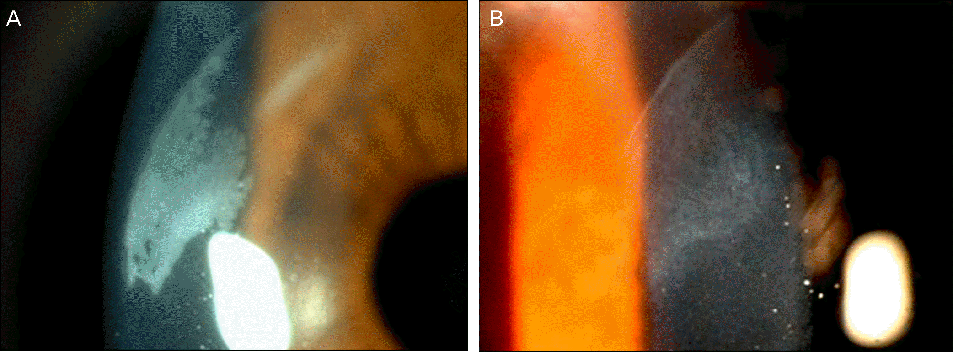 | Figure 3.Epithelial ingrowth does not improve or aggravate. (A) 8 days after LASIK. (B) 22 days after LASIK. |
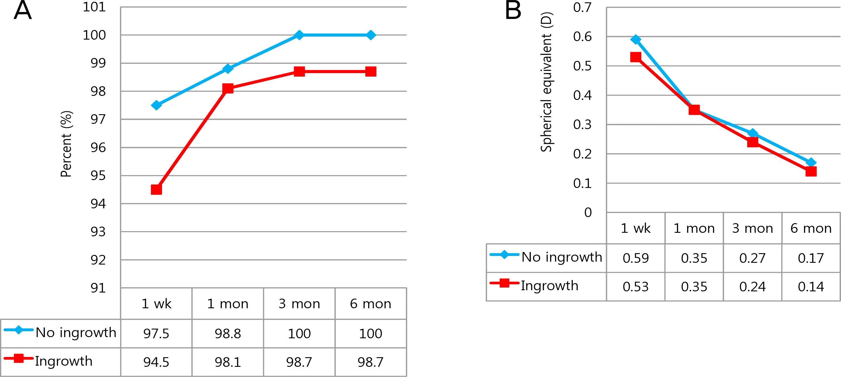
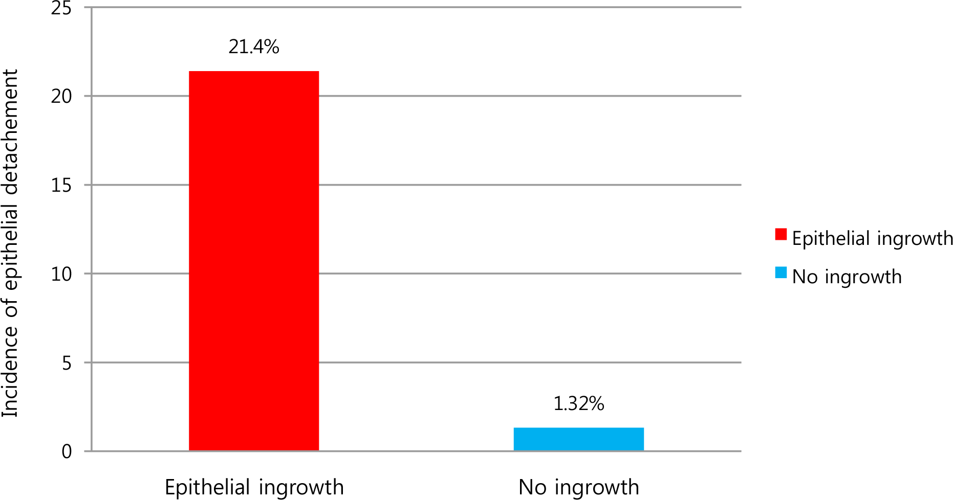 | Figure 5.Incidence of epithelial detachment in epithelial ingrowth group and in no ingrowth group. There was significant relationship between incidence of epithelial detachment and epithelial ingrowth (p=0.00). |
 XML Download