

 PDF
PDF ePub
ePub Citation
 Print
Print


Abstract
Purpose
To evaluate the changes in breakup time (BUT) and corneal sensitivity following LASIK surgery for refractive error correction with presbyopia in patients older than 45 years.
Methods
The authors of the present study measured the BUT and corneal sensitivity of 92 eyes that received LASIK surgery for correcting refractive error with presbyopia. The eyes were divided into groups according to gender and pre-operative refractive error before surgery and 1, 3, 6 and 12 months after LASIK.
Results
The mean age of patients was 52.01 ± 5.51 years, and the male to female eye distribution was 31:61. The value of BUT before surgery and 1, 3, 6, and 12 months postoperative was 5.31 ± 2.03 sec, 4.47 ± 1.67 sec, 4.04 ± 1.58 sec, 4.53 ± 1.51 sec, and 4.87 ± 1.46 sec, respectively; corneal sensitivity was 56.35 ± 5.94 mm, 40.07 ± 14.21 mm, 46.42 ± 10.41 mm, 50.75 ± 8.04 mm, and 52.92 ± 7.51 mm, respectively. BUT was not significantly different relative to refractive error and was significantly shorter in the female group than the male group at 1 month postoperative. Corneal sensation of myopia at 12 months postoperative was statistically higher than at other time points; however, there was no difference between genders. BUT and corneal sensitivity at 12 months postoperative recovered to 91.6% and 93.9% of the preoperative value, respectively.
References
1. Toda I. LASIK and the ocular surface. Cornea. 2008; 27(Suppl 1):S70–6.

2. Konomi K, Chen LL, Tarko RS, et al. Preoperative characteristics and a potential mechanism of chronic dry eye after LASIK. Invest Ophthalmolo Vis Sci. 2008; 49:168–74.
3. Schein OD, Munoz B, Tielsch JM, et al. Prevalence of dry eye among the elderly. Am J Ophthalmol. 1997; 124:723–8.
4. Lin PY, Tsai SY, Cheng CY, et al. Prevalence of dry eye among an elderly Chinese population in Taiwan: the Shihpai Eye Study. Ophthalmology. 2003; 110:1096–101.
5. Brewitt H, Sistani F. Dry eye disease: the scale of the problem. Surv Ophthalmol. 2001; 45:199–202.
6. Rosenfeld SI. Evaluation and management of Post-LASIK dry eye syndrome. Int Ophthalmol Clin. 2010; 50:191–9.
7. Guillon M, Maïssa C. Tear film evaporation-effect of age and gender. Cont Lens Anterior Eye. 2010; 33:171–5.
8. Maissa C, Guillon M. Tear film dynamics and lipid layer characteristics-the effect of aging and gender. Cont Lens Anterior Eye. 2010; 33:176–82.
9. Bragheeth MA, Dua HS. Corneal sensation after myopia and hyperopic LASIK: clinical and confocal microscopic study. Br J Ophthalmol. 2005; 89:580–5.
10. Levinson BA, Rapuano CJ, Cohen EJ, et al. Referrals to the Wills Eye Institute Cornea Service after laser in situ keratomileusis: reasons for patient dissatisfaction. J Cataract Refract Surg. 2008; 34:32–9.
11. Jabbur NS, Sakatani K, O'Brien TP. Survey of complications and recommendations for management in dissatisfied patients seeking a consultation after refractive surgery. J Cataract Refract Surg. 2004; 30:1867–74.
12. Vroman DT, Sandoval HP, Fernández de Castro LE, et al. Effect of hinge location on corneal sensation and dry eye after laser in situ keratomileusis for myopia. J Cataract Refract Surg. 2005; 31:1881–7.
13. Hammond MD, Madigan WP Jr, Bower KS. Refractive surgery in the United States Army, 2000-2003. Ophthalmology. 2005; 112:184–90.
14. Yu EY, Leung A, Rao S, Lam DS. Effect of laser in situ keratomileusis on tear stability. Ophthalmology. 2000; 107:2131–5.
15. Wilson SE. Laser in situ keratomileusis-induced (presumed) neurotrophic epitheliopathy. Ophthalmology. 2001; 108:1082–7.
16. Ambrósio R Jr, Tervo T, Wilson SE. LASIK-associated dry eye and neurotrophic epitheliopathy: pathophysiology and strategies for prevention and treatment. J Refract Surg. 2008; 24:396–407.
17. Salomão MQ, Ambrósio R Jr, Wilson SE. Dry eye associated with laser in situ keratomileusis: Mechanical microkeratome versus femtosecond laser. J Cataract Refract Surg. 2009; 35:1756–60.
18. Kim JS, Kim SH, Kim WS. The change of corneal sensation following LASIK. J Korean Ophthalmol Soc. 1998; 39:1676–82.
19. Mian SI, Li AY, Musch DC, et al. Dry eyes and corneal sensation after laser in situ keratomileusis with femtosecond laser flap creation Effect of hinge position, hinge angle, and flap thickness. J Cataract Refract Surg. 2009; 35:2092–8.
20. Kim JH, Kim JH, Song JS, Kim HM. Factors Associated with the Successful Separation of Corneal Epithelium in Epi-LASIK. J Korean Ophthalmol Soc. 2007; 48:1623–9.
21. Choi W, Park YG, Cho JK, et al. Effect of topical 0.05% cyclo-sporine A in dry eye associated with thyroid ophthalmopathy. J Korean Ophthalmol Soc. 2010; 51:1319–26.
22. Niederer RL, Perumal D, Sherwin T, McGhee CN. Age-related differences in the normal human cornea: a laser scanning in vivo confocal microscopy study. Br J Ophthalmol. 2007; 91:1165–9.
23. Nettun GR, Pflugfelder SC. Post-LASIK tear dysfunction and dysesthesia. Ocul Surf. 2010; 8:135–45.
24. Nejima R, Miyata K, Tanave T, et al. Corneal barrier function, tear film stability, and corneal sensation after photorefractive keratectomy and laser in situ keratomileusis. Am J ophthalmol. 2005; 139:64–71.
25. Goto T, Zheng X, Klyce SD, et al. Evaluation of the tear film stability after laser in situ keratomileusis using the tear film stability analysis system. Am J ophthalmol. 2004; 137:116–20.
26. Lee SJ, Kim JK, Seo KY, et al. Comparison of corneal nerve re-generation and sensitivity between LASIK and laser epithelial keratomileusis. Am J ophthalmol. 2006; 141:1009–15.
27. Pérez-Santoja JJ, Sakla HF, Cardona C, et al. Corneal sensitivity after photorefractive keratectomy and laser in situ keratomileusis for low myopia. Am J Ophthalmol. 1999; 127:497–504.
28. Kalyvianaki MI, Katsanevaki VJ, Kavroulaki DS, et al. Comparison of corneal sensitivity and tear function following Epi-LASIK of laser in situ keratomileusis for myopia. Am J Ophthalmol. 2006; 142:669–71.
29. Shoja MR, Besharati MR. Dry eye after LASIK for myopia: Incidence and risk factors. Eur J Ophthalmol. 2007; 17:1–6.
30. Connor CG, Flockencier LL, Hall CW. The influence of gender on the ocular surface. J Am Opom Assoc. 1999; 70:182–6.
31. Versura P, Campos EC. Menopause and dry eye. A possible relationship. Gynecol Endocrinol. 2005; 20:289–98.
32. du Toit R, Situ P, Simpson T, et al. The effects of six months of contact lens wear on the tear film, ocular surfaces, and symptoms of presbyopes. Optom Vis Sci. 2001; 78:455–62.
Figure 1.
The change of tear breakup time after LASIK. ∗ Significantly different from pre-LASIK value, Wilcoxon signed ranked test.
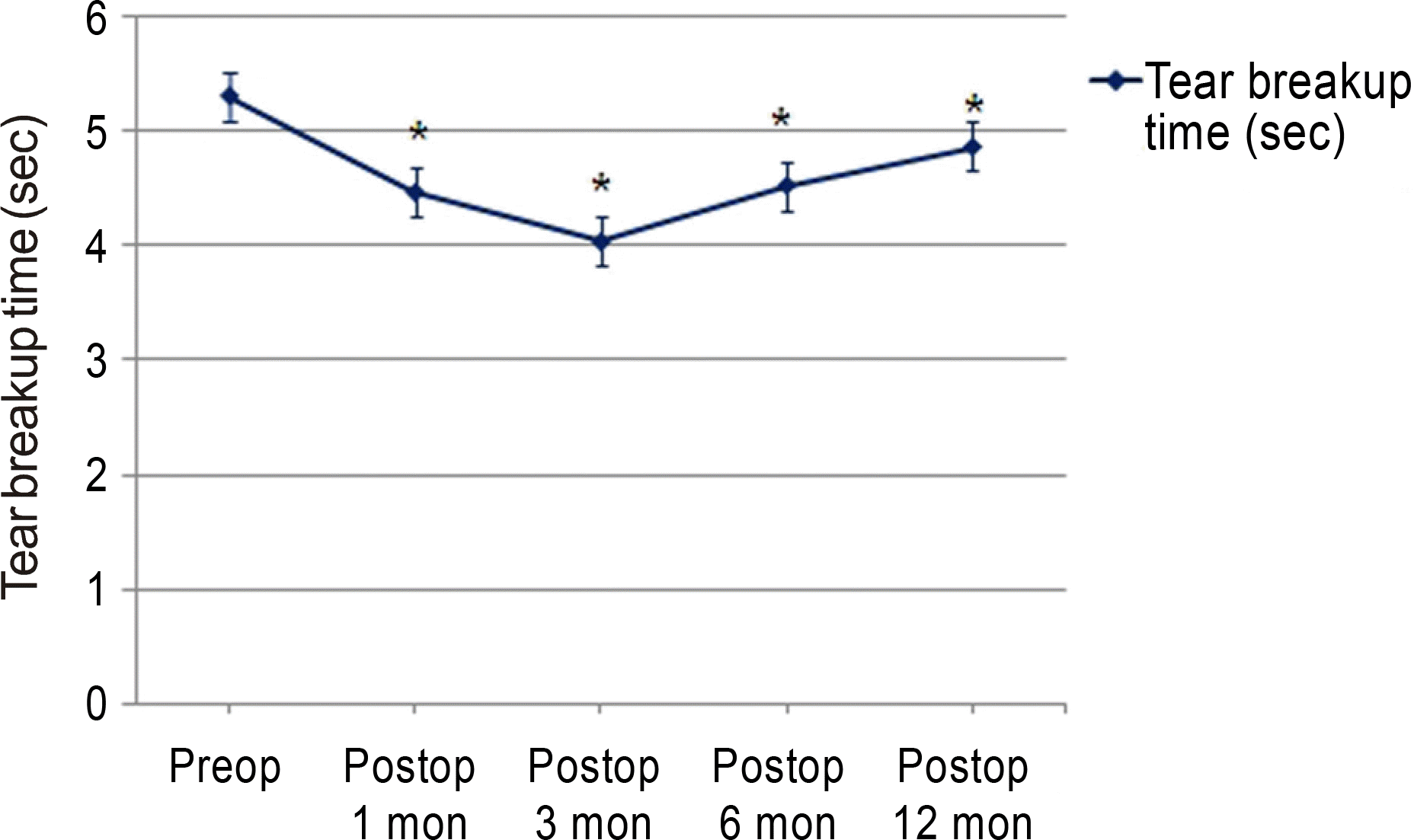
Figure 2.
The change of corneal sensation after LASIK. ∗ Significantly different from pre-LASIK value, Wilcoxon signed ranked test.
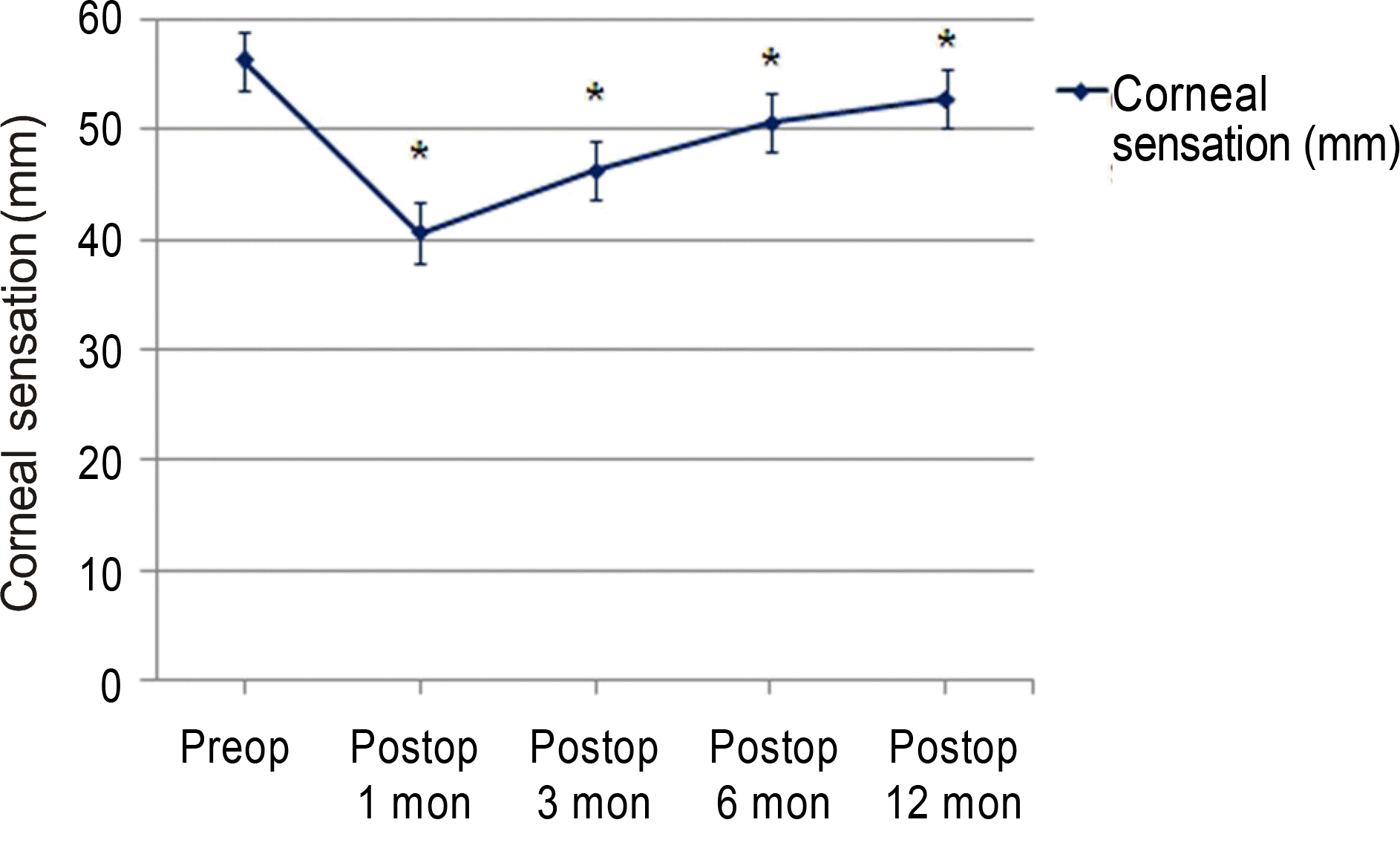
Figure 3.
Time course of changes of tear breakup time after LASIK grouped with pre-LASIK spherical aberration.
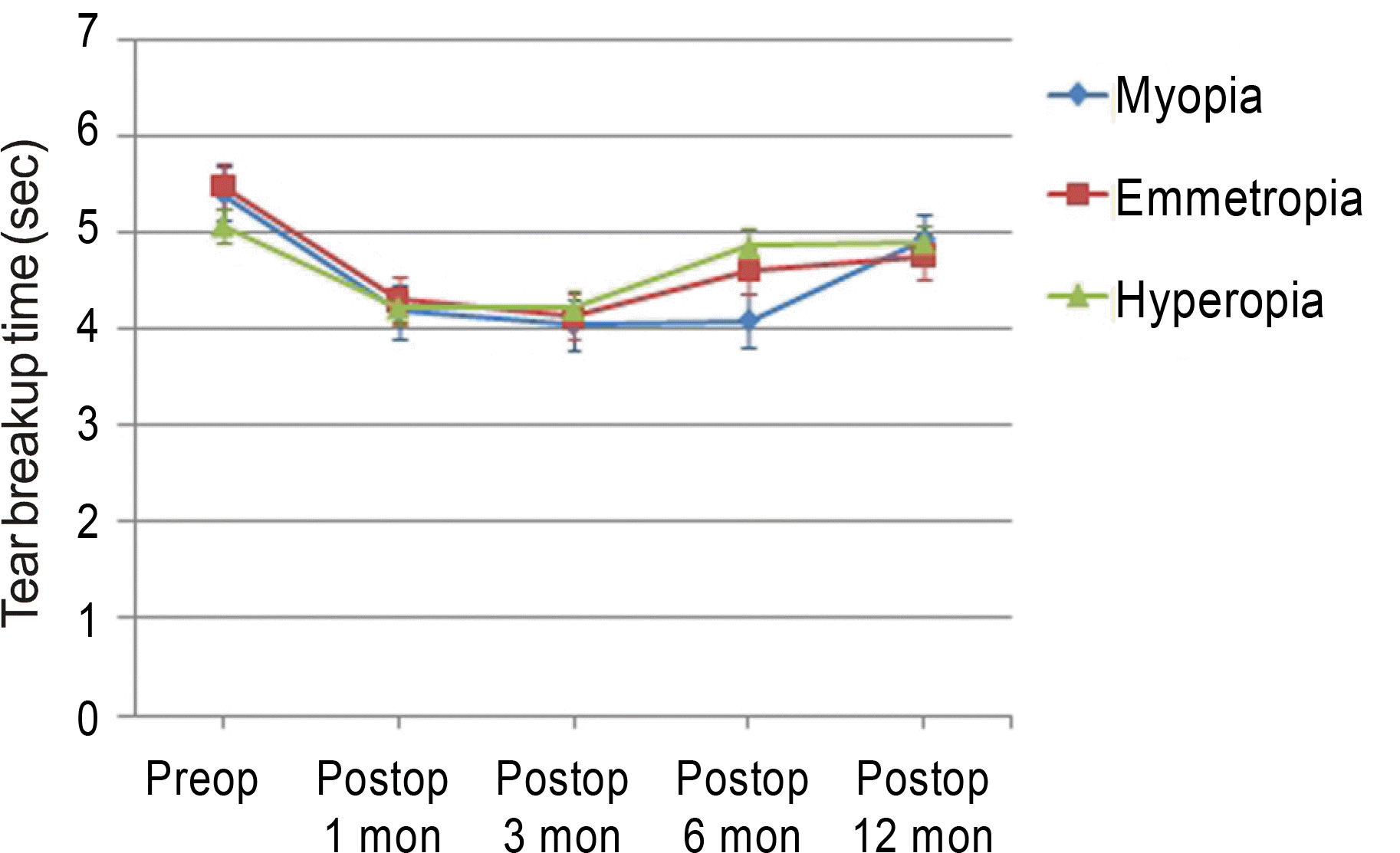
Figure 4.
Time course of changes of corneal sensation after LASIK grouped with pre-LASIK spherical aberration.
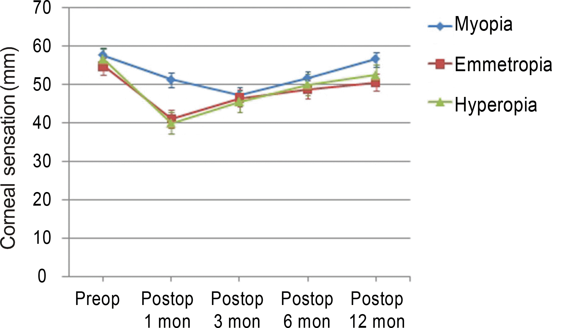
Table 1.
Demographics and clinical characteristics of patients
Table 2.
Mean value of BUT categorized by refractive error after LASIK
|
Tear breakup time (sec) |
|||||
|---|---|---|---|---|---|
| Preop | 1 mon | 3 mon | 6 mon | 12 mon | |
| Myopia | 5.41 ± 1.92 | 4.19 ± 1.51 | 4.05 ± 1.91 | 4.09 ± 1.75 | 4.93 ± 1.94 |
| Emmetropia | 5.50 ± 1.96 | 4.31 ± 1.74 | 4.13 ± 1.36 | 4.62 ± 1.36 | 4.77 ± 1.30 |
| Hyperopia | 5.09 ± 2.21 | 4.23 ± 1.69 | 4.22 ± 1.66 | 4.86 ± 1.36 | 4.91 ± 0.94 |
| p-value∗ | 0.707 | 0.216 | 0.593 | 0.461 | 0.957 |
Table 3.
Mean value of corneal sensation categorized by refractive error after LASIK
|
Corneal sensitivity (mm) |
|||||
|---|---|---|---|---|---|
| Preop | 1 mon | 3 mon | 6 mon | 12 mon | |
| Myopia | 57.69 ± 5.14 | 41.25 ± 13.61 | 47.31 ± 13.43 | 51.66 ± 8.16 | 56.67 ± 7.78 |
| Emmetropia | 54.81 ± 7.53 | 41.11 ± 9.74 | 46.52 ± 7.75 | 48.69 ± 8.69 | 50.57 ± 4.69 |
| Hyperopia | 56.56 ± 4.82 | 40.00 ± 17.58 | 45.62 ± 9.48 | 50.04 ± 6.96 | 52.43 ± 7.07 |
| p-value∗ | 0.207 | 0.933 | 0.831 | 0.322 | 0.039† |
Table 4.
Mean value of BUT categorized by gender after LASIK
|
Tear breakup time (sec) |
|||||
|---|---|---|---|---|---|
| Preop | 1 mon | 3 mon | 6 mon | 12 mon | |
| Female | 5.41 ± 2.12 | 4.17 ± 1.48 | 3.92 ± 1.71 | 4.43 ± 1.60 | 4.81 ± 1.51 |
| Male | 5.12 ± 1.87 | 5.04 ± 1.86 | 4.35 ± 1.91 | 4.84 ± 1.26 | 5.13 ± 1.13 |
| p-value∗ | 0.532 | 0.024† | 0.267 | 0.327 | 0.583 |
Table 5.
Mean value of corneal sensitivity categorized by gender after LASIK
|
Corneal sensitivity (mm) |
|||||
|---|---|---|---|---|---|
| Preop | 1 mon | 3 mon | 6 mon | 12 mon | |
| Female | 55.93 ± 5.99 | 39.10 ± 13.92 | 45.85 ± 10.64 | 51.59 ± 7.75 | 53.33 ± 7.67 |
| Male | 56.79 ± 6.11 | 43.08 ± 14.90 | 48.07 ± 10.21 | 49.52 ± 8.65 | 51.67 ± 7.53 |
| p-value∗ | 0.542 | 0.243 | 0.378 | 0.337 | 0.648 |
 XML Download