

 PDF
PDF ePub
ePub Citation
 Print
Print


Abstract
Purpose
To investigate postoperative outcomes of endonasal dacryocystorhinostomy (DCR) using lacrimal sac flap and silastic sheet in patients with acquired nasolacrimal duct obstruction.
Method
From November 2009 until December 2010, endonasal DCR with lacrimal sac flap was performed in 26 eyes (group 1) and conventional DCR without flap in 28 eyes (group 2). The anatomic and functional success rates and complications were analyzed and compared between the 2 groups.
Results
The anatomical success rate was 96.2% in group 1 and 85.7% in group 2. The functional success rate was 100% in group 1 and 92.9% in group 2. The success rate was higher in group 1 than in group 2, although not being statistically significant. Granuloma was found in 15.4% of patients in group 1 and 32.1% of patients in group 2. Synechia or mem-branous obstruction was not found in group 1, whereas synechia developed in 14.3% of patients in group 2.
Go to : 
References
1. Caldwell GW. Two new operations for obstruction of the nasal duct with preservation of the canaliculi and an incidental description of a new lachrymal probe. NY Med J. 1983; 57:581.
2. McDonogh M, Meiring JD. Endoscopic transnasal dacryocystorhinostomy. J Laryngol Otol. 1989; 103:585–7.

3. Jin HR, Yeon JY, Choi MY. Endoscopic dacryocystorhinostomy: creation of a large marsupialized lacrimal sac. J Korean Med Sci. 2006; 21:719–23.
4. Rebeiz EE. Endoscopic dacryocystorhinostomy. Curr Opin Otolaryngol Head Neck Surg. 1999; 7:44–9.
5. Onerci M, Orhan M, Ogretmenoğ lu O, Irkec M. Long-term results and reasons for failure of intranasal endoscopic dacryocystorhinostomy. Acta Otolaryngol. 2000; 120:319–22.
6. Durvasula VS, Gatland DJ. Endoscopic dacrocystorhinostomy: lont-term results and evolution of surgical technique. J Laryngol Otol. 2004; 118:628–32.
7. Tsirbas A, Wormald PJ. Endonasal dacryocystorhinostomy with mucosal flaps. Am J Ophthalmol. 2003; 135:76–83.
8. Munk PL, Lin DT, Morris DC. Epiphora: treatment by means of dacryocystoplasty with balloon dilatation of the nasolacrimal drainage apparatus. Radiology. 1990; 177:687–90.
9. Zílelíoğ lu G, Tekeli O, Uğ urba SH, et al. Results of endoscopic endonasal non-laser dacryocystorhinostomy. Doc Ophthalmol. 2002; 105:57–62.
10. Mandeville JT, Woog JJ. Obstruction of the lacrimal drainage system. Curr Opin Ophthalmol. 2002; 13:303–9.
11. Lee TS, Shin HH, Hwang SJ, Baek SH. The results of revision surgery for the failed endonasal DCR. J Korean Ophthalmol Soc. 2007; 48:186–92.
12. Sham CL, van Hasselt CA. Endoscopic terminal dacryocystorhinostomy. Laryngoscope. 2000; 110:1045–9.
13. Wormald PJ. Powered endoscopic dacryocystorhinostomy. Laryngoscope. 2002; 112:69–72.
14. Rhee KC, Lee TS. The effect of mitomycin-C eyedrop on pre-vention of internal ostium obstruction after endonasal dacryocystorhinostomy. J Korean Ophthalmol Soc. 1998; 49:2923–7.
15. Weidenbecher M, Hosemann W, Buhr W. Endoscopic endonasal dacryocystorhinostomy: Results in 56 patients. Ann Otol Rhinol Laryngol. 1994; 103:363–7.
16. Tsirbas A, Wormald PJ. Mechamical endonasal dacryocystorhinostomy with mucosal flaps. Br J Ophthalmol. 2003; 87:43–7.
17. Massegur H, Trias E, Ademà JM. Endoscopic dacryocystorhinostomy: Modified technique. Otolaryngol Head Neck Surg. 2004; 130:39–46.
18. Kansu L, Aydin E, Avci S, et al. Comparison of surgical outcomes of endonasal dacryocystorhinostomy with or without mucosal flaps. Auris Nasus Larynx. 2009; 36:555–9.
19. Codère F, Denton P, Corona J. Endonasal dacryocystorhinostomy: a modified technique with preservation of the nasal and lacrimal mucosa. Ophthal Plast Reconstr Surg. 2010; 26:161–4.
20. Yuen KS, Lam LY, Tse MW, et al. Modified endoscopic dacryocystorhinostomy with posterior lacrimal sac flap for nasolacrimal duct obstruction. Hong Kong Med J. 2004; 10:394–400.
21. Dhanasekar G, Umapathy N, Dapling RB, Skinner DW. Short-term results of endonasal dacryocystorhinostomy with a mucosal flap and a bone dissection technique. J Otolaryngol Head Neck Surg. 2009; 38:390–4.
22. Trimarchi M, Giordano Resti A, Bellini C, et al. Anastomosis of nasal mucosal and lacrimal sac flaps in endoscopic dacryocystorhinostomy. Eur Arch Otorhinolaryngol. 2009; 266:1747–52.
Go to :
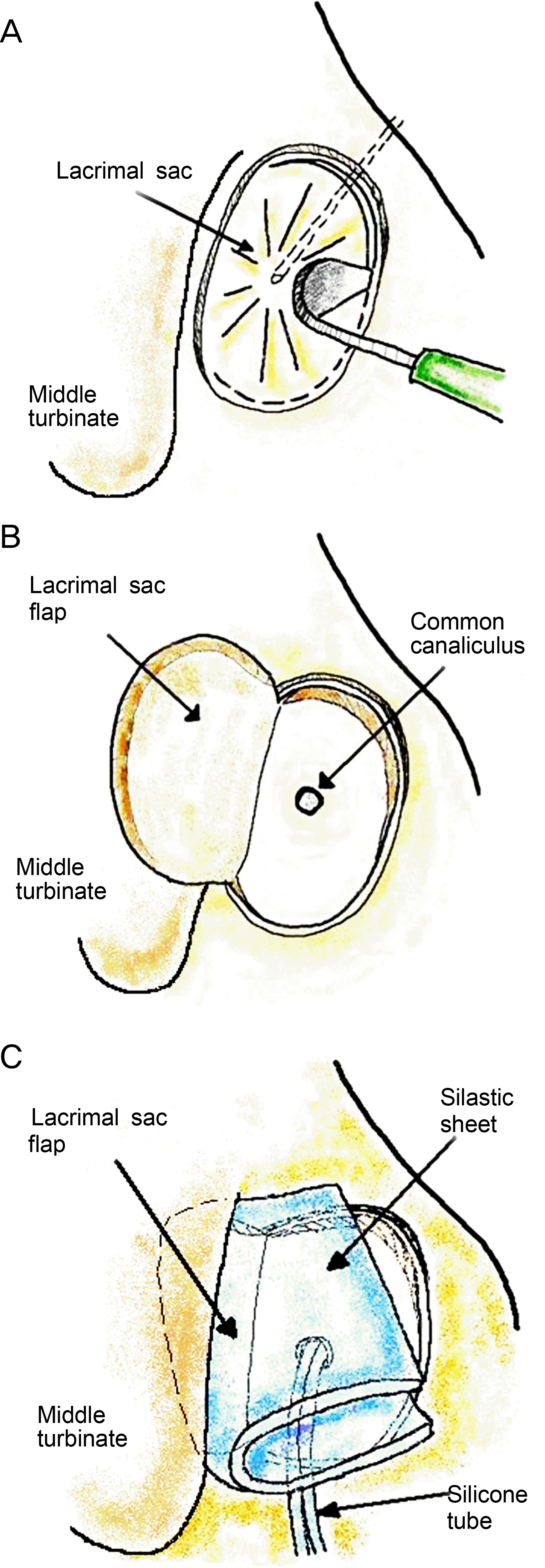 | Figure 1.Schematic illustration of creation and reflection of the U-shaped lacrimal sac flap. (A) In the left nasal cavity, a “U” shaped incision was made on the lacrimal sac using a crescent knife. (B) The exposed portion of bare bone was covered by the lacrimal sac flap. (C) Silastic sheet was inserted between the middle turbinate and lacrimal sac flap to prevent adhesion. |
 | Figure 2.Surgical technique of creating the lacrimal sac flap with silastic sheet. (A) After exposing the lacrimal sac, the extent of the lacrimal sac was identified with a 23G illumination probe. The lacrimal sac is then tented to allow a “U” shaped incision on the lacrimal sac wall using a crescent knife. (B) Lacrimal sac flap is laid back on the exposed posterior part of bony portion after cutting and trimming. (C) Silicone tube is placed through the common canaliculi in the lateral nasal wall. (D) Silastic sheet is inserted to stabi-lize the lacrimal sac flap. (E) Silastic sheet and lacrimal sac flap are well positioned in the lateral nasal wall. The lacrimal sac flap covers the retained bony spicles. CC = common canalicular opening; LF = lacrimal sac flap; SS = silastic sheet. |
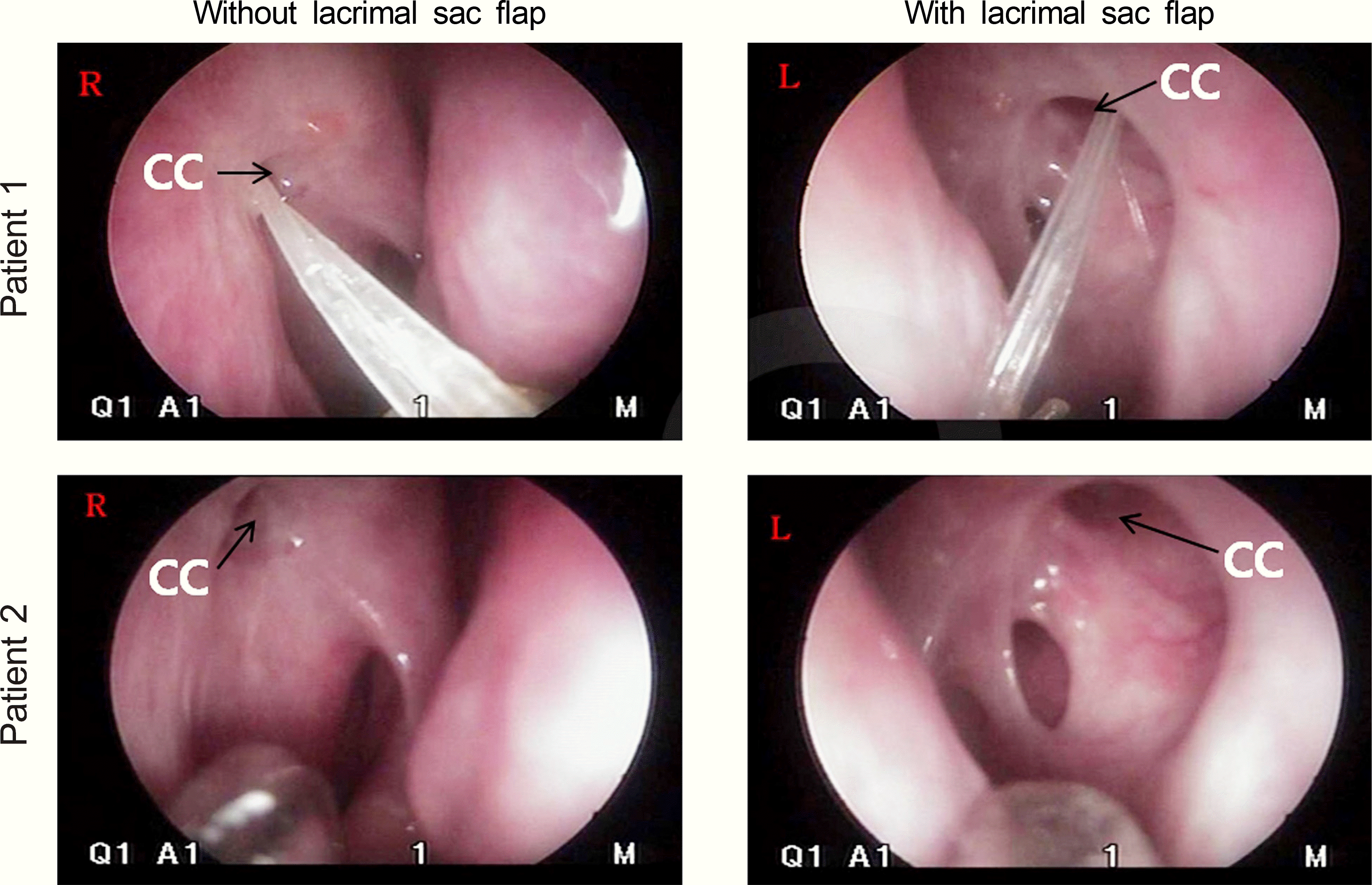 | Figure 3.Endoscopic findings of healed nasal ostium with and without lacrimal sac flap. The healed ostium with the assisted lacrimal sac flap was larger and clearer compared to that without the lacrimal sac flap. CC = common canalicular opening; R = right healed nasal ostium; L = left healed nasal ostium. |
Table 1.
Characteristics of group1 and group 2
Table 2.
The success rates of mucosal flap assisted endonasal dacryocystorhinostomy
 XML Download