Abstract
Purpose
The authors of the present case report observed a bilateral retinal racemose hemangioma which was located within the peripapillary area.
Case summary
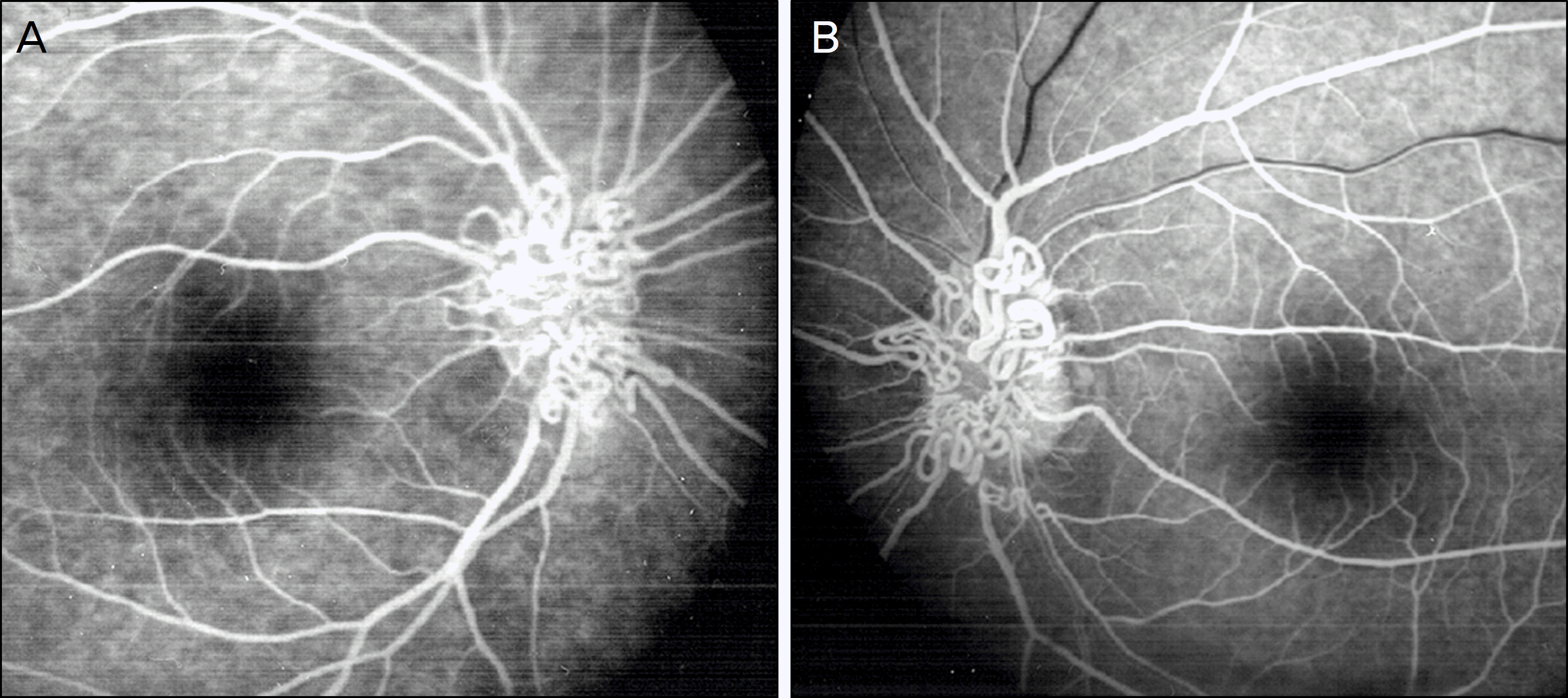
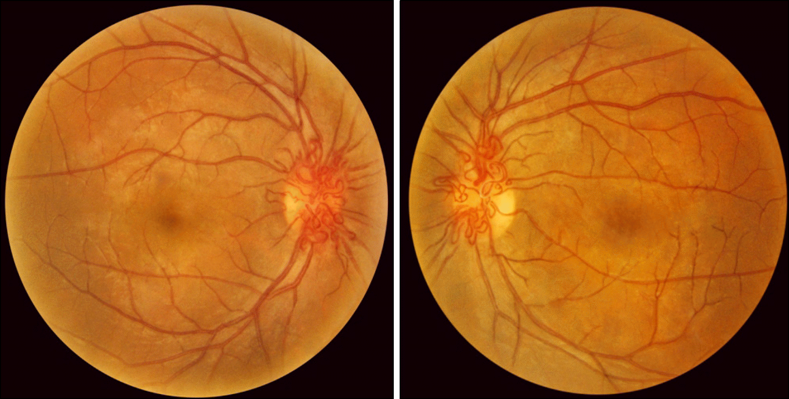
A 17-year-old man presented with floaters in both eyes. Fundus revealed tortuous and anastomosed retinal vasculature around the optic disc. In addition, fluorescein angiography showed a non-leaking retinal arteriovenous anastomosis. Seven years after the initial visit, vitreous hemorrhage occurred in the patient's left eye, and then 1 year later, subretinal hemorrhage was found in his left eye.
References
1. Soliman W, Haamann P, Larsen M. Exudation, response to photocoagulation and spontaneous remission in a case of bilateral racemose haemangioma. Acta Ophthalmol Scand. 2006; 84:429–31.

2. Archer DB, Deutman A, Ernest JT, Krill AE. Arteriovenous communications of the retina. Am J Ophthalmol. 1973; 75:224–41.

3. Jang HS, Lee TK, Kim JW. A case of retinal racemose hemangioma. J Korean Ophthalmol Soc. 2001; 42:1232–5.
5. Magnus H. Aneurysma arteriosovenosum retinale. Virchows Arch Pathol Anat Physiol. 1874; 60:38–43.

6. Mansour AM, Walsh JB, Henkind P. Arteriovenous anastomoses of the retina. Ophthalmology. 1987; 94:35–40.

7. Ferry AP. Other phakomatoses. Ryan SJ, editor. Retina. 2nd ed.St. Louis: Mosby;1994. p. 650–9.
8. Chopdar A. Racemose haemangioma. Chopdar A, editor. Fundus Fluorescein Angiography. 1st ed.Iowa City: Butterworth Heinemann;1996. p. 126–7.
9. Richard G. Racemose hemangioma. Richard G, editor. Fluorescein Angiography, Textbook and Atlas. 1st ed.New York: Thieme Medical Publishers;1990. p. 218–9.
10. Ebert EM, Albert DM. The phacomatoses. Jacobiec FA, Albert DM, editors. Principles and Practice of Ophthalmology. 2nd ed.Madison: Saunders W.B;2000. p. 5120–46.
11. Matsui M. Atlas of the Ocular Fundus. 1st ed.Seoul: Koonja;1990. p. 105–8.
12. Hsieh YT, Yang CM. The clinical study of congenital loop-ed/coiled peripapillary retinal vessels. Eye. 2005; 19:906–9.

13. Wyburn-Mason R. Arteriovenous aneurysm of midbrain and retina, facial nevi and mental changes. Brain. 1943; 66:163–203.
14. Bech K, Jensen OA. On the frequency of coexisting racemose haemangiomata of the retina and brain. Acta Psychiatr Scand. 1961; 36:47–56.

15. Shields JA, Shields CL. Other phakomatoses. Ryan SJ, editor. Retina. 4th ed.St Louis: Mosby;2006. v.1. chap.26.

16. Papageorgiou KI, Ghazi-Nouri SM, Andreou PS. Vitreous and subretinal haemorrhage: an unusual complication of retinal racemose haemangioma. Clin Experiment Ophthalmol. 2006; 34:176–7.

Figure 1.
Fluorescein angiograms of both eyes show rapid filling of vessels, no leakage and arteriovenous anastomosis.


 PDF
PDF ePub
ePub Citation
 Print
Print


 XML Download