Abstract
Purpose
To report a case of macular injury after exposure to a high energy laser beam used in a laser show.
Case
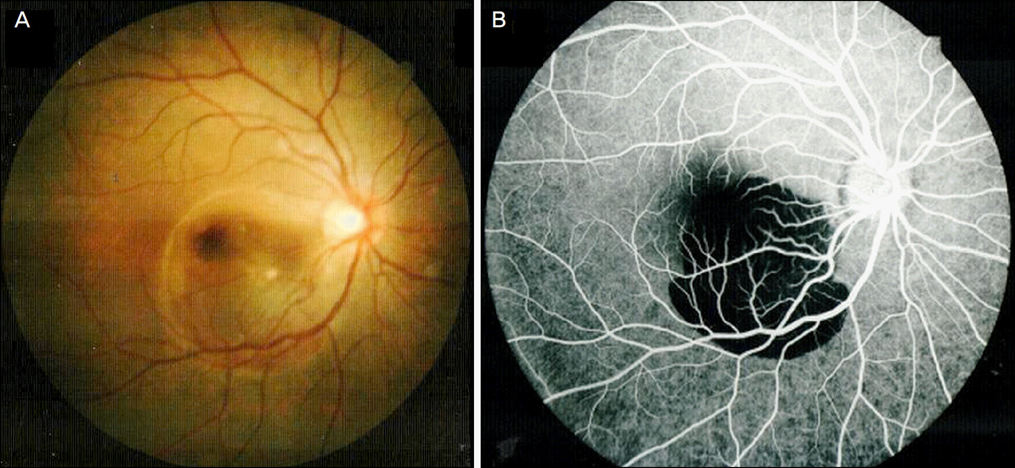
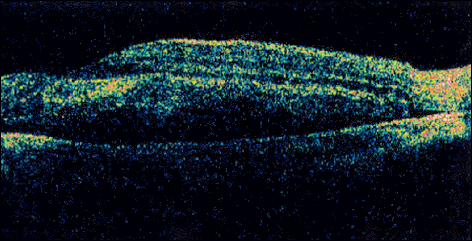
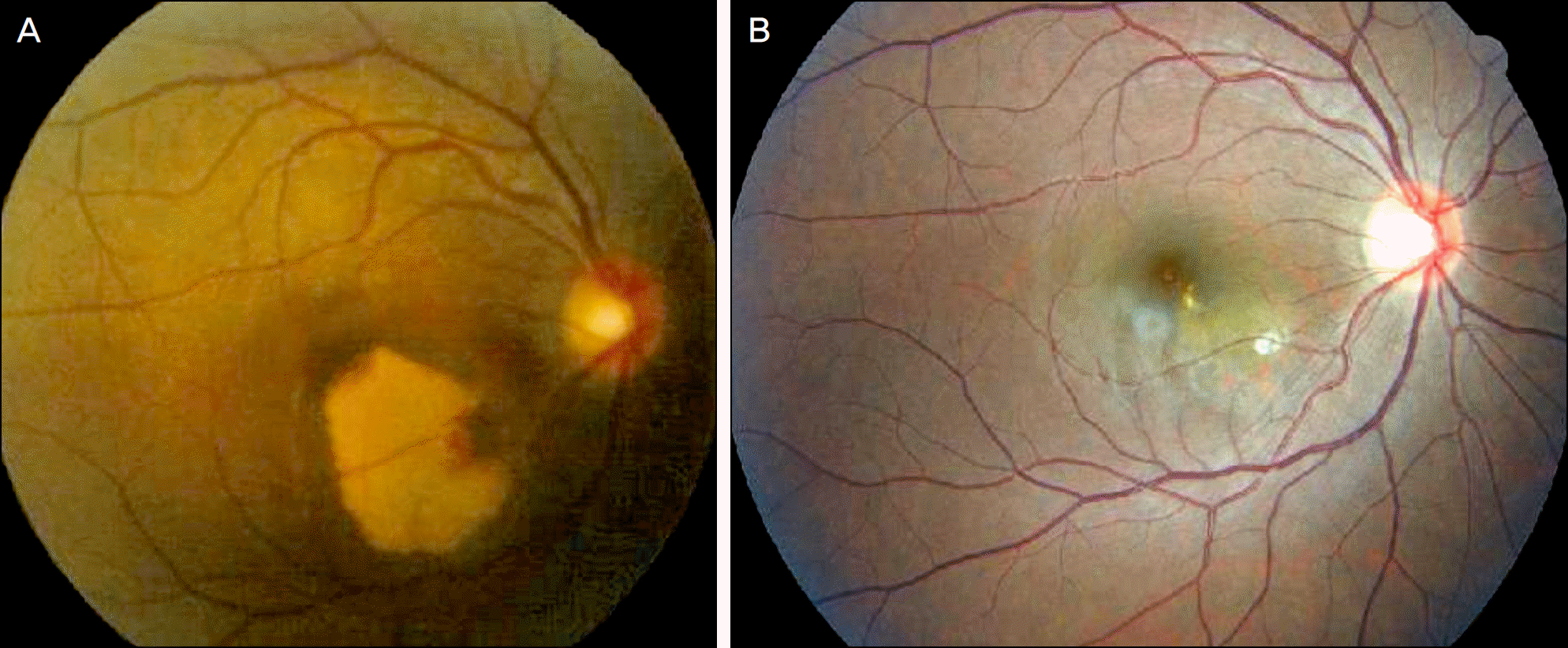
A 19-year-old female presented 2 days after exposure to a high energy laser beam at a laser show in a night club with decreased vision in her right eye. The patient's best corrected visual acuity of the right eye was hand motion. Fundus examination reveald a retinal swelling in the macular area approximately 5 disc diameter in size and a submacular hemorrhage. Fluorescein angiography of the right eye showed marked hypofluorescence in the macular area and optical coherence tomography (OCT) showed a neurosensory retinal detachment with a macular edema. Three years after exposure, the visual acuity of the right eye improved to 20/600. The fundus revealed scar and depigmented area at the macula.
References
2. Cai YS, Xu D, Mo X. Clinical, pathological and photochemical studies of laser injury of the retina. Health Phys. 1989; 56:643–6.
4. Gabel VP, Birngruber R, Lorenz B, Lang GK. Clinical observations of six cases of laser injury to the eye. Health Phys. 1989; 56:705–10.

6. Mainster MA, Timberlake GT, Warren KA, Sliney DH. Pointers on laser pointers. Ophthalmology. 1997; 104:1213–4.

8. Yolton RL, Citek K, Schmeisser E, et al. Laser pointers: toys, nui-sances, or significant eye hazards? J Am Optom Assoc. 1999; 70:285–9.
9. Zamir E, Chowers I. Concerns about laser pointers and macular damage. Arch Ophthalmol. 2001; 119:1731–2.
10. Jeong WD, Hwang YH, Kim JS, Lee JH. Maculopathy from red laser pointer. J Korean Ophthalmol Soc. 2007; 48:1007–11.
11. Kim M, Kwon JW, Han YK. A case of green laser pointer injury to the macula. J Korean Ophthalmol. 2008; 49:681–4.

12. Ryan S. Photic Retinal Injury and Safety. RETINA. 3th ed.2. Los Angeles: Elsever Mosby;2001. p. 1797–805.
Figure 1.
(A) Initial fundus photograph of the right eye, two days after exposure showed about a five disc diameter sized retinal swelling at the macula and a submacular hemorrhage. (B) Initial fluorescein angiograph of the right eye, two days after exposure to laser beam showed a marked hypofluorescence area at the macula.


 PDF
PDF ePub
ePub Citation
 Print
Print


 XML Download