

 PDF
PDF ePub
ePub Citation
 Print
Print


Abstract
Purpose
To report the results of scleral suture fixation using a hydrophilic acrylic intraocular lens (IOL) with 3 hollow haptics through a small corneal incision, the Triple Cow-Hitch Method.
Case summary
Three-point suture fixation of a XL Stabi ZO IOL was performed in 5 eyes of 5 patients with aphakia after penetrating keratoplasty (PKP), vitrectomy and subluxated lens extraction. Postoperatively, the corrected distance visual acuity and spherical equivalent improved in all measured eyes. There were no cases of pigment dispersion or cystoid macular edema (CME).
Go to : 
References
1. Por YM, Lavin MJ. Techniques of intraocular lens suspension in the absence of capsular/zonular support. Surv Ophthalmol. 2005; 50:429–62.

2. Teichmann KD, Teichmann IA. The torque and tilt gamble. J Cataract Refract Surg. 1997; 23:413–8.
3. Sasahara M, Kiryu J, Yoshimura N. Endoscope-assisted transscleral suture fixation to reduce the incidence of intraocular lens dislocation. J Cataract Refract Surg. 2005; 31:1777–80.
4. Ahn JK, Yu HG, Chung H, et al. Transscleral fixation of foldable intraocular lens in aphakic vitrectomized eyes. J Cataract Refract Surg. 2003; 29:2390–6.
5. Sabti K, Lindley SK, Mansour M, Discepola M. Uveal effusion after cataract surgery: an echographic study. Ophthalmology. 2001; 108:100–3.
6. Beatty S, Lotery A, Kent D, et al. Acute intraoperative suprachoroidal haemorrhage in ocular surgery. Eye (Lond). 1998; 12:815–20.
7. Fass ON, Herman WK. Sutured intraocular lens placement in aphakic postvitrectomy eyes via smallin-cision surgery. J Cataract Refract Surg. 2009; 35:1492–7.
8. Chen SX, Lee LR, Sii F, Rowley A. Modified cowhitch suture fixation of transscleral sutured posterior chamber intraocular lenses: long-term safety and efficacy. J Cataract Refract Surg. 2008; 34:452–8.
9. Grigorian R, Chang J, Zarbin M, Del Priore L. A new technique for suture fixation of posterior chamber intraocular lenses that elimi-nates intraocular knots. Ophthalmology. 2003; 110:1349–56.
10. Fass ON, Herman WK. Sutured intraocular lens placement in aphakic postvitrectomy eyes via small-incision surgery. J Cataract Refract Surg. 2009; 35:1492–7.
11. Uy HS, Chan PT. Pigment release and secondary glaucoma after implantation of single-piece acrylic intraocular lenses in the ciliary sulcus. Am J Ophthalmol. 2006; 142:330–2.
Go to :
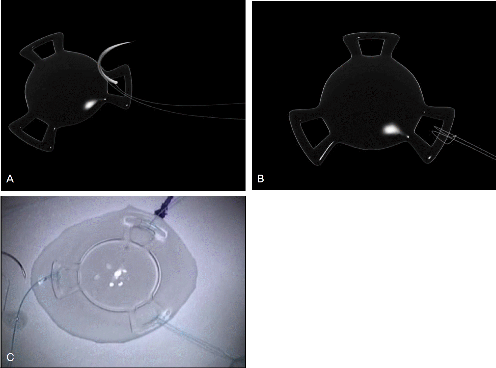 | Figure 1.Anchoring the 10-0 prolene needle to the XL Stabi ZO IOL using the triple cow-hitch scleral fixation method. (A) Illustration of the needle attached to the 10-0 prolene going through the haptic of the XL Stabi ZO IOL from underneath. (B) Illustration of the 10-0 prolene attached to the IOL in the cow-hitch manner. (C) Operation room view of the 10–0 prolene needles attached to all 3 IOL haptics: The Triple cow-hitch method. |
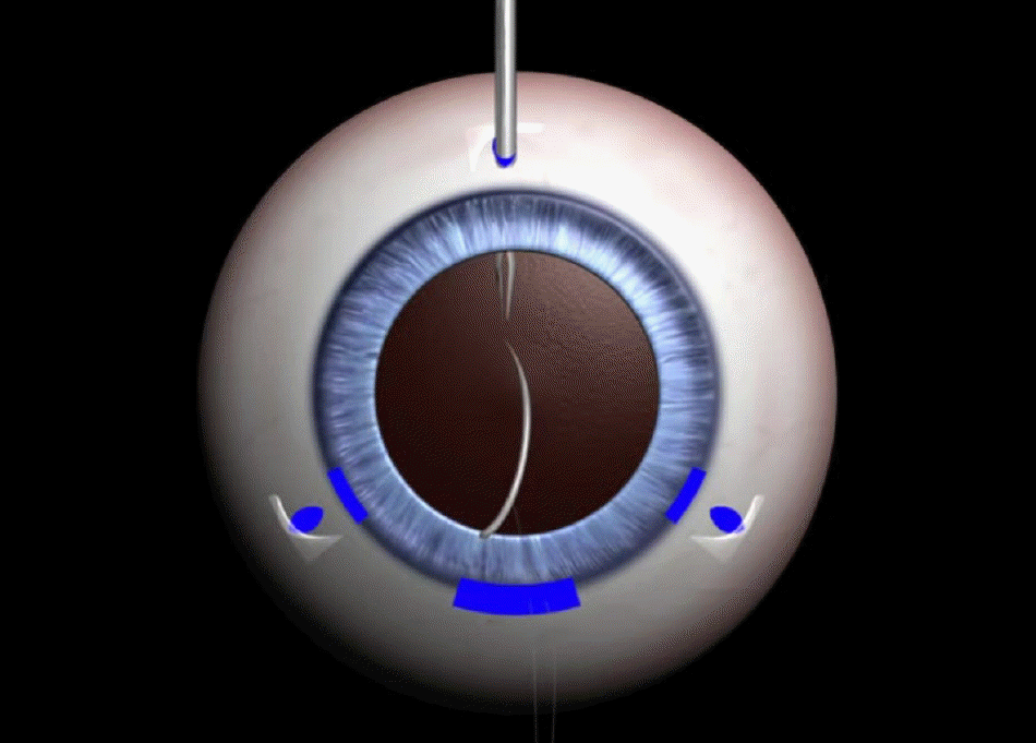 | Figure 2.Illustration of the needle prior to being pulled out of the globe using the Grieshaber forceps. |
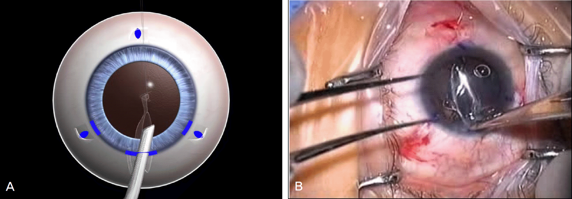 | Figure 3.Grabbing and folding the IOL, pushing it through the corneal tunnel into the globe. (A) Illustration of the Stabi IOL folded in half using the IOL forceps and placed into the anterior chamber through the corneal incision site. (B) Operation room view of the Stabi IOL placed into the anterior chamber. |
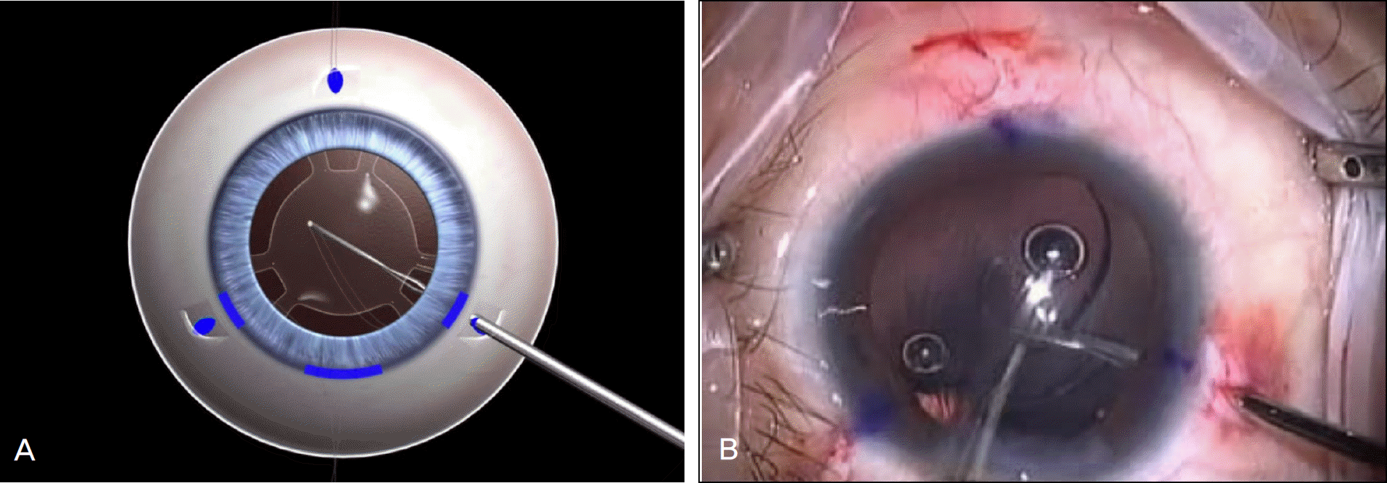 | Figure 4.Pulling the prolene needle out of the globe. (A) Illustration of grasping the 10–0 prolene needle inside the globe using Grieshaber forceps and pulling it out through the scleral tunnel. (B) Operation room view of the needle parallel to the Grieshaber forceps. |
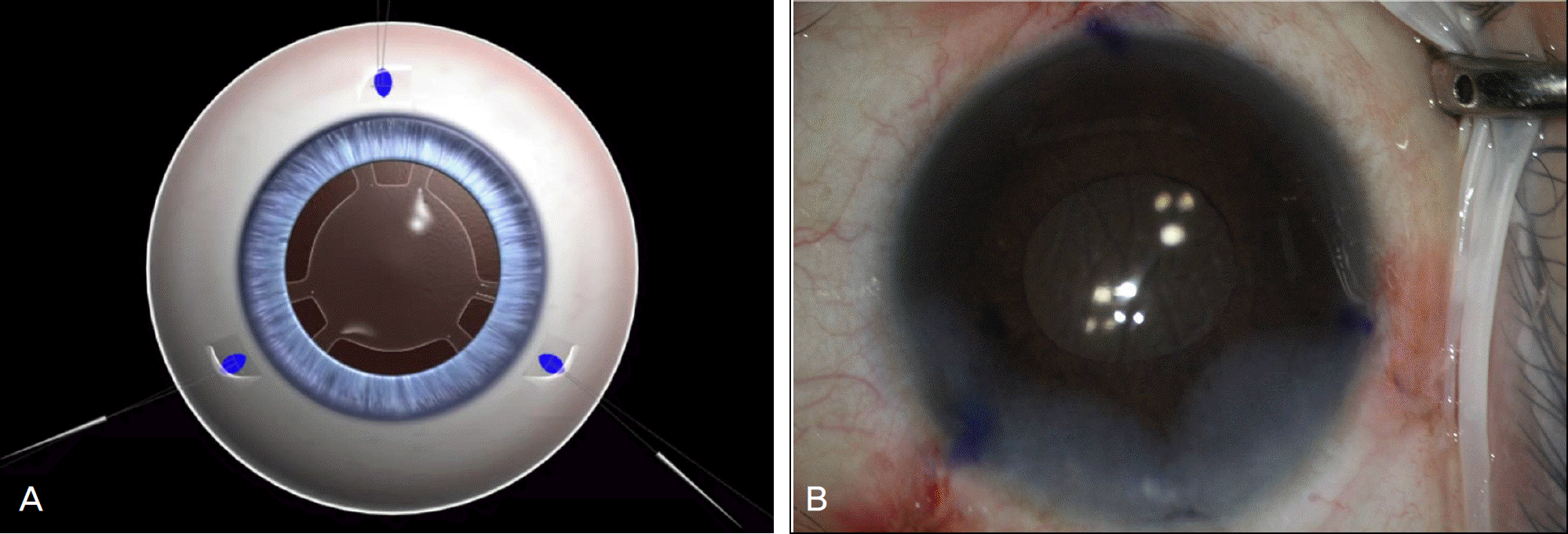 | Figure 5.Final adjustment of the IOL. (A) Illustration of the IOL prior to being fixed to the scleral wall. (B) Operation room view after centering the IOL. |
Table 1.
Preoperative and postoperative CDVA, SE, and change
Table 2.
Complications and additional procedures
 XML Download