

 PDF
PDF ePub
ePub Citation
 Print
Print


Abstract
Case summary
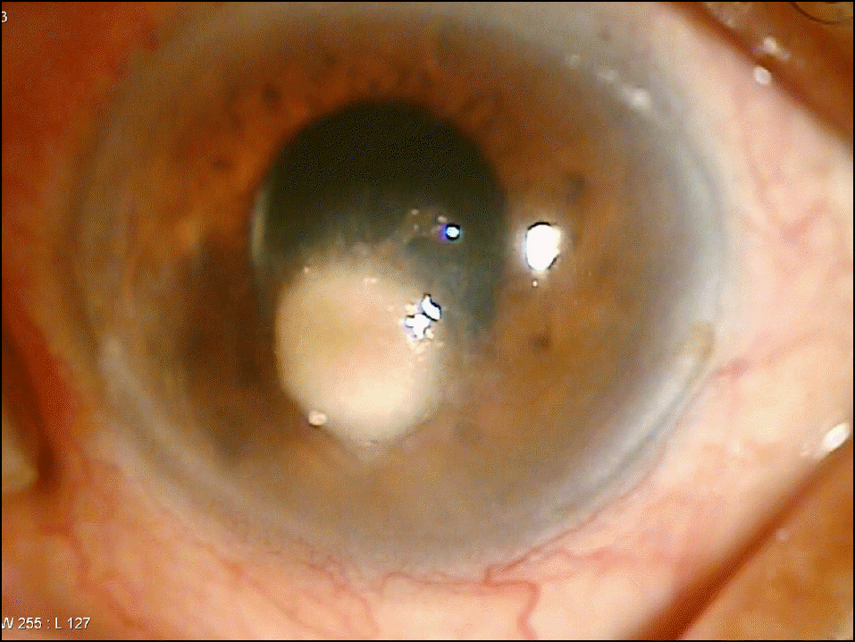
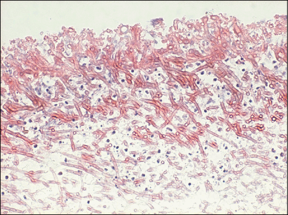
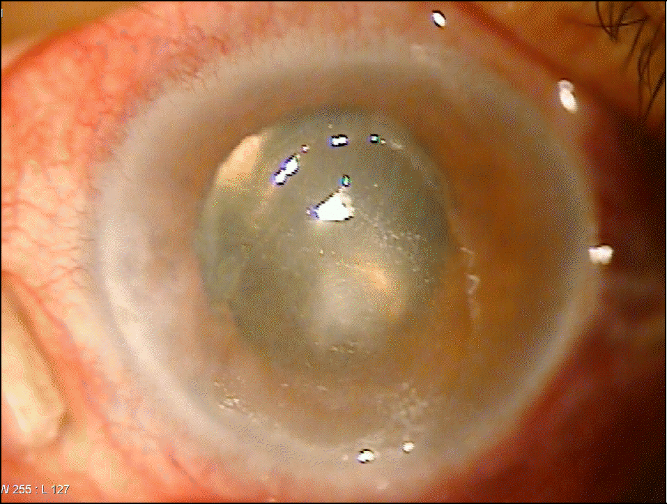
A 77-year-old man visited our clinic complaining of left ocular pain and decrease of visual acuity for 1 week On slit-lamp examination, epithelial defect and stromal infiltration on the corneal center with numerous inflammatory cells in the anterior chamber were found. There was no improvement after routine antibiotic treatment. A corneal biopsy and culture were performed on the corneal lesion. The KOH smear study reported hyphae, thus the patient was treated with 0.25% amphotericin B, 0.2% fluconazole and 5% natamycin eye drops. A clinical improvement was observed on the corneal lesion and Drechslera species was identified by the culture study.
References
1. Leber TH. Keratomycosis aspergillina als ursache von hypopyonkeratitis. Graefes Arch Ophthalmol. 1879; 25:285–301.

2. Jurkunas U, Behlau I, Colby K. Fungal keratitis: Changing pathogens and risk factors. Cornea. 2009; 28:638–43.
3. Thomas PA. Fungal infections of the cornea. Eye. 2003; 17:852–62.
4. Rosa RH Jr, Miller D, Alfonso EC. The changing spectrum of fungal keratitis in south Florida. Ophthalmology. 1994; 101:1005–13.
5. Alfonso EC, Cantu-Dibildox J, Munir WM, et al. Insurgence of Fusarium keratitis associated with contact lens wear. Arch Ophthalmol. 2006; 124:941–7.
6. Bernal MD, Acharya NR, Lietman TM, et al. Outbreak of Fusarium keratitis in soft contact lens wearers in San Francisco. Arch Ophthalmol. 2006; 124:1051–3.
7. Afonso EC, Rosa RH, Miller D. Fungal Keratitis Cornea. 2nd ed.2. Philadelphia: Elsevier Mosby;2005. p. 1101–22.
8. Kim YK, Kim TY, Kim BC, et al. Clinical Mycology. 3rd ed.Seoul: Korea Medicine;2008. p. 202–3.
9. Seo JH, Wee WR, Lee JH, Kim MK. Risk factors affecting efficacy of intracameral amphotericin injection in deep keratomycosis. J Korean Ophthalmol Soc. 2007; 48:1202–11.
10. Tanure MA, Cohen EJ, Sudesh S, et al. Spectrum of fungal keratitis at Wills Eye Hospital, Philadelphia, Pennsylvania. Cornea. 2000; 19:307–12.
11. Hahn YH, Lee DJ, Kim MS, et al. Epidemiology of fungal keratitis in Korea: A multicenter study. J Korean Ophthalmol Soc. 2000; 41:1499–508.
12. O'Day DM. Selection of appropriate antifungal therapy. Cornea. 1987; 6:238–45.
13. Srinivasan M. Fungal keratitis. Curr Opin Ophthalmol. 2004; 15:321–7.
14. O'Day DM, Head WS, Robinson RD, Clanton JA. Corneal penetration of Topical amphotericin B and natamycin. Curr Eye Res. 1986; 5:877–82.
15. Zapater RC, Albesi EJ, Garcia GH. Mycotic keratitis by Drechslera spicifera. Sabouraudia. 1975; 13:295–8.
16. Sundaram BM, Badrinath S, Subramanian S. Studies on mycotic keratitis. Mycoses. 1989; 32:568–72.
17. Fitzsimons R, Peters AL. Miconazole and ketoconazole as a satisfactory first-line treatment for keratomycosis. Am J Ophthalmol. 1986; 101:605–8.
18. Chander J, Sharma A. Prevalence of fungal corneal ulcers in Northern India. Infection. 1994; 22:207–9.
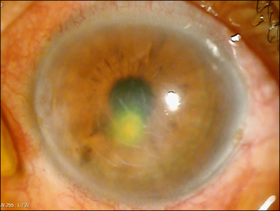
Figure 1.
On the first day the patient visited our clinic epithelial defect and corneal stromal infiltration is located in the center of the cornea. There is no hypopyon.
 XML Download