

 PDF
PDF ePub
ePub Citation
 Print
Print


Abstract
Purpose
To report a case of idiopathic orbital inflammation presenting as unilateral acute dacryoadenitis in a child.
Case summary
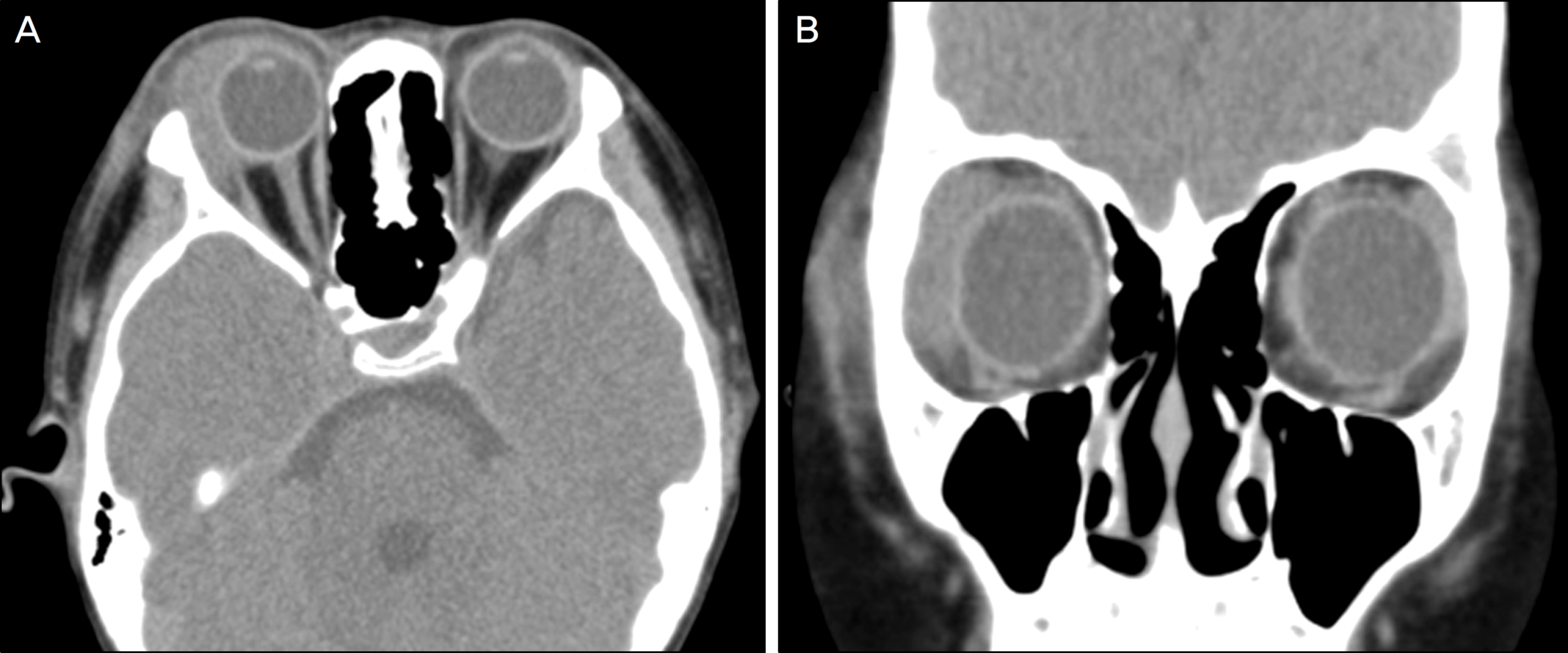
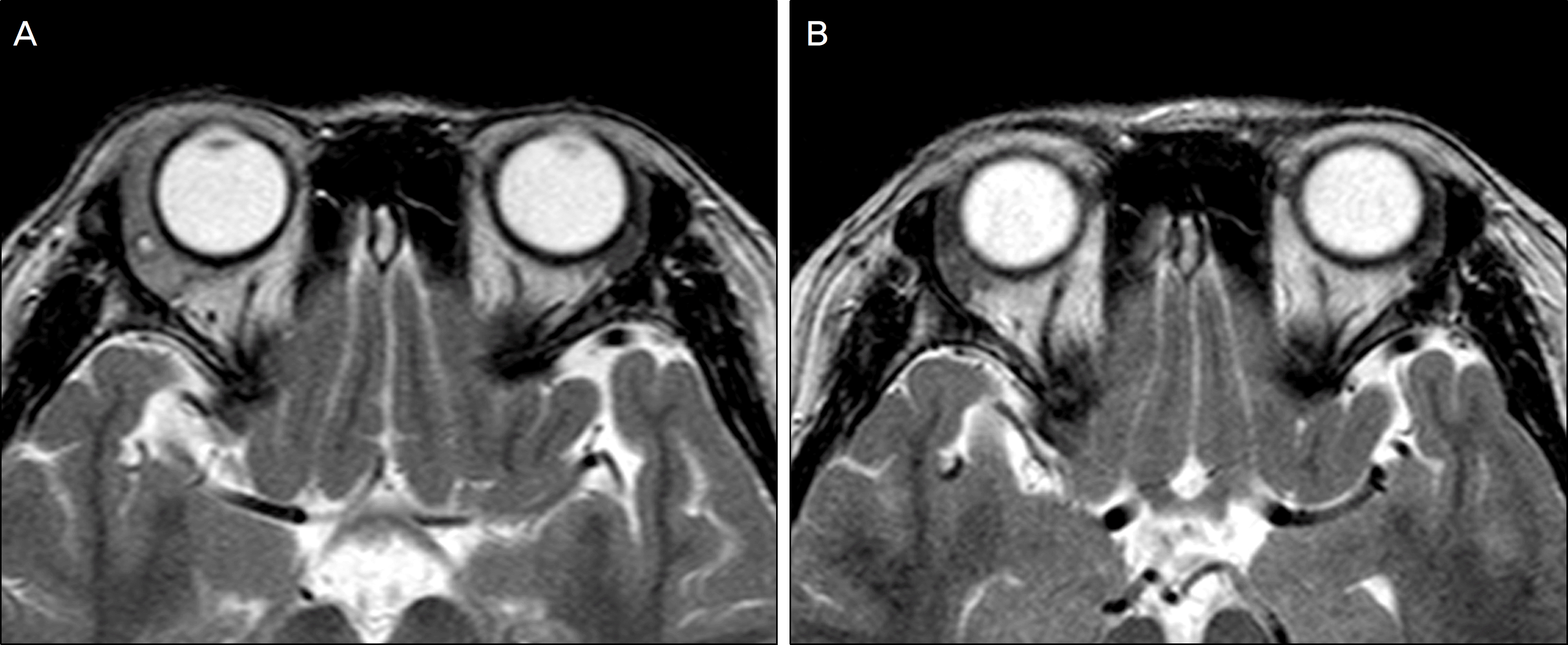
A nine-year-old boy presented with painful swelling and redness in the right upper eyelid and temporal conjunctiva without systemic symptoms for five days. Acute orbital cellulitis was suspected but did not respond to systemic antibiotics. An orbital computed tomogram and magnetic resonance imaging revealed a diffusely enlarged and inflamed right lacrimal gland. The patient showed dramatic response to systemic corticosteroids, and acute dacryoadenitis due to idiopathic orbital inflammation was diagnosed. Treatment with oral corticosteroids for two months resolved the inflammation. No relapse was observed during four months of follow-up.
References
1. Snebold NG. Noninfectious orbital inflammations and vasculitis. Albert DM, Jacobiec FA, editors. Principles and Practice of Ophthalmology. 2nd ed.Philadelphia: Saunders;2000. p. 3103.
2. Sutula FC. Tumors of the lacrimal gland and sac. Albert DM, Jacobiec FA, editors. Principles and Practice of Ophthalmology. 2nd ed.Philadelphia: Saunders;2000. p. 3131–2.

3. Duke-Elder S, MacFaul P. The Ocular Adnexa. In System of Ophthalmology. St Louis: CV Mosby;1974. p. 638–72.
4. Wilhelmus KR. Mumps. Gold DH, Weingeist TA, editors. The Eye in Systemic Disease. Philadelphia: JB Lippincott;1990. p. 262–5.
5. Tanner OR. Ocular manifestations of infectious mononucleosis. AMA Arch ophthalmol. 1954; 51:229–41.
6. Aburn NS, Sullivan TJ. Infectious mononucleosis presenting with dacryoadenitis. Ophthalmology. 1996; 103:776–8.
7. Rhem MN, Wilhelmus KR, Jones DB. Epstein-barr virus dacryoadenitis. Am J Ophthalmol. 2000; 129:372–5.
8. Marchese-Ragona R, Marioni G, Staffieri A, de Filippis C. Acute infectious mononucleosis presenting with dacryoadenitis and tonsillitis. Acta Ophthalmol Scand. 2002; 80:345–6.
9. Fitzsimmons TD, Wilson SE, Kennedy RH. Infectious dacryoadenitis. Pepose JS, Holland GN, Wilhelmus KR, editors. Ocular Infection & Immunity. St Louis: CV Mosby;1996. p. 1341–5.
10. Kiratli H, Ş ekeroglu MA, Söylemezoğ lu F. Unilateral dacryoadenitis as the sole presenting sign of Wegener's granulomatosis. Orbit. 2008; 27:157–60.
11. Colegrove JA. Localized orbital inflammation: A case of dacryoadenitis. Optom Vis Sci. 2000; 77:121–4.
12. Kapamajian MA, Ahmad A, Burnett JW, et al. Unilateral Orbital inflammation in a child after a jellyfish sting to the lower extremities. Ophthal Plast Reconstr Surg. 2009; 25:239–41.
13. Belanger C, Zhang KS, Reddy AK, et al. Inflammatory disorders of the orbit in childhood: a case series. Am J Ophthalmol. 2010; 150:460–3.
14. Rootman J, Nugent R. The classification and management of acute orbital pseudotumors. Ophthalmology. 1982; 89:1040–8.
15. Henderson JW. Orbital Tumors. 3rd ed.New York: Raven Press;1994. p. 391–411.
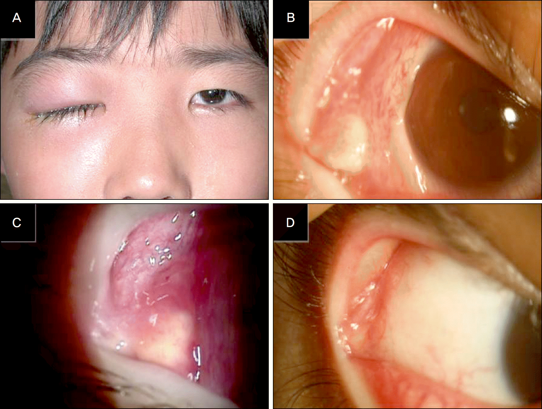
Figure 1.
(A) Erythematous swelling and mechanical ptosis of the right upper eyelid. (B) Inflammatory enlargement of the palpebral lobe of right lacrimal gland with yellowish nodular lesion and mild chemosis and injection of the temporal conjunctiva. (C) Markedly enlarged and inflammed palpebral lobe in the upper fornix. (D) Two months after treatment, inflammatory enlargement of the lacrimal gland and injection of the conjunctiva was subsided.
 XML Download