

 PDF
PDF ePub
ePub Citation
 Print
Print


Abstract
Purpose
To report a case of a neurosyphilis patient with cilioretinal artery occlusion as the first manifestation.
Case summary
A 58-year old man presented with a visual impairment which developed 5 days earlier in his right eye. During initial evaluation, the visual acuity in the right eye was 0.02. On fundus examination, a whitening adjacent to the macula was observed and fluorescein angiography showed filling delay and late leakage of the cilioretinal arteries. The patient was diagnosed with cilioretinal artery occlusion. Two weeks after his initial visit, visual acuity in the patient's right eye improved to 0.7 and retinal whitening was decreased on fundus examination. Three months later, a visual decrease in the right eye reoccurred and the visual acuity was 0.01. Anterior chamber cell reaction with hypopyon was observed on slit lamp examination and vitreal opacity was noted during fundus examination. Among the numerous tests performed in order to determine the cause of uveitis, RPR antibody and treponemal antibody were found positive. The patient was diagnosed with neurosyphylis and 2 weeks after intravenous administration of penicillin, visual acuity improved to 0.7 and anterior chamber cell reaction as well as vitreal opacity decreased.
References
1. Song YM, Choi O. A study of the cilioretinal artery in Korean. J Korean Ophthalmol Soc. 1981; 22:363–6.
2. Wood AC. Syphilis of the eye. Am J Syph Gonor Vener Dis. 1943; 27:133–86.
3. Deschenes J, Seamone CD, Baines MG. Acquired ocular syphilis: diagnosis and treatment. Ann Ophthalmol. 1992; 24:134–8.
4. Schlaegel TF Jr, Kao SF. A review (1970–1980) of 28 presumptive cases of syphilitic uveitis. Am J Ophthalmol. 1982; 93:412–4.

5. Aldave AJ, King JA, Cunningham ET Jr. Ocular syphilis. Curr Opin Ophthalmol. 2001; 12:433–41.
6. Barile GR, Flynn TE. Syphilis exposure in patients with uveitis. Ophthalmology. 1997; 104:1605–9.
7. Hong MC, Sheu SJ, Wu TT, Chuang CT. Ocular uveitis as the initial presentation of syphilis. J Chin Med Assoc. 2007; 70:274–80.
8. Morgan CM, Webb RM, O'Connor GR. Atypical syphilitic cho-rioretinitis and vasculitis. Retina. 1984; 4:225–31.
9. Savir H, Kurz O. Fluorescein angiography in syphilitic retinal vasculitis. Ann Ophthalmol. 1976; 8:713–6.
10. Lobes LA Jr, Folk JC. Syphilitic phlebitis simulating branch vein occlusion. Ann Ophthalmol. 1981; 13:825–7.
11. Yokoi M, Kase M. Retinal vasculitis due to secondary syphilis. Jpn J Ophthalmol. 2004; 48:65–7.
12. Lee MJ, Park YG, Ahn JK. Isolated cilioretinal artery occlusion in pseudoexfoliation syndrome: A case report. J Korean Ophthalmol Soc. 2006; 47:1686–90.
Figure 1.
Fundus photography of the right eye of the patients at the initial visit and 2 months. (A) The figure shows retinal whitening nearby the macula at the initial visit (black arrow heads indicate the affected vessel). (B) 2 months later, retinal whitening is not seen.
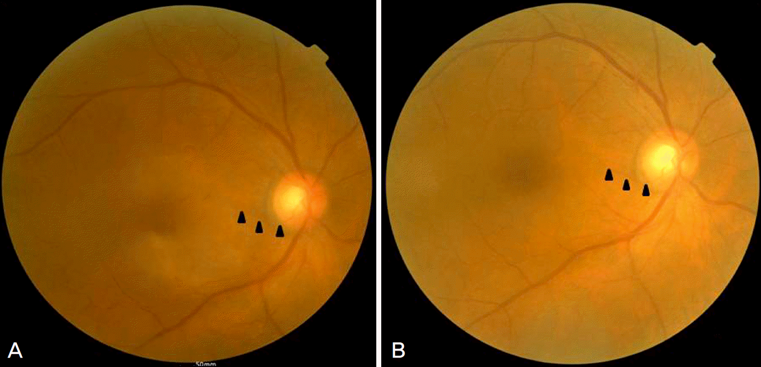
Figure 2.
Fluorescein angiogrphy of the right eye of the patients at the initial visit. The photos show delayed filling of the cilioretinal artery and late leakage (black arrow heads indicate the affected vessel). (A) 00:17, (B) 02:54, (C) 09:22.
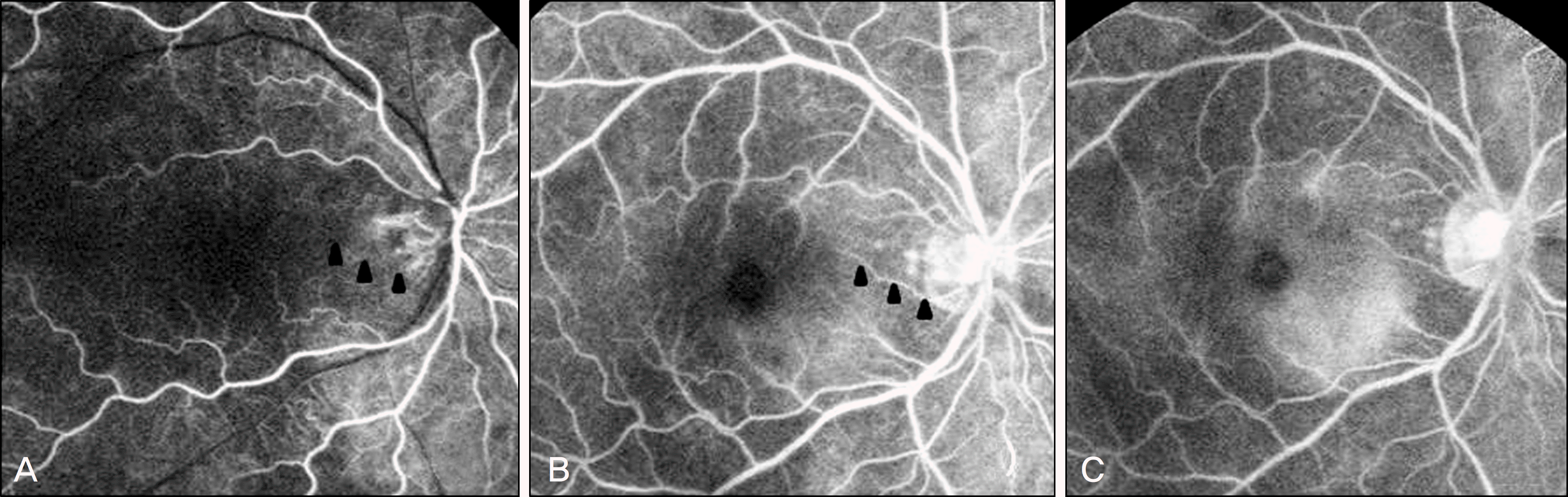
Figure 3.
Optical coherence tomogrphy of the right eye of the patients at the initial visit and 2 months later. (A) The figure shows macular edema with subretinal fluid at the initial visit. (B) 2 months later, macular edema and subretinal fluid disappeared.
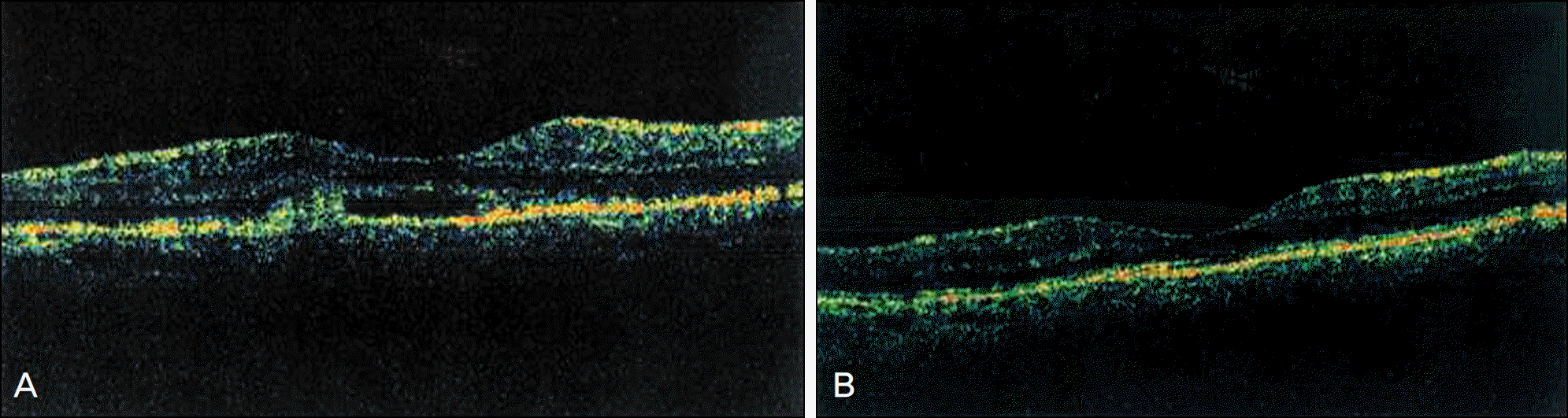
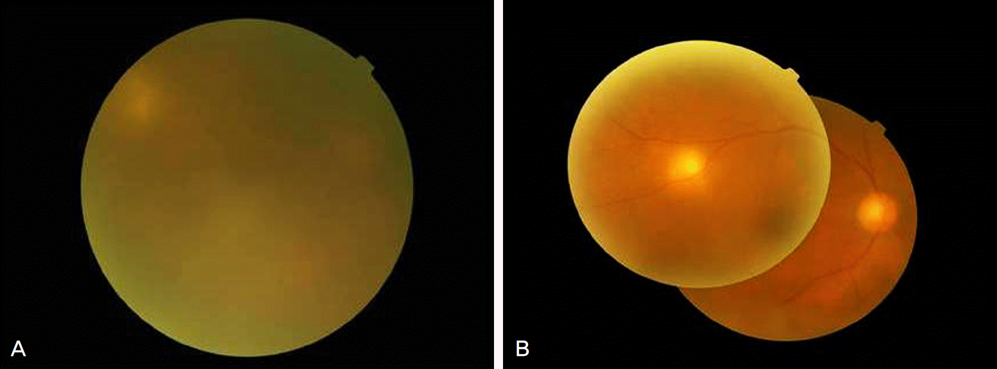
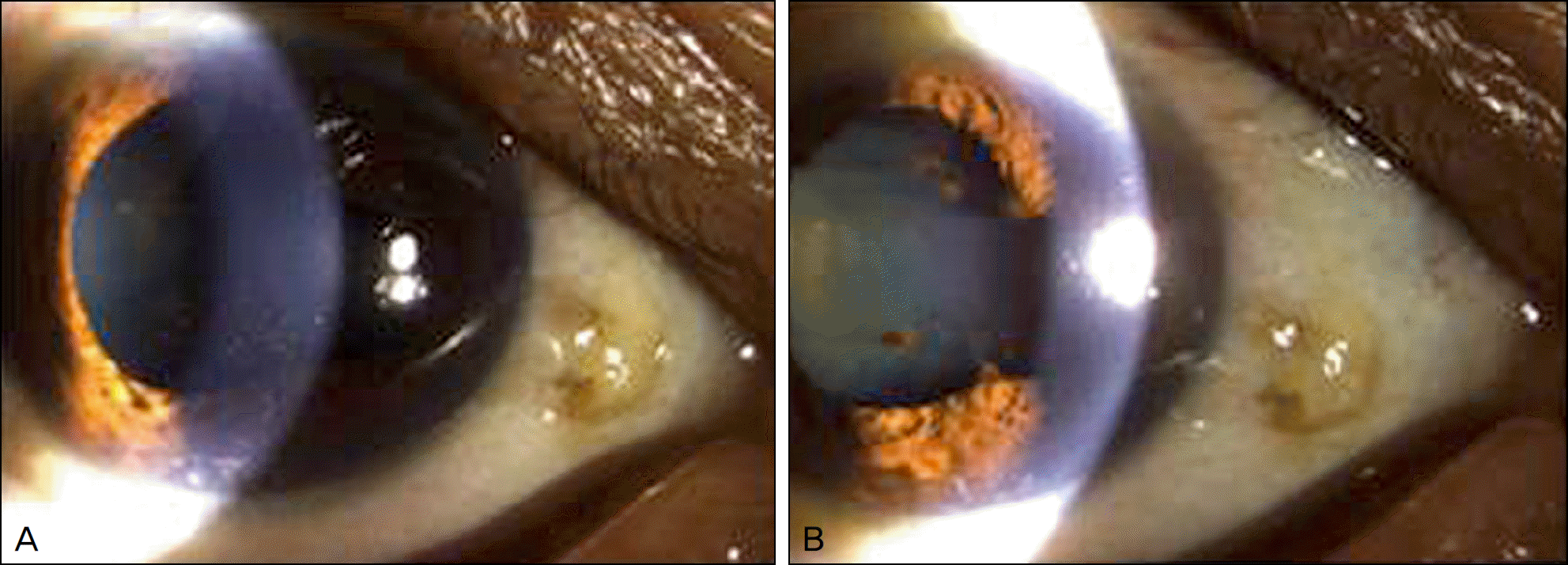
Figure 4.
Fundus photography of the right eye 5 months later after the initial visit and 2 months later after penicillin G IV. (A) The figure shows vitreal opacity and focal whitish lesion on the retina of superonasal area 5 months later after the initial visit. (B) The figure shows decreased vitreal opacity and apparent focal whitish lesion on the superonasal area 2 weeks after penicillin G IV.
 XML Download