Abstract
Purpose
The authors report a case of a giant subretinal nodular mass following surgical vitreous opacity removal.
Case summary
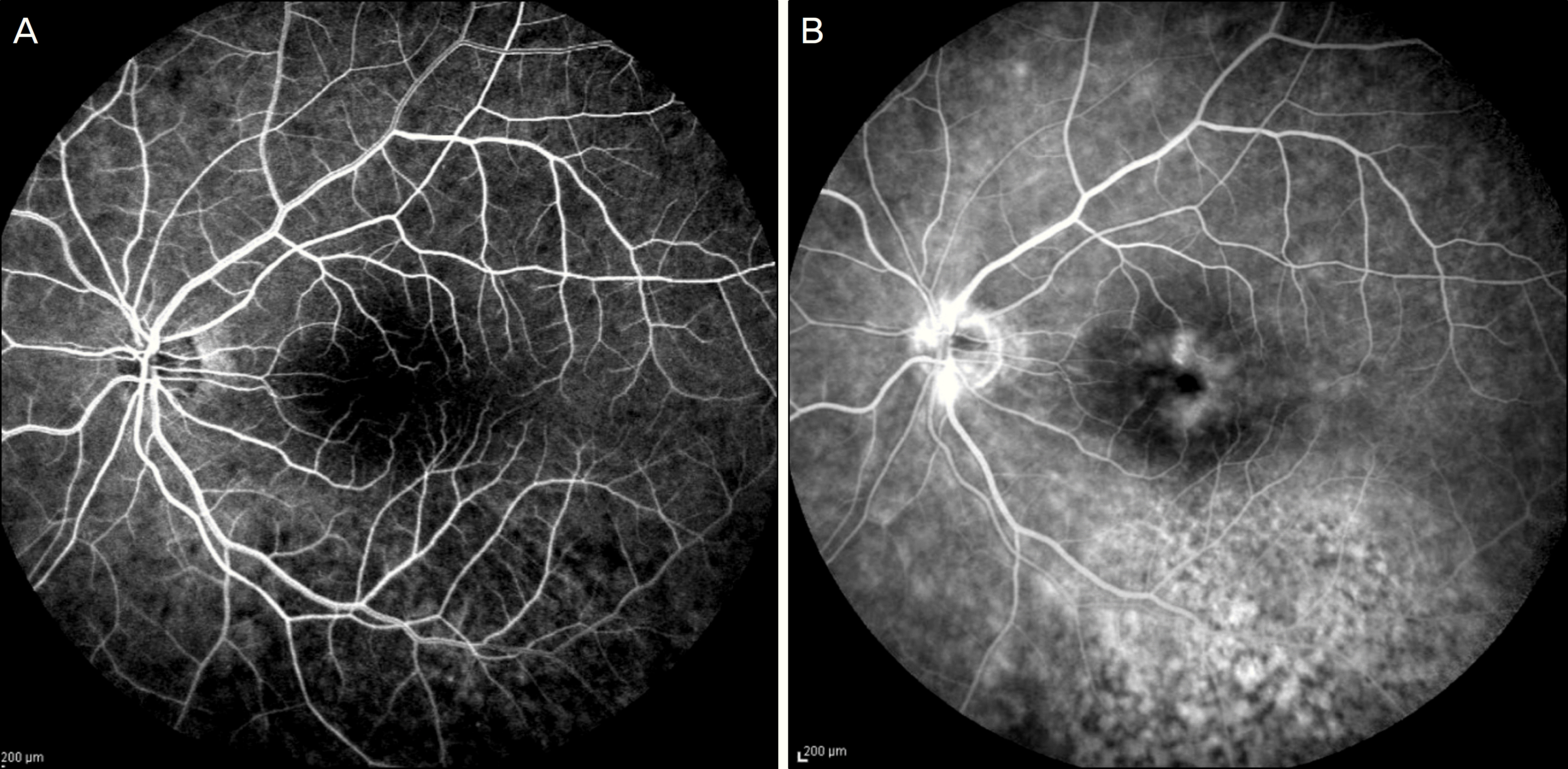
A 72-year-old man complained of visual loss and vitreous floaters in the left eye for 3 months (best corrected visual acuity: 0.1). The fundus was not clearly visualized due to vitreous opacity. Vitrectomy, phacoemulsification, and posterior chamber intraocular lens implantation were performed in the left eye. After removing the vitreous opacity, fundus examination revealed a creamy yellowish-white subretinal nodular mass (4 disc in size) inferotemporal to the fovea with adjoining chorioretinal folds and exudative retinal detachment. Fluorescein angiography (FA) of the left eye showed hypofluorescence of the nodule surrounded by a well-demarcated hyperfluorescent margin. B-scan ultrasonography revealed a prominent dome-shaped large echogenic nodule bordered by hypoechoic signal at the sclerochoroidal level. Giant nodular posterior scleritis was suspected and the patient was treated with oral corticosteroids. The large nodular lesion resolved completely within 3 months after initiation of the treatment and the best corrected visual acuity improved to 0.5.
References
2. Demirci H, Shields CL, Honavar SG, et al. Long-term follow-up of giant nodular posterior scleritis simulating choroidal melanoma. Arch Ophthalmol. 2000; 118:1290–2.

3. Shukla D, Kim R. Giant nodular posterior scleritis simulating choroidal melanoma. Indian J Ophthalmol. 2006; 54:120–2.

4. Finger PT, Perry HD, Packer S, et al. Posterior scleritis as an intraocular tumour. Br J Ophthalmol. 1990; 74:121–2.

5. Arevalo JF, Shields CL, Shields JA. Giant nodular posterior scleritis simulating choroidal melanoma and birdshot retinochoroidopathy. Ophthalmic Surg Lasers Imaging. 2003; 34:403–5.

6. McCluskey PJ, Watson PG, Lightman S, et al. Posterior scleritis: Clinical features, systemic associations and outcome in a large series of patients. Ophthalmology. 1999; 106:2380–6.
7. Watson PG, Hazelman BL, Pavesio C, Green WR. The Sclera and Systemic Disorders. 2nd ed.Butterworth-Heinmann: Edinburgh;2004. p. 59–89.
Figure 1.
Postoperative fundus photograph of the left eye showing a large subretinal nodular mass inferotemporal to the fovea with adjoining choroidal folds and exudative retinal detachment. (A) Central view. (B) Inferior view.
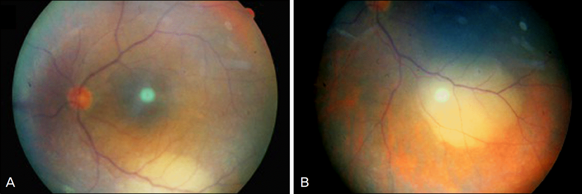
Figure 2.
Immediate post-operative fluorescein angiography (FA). (A) Early-stage FA of the left eye shows hypofluorescence corresponding to the nodule. (B) Late-stage FA shows hypofluorescence of the nodule surrounded by a well demarcated hyperfluorescent margin.
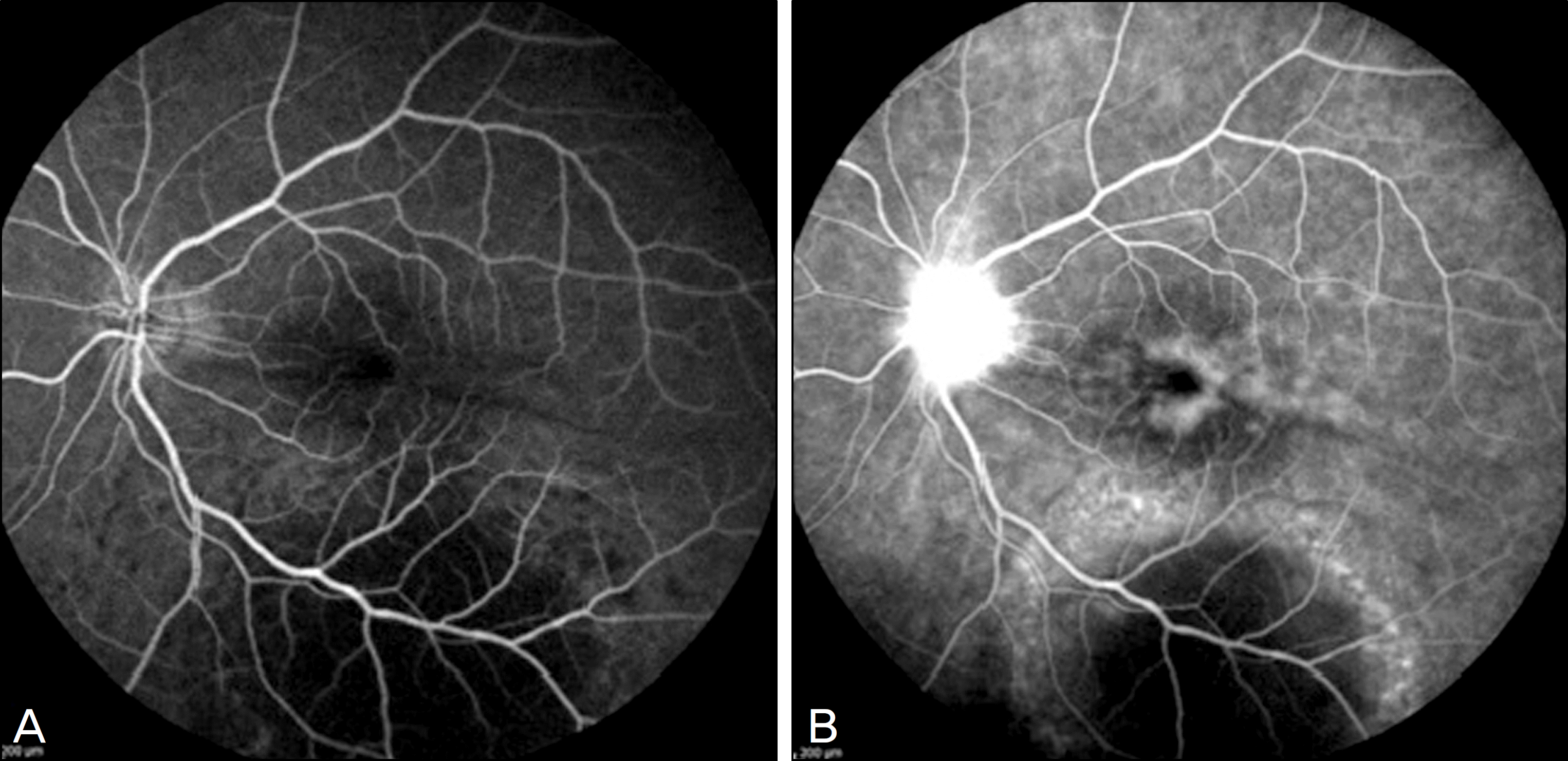
Figure 3.
(A) Before treatment, B-scan ultrasonography reveals a prominent dome-shaped hyperechoic nodule (asterisk) bordered posteriorly by hypoechoic signals (arrow) at the sclerochoroidal level. (B) 3 months later after treatment, B-scan ultrasonography shows regression of the hyperechoic nodular lesion.
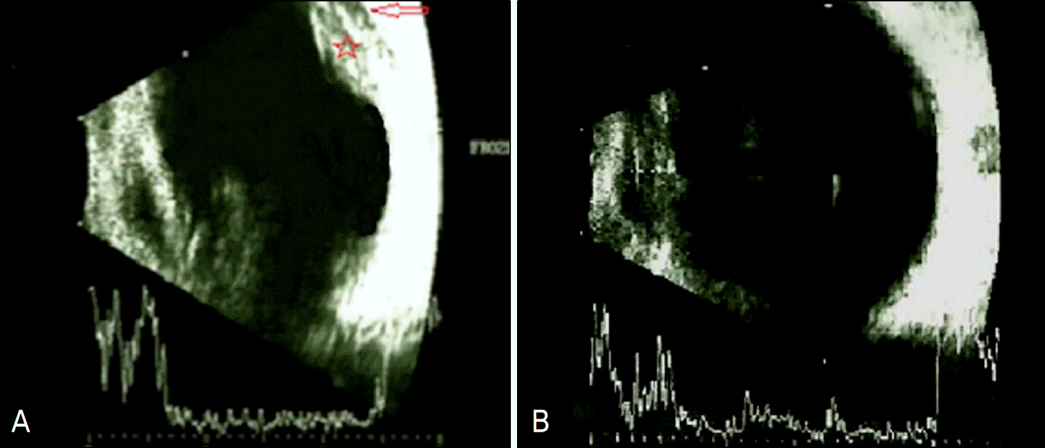
Figure 4.
(A) Magnetic resonance imaging (T1-weighted, fat suppression, gadolinium-enhanced) shows a curvilinear nodular lesion along the posterior portion of the left eye. This lesion (arrow) reveals enhancement. (B) Magnetic resonance imaging (T2-weighted) shows hypointense signals (arrow).
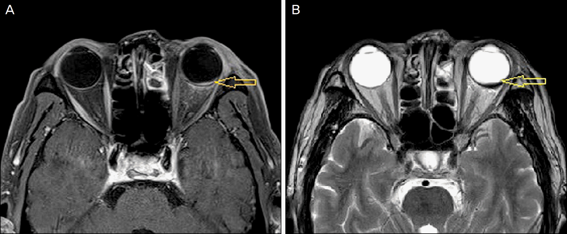
Figure 5.
Fluorescein angiography (FA) taken at 3 months after treatment. (A) Early-stage FA of the left eye shows hypofluorescence with an indistinct border corresponding to the nodular lesion. (B) Late-stage FA shows poorly defined hyperfluorescent peripapillary area with hyperfluorescent spots at the macula and mottled hyperfluorescence in the area of the regressed nodular lesion.


 PDF
PDF ePub
ePub Citation
 Print
Print


 XML Download