

 PDF
PDF ePub
ePub Citation
 Print
Print


Abstract
Case summary
One patient, who previously underwent three rounds of penetrating keratoplasty and limbal transplantation for uncontrollable peripheral and central corneal melting, received total corneolimbal transplantation. The other patient who underwent penetrating keratoplasty with limbal transplanation for a chemical burn and who did not experience corneal perforation also received total corneolimbal transplantation. During the average 19 months of follow-up, cyclophotocoagulation was performed to control high intraocular pressure in both patients. Both eyes were tectonically maintained without further corneal destruction despite poor visual acuity and rejection.
References
1. Castroviejo R. Total penetrating keratoplasty; a preliminary report. Am J Ophthalmol. 1951; 34:1697–706.
2. Burke JW. Total keratoplasty. Trans Am Ophthalmol Soc. 1920; 18:440–7.
3. Tsubota K, Satake Y, Kaido M, et al. Treatment of severe ocular-surface disorders with corneal epithelial stem-cell transplantation. N Engl J Med. 1999; 340:1697–703.

4. Tsai RJ, Tseng SC. Human allograft limbal transplantation for corneal surface reconstruction. Cornea. 1994; 13:389–400.
5. Kenyon KR, Tseng SC. Limbal autograft transplantation for ocular surface disorders. Ophthalmology. 1989; 96:709–22.
6. Lee S. Ophthalmic Plastic and Reconstructive Surgery. 1st ed.Seoul: Korean Society of Ophthalmic Plastic and Reconstructive Surgery;2004. p. 4.
7. Kuckelkorn R, Redbrake C, Schrage NF, Reim M. Keratoplasty with 11–12 mm diameter for management of severely chem-ical-burned eyes. Ophthalmologe. 1993; 90:683–7.
8. Vajpayee RB, Thomas S, Sharma N, et al. Large-diameter lamellar keratoplasty in severe ocular alkali burns: A technique of stem cell transplantation. Ophthalmology. 2000; 107:1765–8.
9. Kwitko S, Marinho D, Barcaro S, et al. Allograft conjunctival transplantation for bilateral ocular surface disorders. Ophthalmology. 1995; 102:1020–5.
10. Jager M. Corneal Langerhans cells and ocular immunology. Regional Immunol. 1992; 4:186–95.
11. Skeens HM, Holland EJ. Large-diameter penetrating keratoplasty: indications and outcomes. Cornea. 2010; 29:296–301.
12. Cobo M, Ortiz JR, Safran SG. Sclerokeratoplasty with maintenance of the angle. Am J Ophthalmol. 1992; 113:533–7.
13. Panda A, Sharma N, Angra SK, Singh R. Therapeutic sclerokeratoplasty versus therapeutic penetrating keratoplasty in refractory corneal ulcers. Aust N Z J Ophthalmol. 1999; 27:15–9.
14. Jonas JB, Rank RM, Budde WM. Tectonic sclerokeratoplasty and tectonic penetrating keratoplasty as treatment for perforated or pre-descemetal corneal ulcers. Am J Ophthalmol. 2001; 132:14–8.
15. Zerve BL, Belin MW, Ciolino JB. Boston Type 1 Keratoprosthesis study group. Results from the multicenter Boston type 1 keratoprosthesis study. Ophthalmology. 2006; 113:1779.
16. Greiner MA, Li J-Y, Mannis MJ. Longer term vision outcomes and complications with the Boston type 1 keratoprosthesis at the University of California, Davis. Ophthalmology. 2011; 118:1543–50.
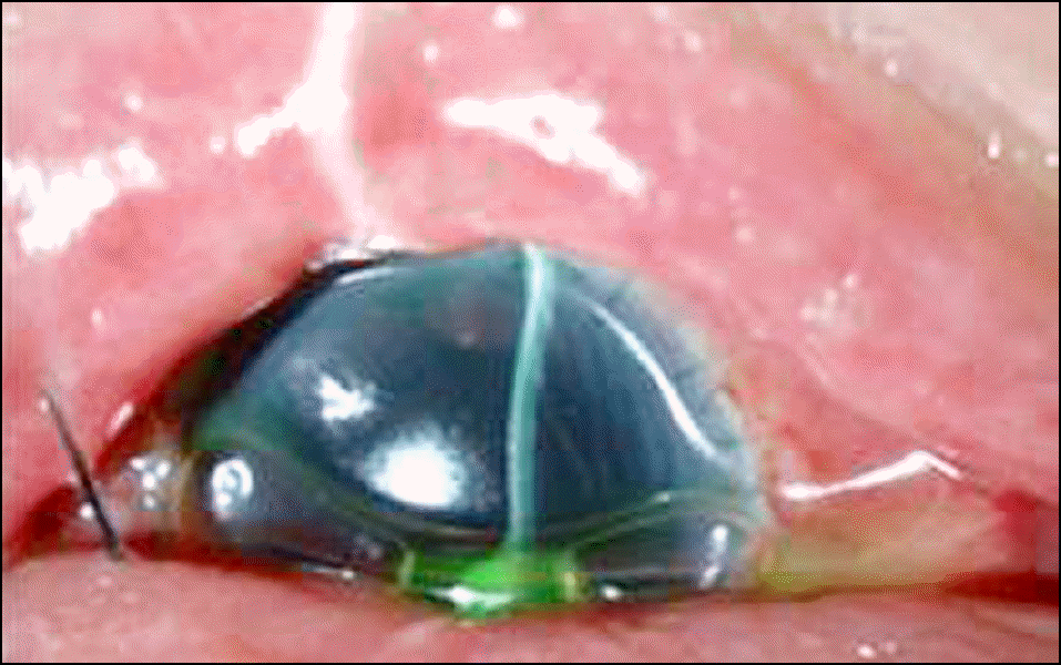
Figure 1.
Preoperative anterior segment photographs show diffuse graft edema, limbal insuffiency (A), and loosened suture due to peripheral cornea thinning (B) (case 1).
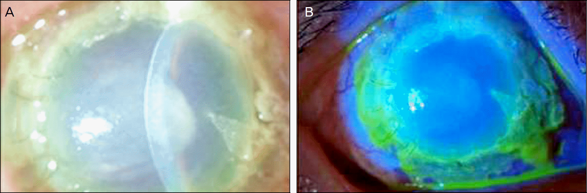
Figure 2.
Anterior segment photograph 1 day after total cornea transplantation showing no leakage (case 1).
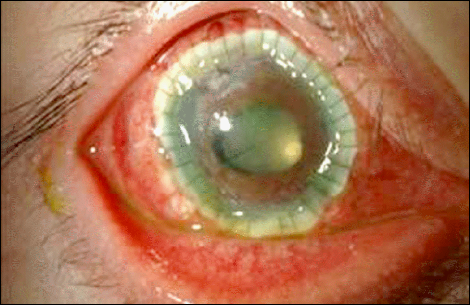
Figure 3.
Anterior segment photographs 21 months after total cornea transplantation with limbus shows mild corneal edema due to chronic rejection (A) and mild corneal epithelial defects (B), but no recurrence of peripheral corneal thinning (case 1).
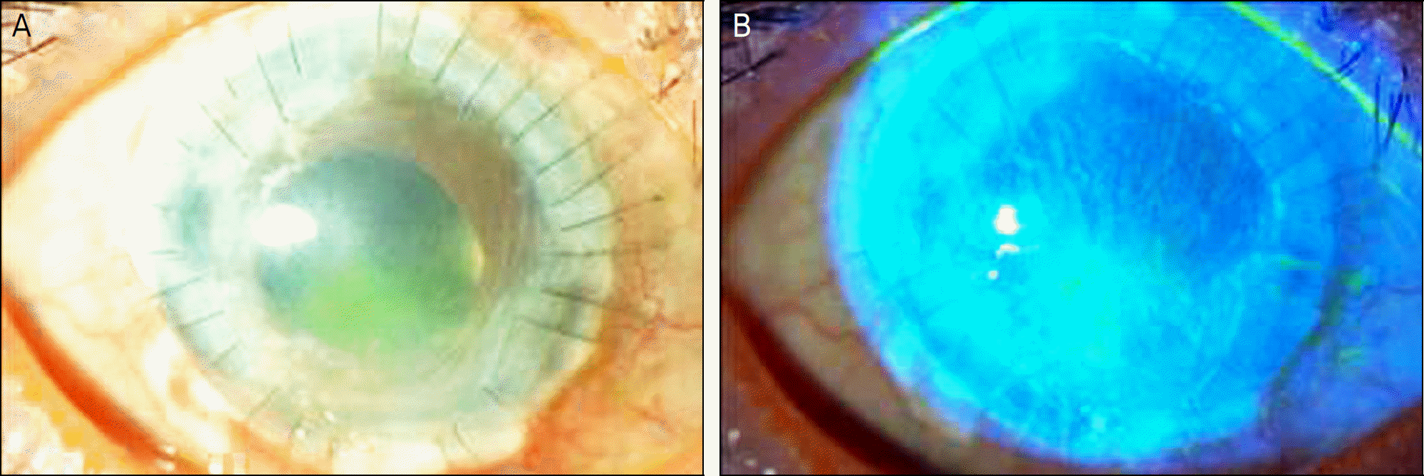
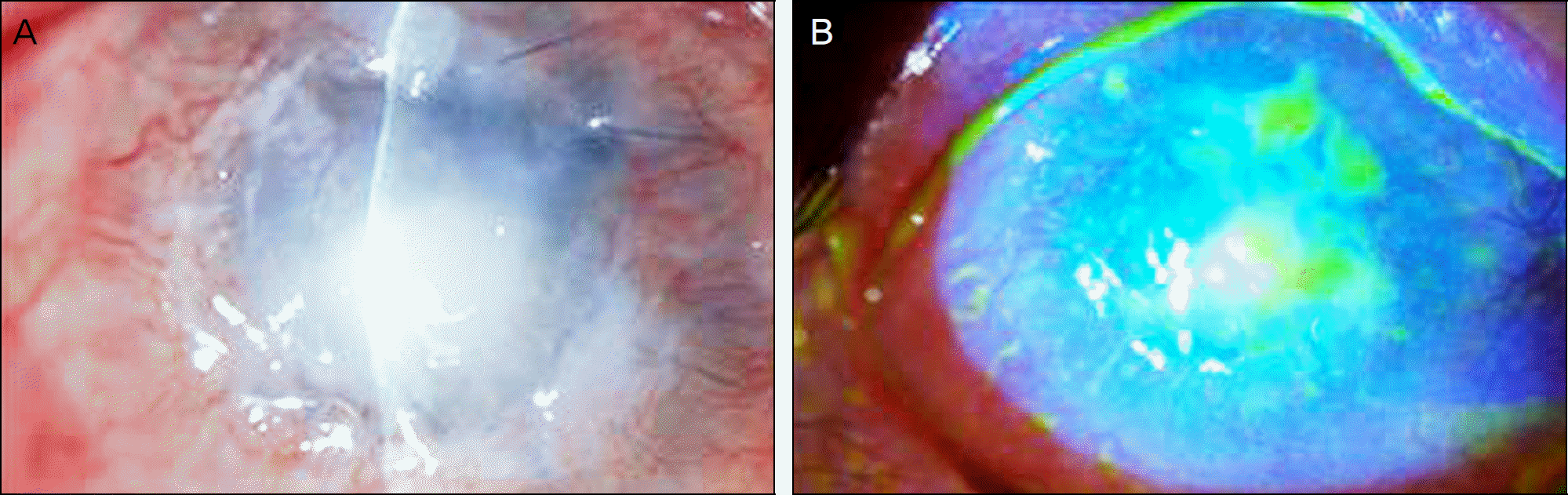
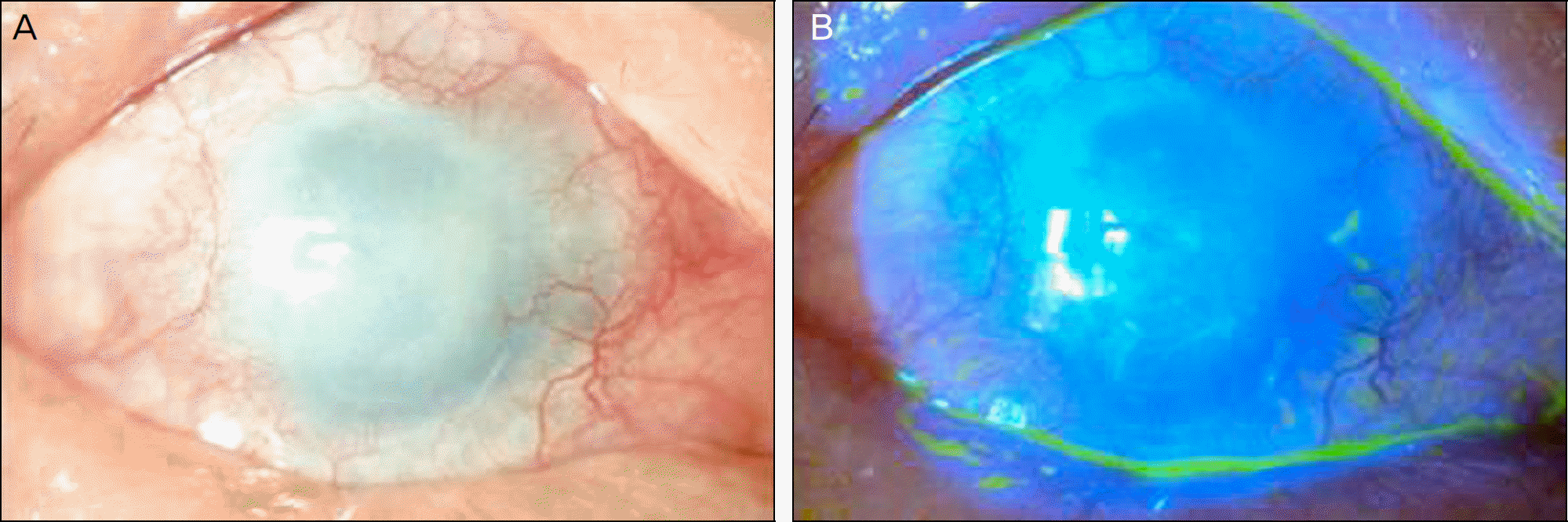
Figure 4.
Preoperative anterior segment photograph shows dense inferior cornea infiltration and severe thinning especially in the superior and inferior cornea with loosened suture, severe limbal insuffiency with neovascularization (A), and corneal epithelial defects (B) (case 2).
 XML Download