Abstract
Purpose
To evaluate the efficacy of five-layered reinforced amniotic membrane (AM) transplantation for treating deep corneal ulcer or perforation.
Case summary
We performed a five-layered reinforced AM transplant in three cases of corneal ulceration or perforation using mechanical compressed lyophilized AMs. In all cases, the perforated cornea healed with reepithelization within 2 weeks and the ocular surface was stabilized for more than 1 year.
References
1. Kenyon KR. Inflammatory mechanisms in corneal ulceration. Trans Am Ophthalmol Soc. 1985; 83:610–63.
2. Prabhasawat P, Tesavibul N, Komolsuradej W. Single and multilayer amniotic membrane transplantation for persistent corneal epithelial defect with and without stromal thinning and perforation. Br J Ophthalmol. 2001; 85:1455–63.

3. Kim JC, Kim JH, Cheong TB, Kim JC. Amniotic membrane transplantation in perforation or impending perforation of cornea. J Korean Ophthalmol Soc. 1999; 40:1487–95.
4. Portnoy SL, Insler MS, Kaufman HE. Surgical management of corneal ulceration and perforation. Surv Ophthalmol. 1989; 34:47–58.

5. Bernauer W, Ficker LA, Watson PG, Dart JK. The management of corneal perforations associated with rheumatoid arthritis. An analysis of 32 eyes. Ophthalmology. 1995; 102:1325–37.
6. Abel R Jr, Binder PS, Polack FM, Kaufman HE. The results of penetrating keratoplasty after chemical burns. Trans Am Acad Ophthalmol Otolaryngol. 1975; 79:584–95.
7. Azuara-Blanco A, Pillai CT, Dua HS. Amniotic membrane transplantation for ocular surface reconstruction. Br J Ophthalmol. 1999; 83:399–402.

8. Tseng SC, Prabhasawat P, Barton K, et al. Amniotic membrane transplantation with or without limbal allografts for corneal surface reconstruction in patients with limbal stem cell deficiency. Arch Ophthalmol. 1998; 116:431–41.

9. Shimazaki J, Shinozaki N, Tsubota K. Transplantation of amniotic membrane and limbal autograft for patients with recurrent pterygium associated with symblepharon. Br J Ophthalmol. 1998; 82:235–40.

10. Talmi YP, Finckelstein Y, Zohar Y. Use of human amniotic membrane as a biologic dressing. Eur J Plast Surg. 1990; 13:160–2.

11. Rodríguez-Ares MT, Touriño R, López-Valladares MJ, Gude F. Multilayer amniotic membrane transplantation in the treatment of corneal perforations. Cornea. 2004; 23:577–83.

12. Ma DH, Wang SF, Su WY, Tsai RJ. Amniotic membrane graft for the management of scleral melting and corneal perforation in recalcitrant infectious scleral and corneoscleral ulcers. Cornea. 2002; 21:275–83.

13. Hanada K, Shimazaki J, Shimmura S, Tsubota K. Multilayered amniotic membrane transplantation for severe ulceration of the cornea and sclera. Am J Ophthalmol. 2001; 131:324–31.

14. Prabhasawat P, Tesavibul N, Komolsuradej W. Single and multilayer amniotic membrane transplantation for persistent corneal epithelial defect with and without stromal thinning and perforation. Br J Ophthalmol. 2001; 85:1455–63.

15. Dekaris I, Gabrić N, Mravicić I, et al. Multilayer vs. monolayer amniotic membrane transplantation for deep corneal ulcer treatment. Coll Antropol. 2001; 25:23–8.
16. Tsubota K, Satake Y, Ohyama M, et al. Surgical reconstruction of the ocular surface in advanced ocular cicatricial pemphigoid and Stevens-Johnson syndrome. Am J Ophthalmol. 1996; 122:38–52.

17. Ahn YS, Cho MS, Lee JH. Multilayer amniotic membrane transplantation for corneal ulcer perforation. J Korean Ophthalmol Soc. 2003; 44:1290–6.
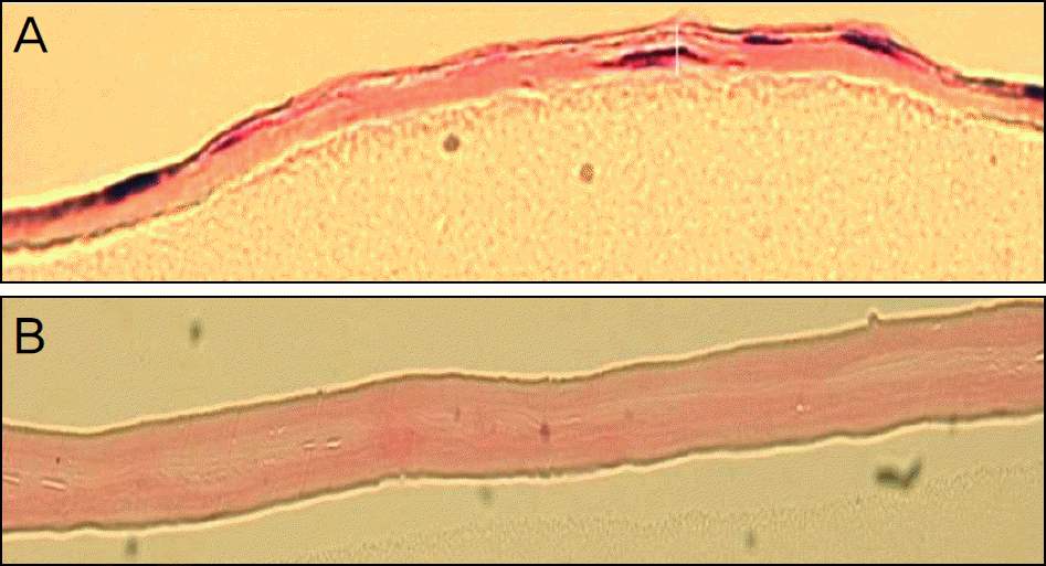
Figure 1.
Light microscopic findings of amniotic membrane. (A) Single-layered lyophilized AM, (B) 5-layered reinforced AM (hematoxylin-eosin, original magnification, ×200). AM = amniotic membrane.

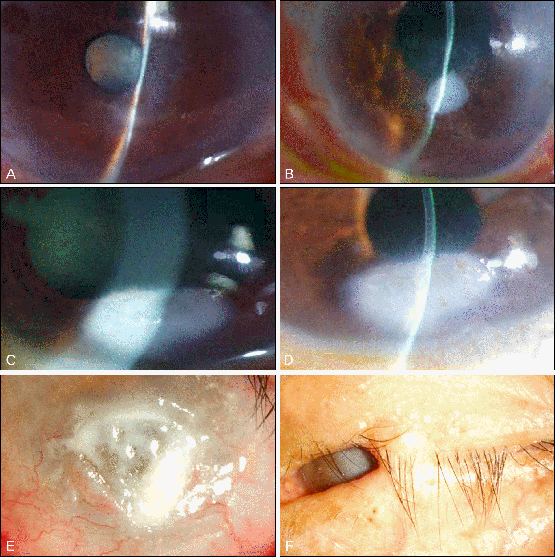
Figure 2.
Case 1 (A) Initial photographs. The cornea was perforated and the anterior chamber was shallow. (B) Anterior chamber was well formed and no epithelial defects were seen 18 months after 5-layered reinforcing AM trasplantation. Case 2 (C) Initial photographs. Corneal ulcer (2 × 3 mm) with peripheral opacity was seen preoperatively. (D) The ocular surface was stabilized until postoperative 5 months. Case 3 (E) Initial photographs. Corneal ulcer (5 × 5 mm) and total limbal deficiency with neovascularization were seen. (F) To prevent complication of exposure keratitis, tarsorrhaphy has been maintained until post operative 15 months.

Figure 3.
Representative AS-OCT imaging of case 2. Recovery of corneal thickness and anterior chamber stability was showed with AS-OCT at 12 months after 5-layered reinforced AM transplantation. AS-OCT = anterior segment Optical coherence tomography; AM = amniotic membrane.

Table 1.
Characteristics and clinical outcomes of patients receiving transplantation of 5-layered reinforced AM for corneal perforation or ulceration


 PDF
PDF ePub
ePub Citation
 Print
Print


 XML Download