Abstract
Purpose
To report the clinical outcomes and utility of 23-gauge (G) transconjunctival sutureless vitrectomy (TSV) in patients with postoperative endophthalmitis following cataract surgery.
Methods
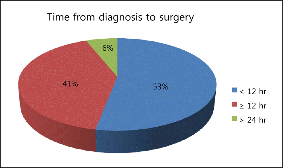
The medical records of 16 patients (17 eyes) who underwent 23-G TSV between January 2008 and December 2009 at Konyang University Hospital due to postoperative endophthalmitis following cataract surgery were retrospectively analyzed. The pre- and post-operative best-corrected visual acuities (BCVA), changes in intraocular pressure, the time from diagnosis to surgery, the intraoperative and postoperative complications, and the average duration of hospitalization were investigated.
Results
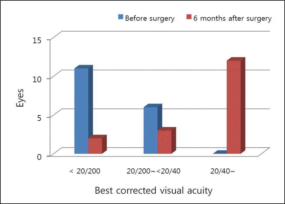
The mean BCVA significantly improved from log MAR 1.89 ± 1.03 to log MAR 0.42 ± 0.82 (p = 0.001), and the mean intraocular pressure changed from 16.1 ± 4.1 mm Hg at baseline to 16.2 ± 3.3 mm Hg on the first postoperative day without any significant difference (p = 0.955). In addition, none of the patients required sutures to treat wound leakage or showed hypotony on follow-up observation. The average operation time was 64.7 ± 22.5 minutes, and the average duration of hospitalization was 5.4 ± 4.5 days.
Go to : 
References
1. Puliafito CA, Baker AS, Haaf J, Foster CS. Infectious endophthalmitis. Review of 36 cases. Ophthalmology. 1982; 89:921–9.
3. Aaberg TM Jr, Flynn HW Jr, Schiffman J, Newton J. Nosocomial acute-onset postoperative endophthalmitis survey. A 10 year review of incidence and outcomes. Ophthalmology. 1998; 105:1004–10.
4. Schmitz S, Dick HB, Krummenauer F, Pfeiffer N. Endophthalmitis in cataract surgery; results of a German survey. Ophthalmology. 1999; 106:1869–77.
5. Barry P, Seal DV, Gettinby G, et al. ESCRS study of prophylaxis of postoperative endophthalmitis after cataract surgery: preliminary report of principal results from a European multicenter study. J Cataract Refract Surg. 2006; 32:407–10.
6. Maquire JI. Postoperative endophthalmitis: optimal management and the role and timing of vitrectomy surgery. Eye. 2008; 22:1290–300.

7. Nagaki Y, Hayasaka S, Kadoi C, et al. Bacterial endothalmitis after small-incision cataract surgery: effect of incision placement and intraocular lens type. J Cataract Refract Surg. 2003; 29:20–6.
8. Cooper BA, Holekamp NM, Bohigian G, Thompson PA. Case-control study of endophthalmitis after cataract surgery comparing scleral tunnel and clear corneal wounds. Am J Ophthalmol. 2003; 136:300–5.

9. Endophthalmitis Vitrectomy Study Group. Results of the Endophthalmitis Vitrectomy Study. A randomized trial of immediate vitrectomy and of intravenous antibiotics for the treatment of postoperative bacterial endophthalmitis. Arch Ophthalmol. 1995; 113:1479–96.
10. Doft BH, Kelsey SF, Wisniewski S, et al. Treatment of endophthalmitis after cataract extraction. Retina. 1994; 14:297–304.

11. Han JI, Cho SW, Lee TG, et al. The clinical results of sutureless vitrectomy using 23-gauge surgical system. J Korean Ophthalmol Soc. 2008; 49:911–6.

12. Woo SJ, Park KH, Hwang JM, et al. Risk factors associated with sclerotomy leakage and postoperative hypotony after 23-gauge transconjunctival sutureless vitrectomy. Retina. 2009; 29:456–63.

13. Laatikainen L, Tarkkanen A. Early vitrectomy in the treatment of postoperative purulent endophthalmitis. Acta Ophthalmol. 1987; 65:455–60.

14. Kuhn F, Gini G. Ten years after. Are findings of the Endophthalmitis Vitrectomy Study still relevant today? Graefes Arch Clin Exp Ophthalmol. 2005; 243:1197–9.
15. Kim SH, Kim JS. A case of Agrobacterium radiobacter endophthalmitis following cataract surgery. J Korean Ophthalmol Soc. 2001; 42:1117–21.
16. Johnson MW, Doft BH, Kelsey SF, et al. The endophthalmitis Vitrectomy Study, Relationship between clinical presentation and microbiologic spectrum. Ophthalmology. 1997; 104:261–72.
17. Lee SB, Han JW, Chung SK, Baek NH. Factors associated with visual outcomes of postoperative endophthalmitis following cataract surgery. J Korean Ophthalmol Soc. 2005; 46:1618–23.
Go to : 
Table 1.
Distribution and demographic characteristics of the patients
| Case No. | Age | Sex | Culture | PreOP* BCVA† | PostOP‡ BCVA (6 mon) | PreOP IOP§ (mm Hg) | PostOP IOP (mm Hg) | PreOP INJ∏ | PostOP INJ | Hospitalization (day) | Operation time (min) | Remark |
|---|---|---|---|---|---|---|---|---|---|---|---|---|
| 1 | 57 | F | No growth | HM | 0.4 | 11 | 15 | 0 | 0 | 4 | 90 | |
| 2 | 57 | F | No growth | FC | 1.0 | 10 | 16 | 0 | 0 | 4 | 105 | |
| 3 | 74 | F | | 0.06 | 0.8 | 21 | 16 | 0 | 0 | 2 | 40 | |
| 4 | 39 | F | No growth | 0.1 | 1.0 | 25 | 26 | 1 | 0 | 8 | 70 | |
| 5 | 58 | F | | 0.2 | 1.0 | 18 | 18 | 1 | 0 | 2 | 55 | Endolaser |
| 6 | 68 | M | | HM | 1.0 | 15 | 15 | 1 | 0 | 5 | 115 | Post capsulotomy, Endolaser |
| 7 | 70 | F | Staphylococcus epidermidis | 0.2 | 1.0 | 16 | 13 | 0 | 0 | 3 | 55 | |
| 8 | 26 | M | | HM | 1.0 | 16 | 20 | 1 | 0 | 3 | 75 | |
| 9 | 78 | F | No growth # | 0.03 | 0.3 | 13 | 11 | 1 | 1 | 3 | 52 | Previous posterior capsular rupture |
| 10 | 52 | F | Pantoea spp.# | 0.1 | 0.8 | 10 | 13 | 0 | 0 | 6 | 65 | |
| 11 | 83 | M | No growth | HM | FC | 20 | 16 | 1 | 1 | 6 | 80 | Vitreous flushing Macular degeneration |
| 12 | 55 | F | No growth | 0.1 | 0.6 | 11 | 15 | 1 | 0 | 7 | 60 | |
| 13 | 78 | M | | HM | 0.3 | 16 | 15 | 1 | 0 | 5 | 45 | |
| 14 | 50 | M | | FC | 1.0 | 23 | 18 | 0 | 0 | 1 | 43 | |
| 15 | 43 | M | | 0.1 | 1.0 | 18 | 17 | 0 | 0 | 2 | 35 | |
| 16 | 77 | M | Staphylococcus epidermidis | HM | HM | 16 | 14 | 0 | 0 | 18 | 50 | Macular infarction |
| 17 | 63 | M | Staphylococcus epidermidis | HM | 0.5 | 15 | 17 | 0 | 0 | 14 | 65 | Core vitrectomy |


 PDF
PDF ePub
ePub Citation
 Print
Print


 XML Download