Abstract
Purpose
To investigate the prevalence, clinical manifestations, and risk factors of dry eye syndrome (DES) among people over 50 years old in the Incheon area.
Methods
A cross-sectional prevalence study was performed on 462 people over 50 years old in Dong-gu, Incheon. DES was defined as the constant or frequent presence of symptoms of both dryness and irritation. Symptoms and past medical history were assessed by a survey. Eye examination included slit lamp examination, Schirmer test, and tear breakup time (T-BUT). Age, sex, living habits, systemic and eye diseases were also analyzed to determine the risk factors of DES.
Results
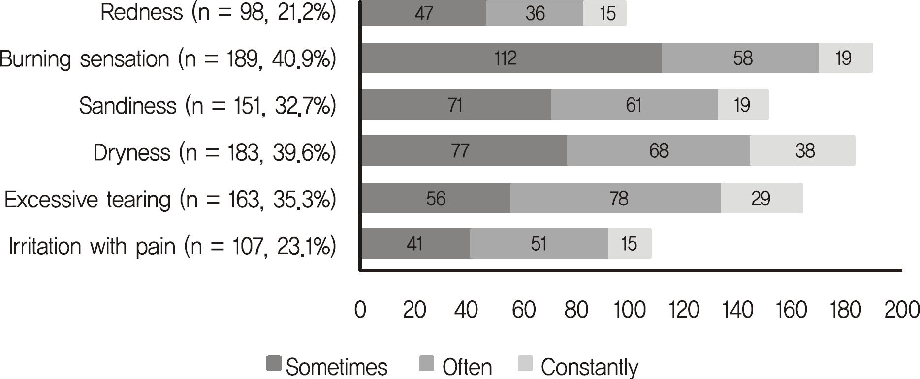
The prevalence of DES was 26.2%. The major symptoms were as follows in descending order: dryness (77.9%), tearing (75.2%), and sandiness (72.7%). An average of 12.1 ± 6.0 mm in the Schirmer test and 6.7 ± 2.4 seconds in the T-BUT were significantly different in the DES group from the normal group (p < 0.001). Variables such as age, sex, living habits, and eye diseases were not related to the diagnosis of DES, whereas diabetes was the only risk factor of DES with statistical significance (p = 0.03).
References
1. The definition and classification of dry eye disease: report of the definition and classification subcommittee of the International Dry Eye Workshop (2007). Ocul Surf. 2007; 5:75–92.
2. Pflugfelder SC, Solomon A, Stern ME. The diagnosis and management of dry eye: a twenty-five-year review. Cornea. 2000; 19:644–9.
3. Sall K, Stevenson OD, Mundorf TK, Reis BL. Two multicenter, randomized studies of the efficacy and safety of cyclosporine ophthalmic emulsion in moderate to severe dry eye disease. CsA Phase 3 study Group. Ophthalmology. 2000; 107:631–9.
4. Stevenson D, Tauber J, Reis BL. Efficacy and safety of cyclosporin A ophthalmic emulsion in the treatment of moderate-to-severe dry eye disease: a dose-ranging, randomized trial. The cyclosporin A phase 2 study group. Ophthalmology. 2000; 107:967–74.
5. Stern ME, Gao J, Siemasko KF, et al. The role of the lacrimal functional unit in the pathophysiology of dry eye. Exp Eye Res. 2004; 78:409–16.

6. Schein OD, Munoz B, Tielsch JM, et al. Prevalence of dry eye among the elderly. Am J Ophthalmol. 1997; 124:723–8.

7. Lin PY, Tsai SY, Cheng CY, et al. Prevalence of dry eye among an elderly Chinese population in Taiwan: The Shihpai Eye Study. Ophthalmology. 2003; 110:1096–101.
8. Brewitt H, Sistani F. Dry eye disease: the scale of the problem. Surv Ophthalmol. 2001; 45:S199–202.
9. The epidemiology of dry eye disease: report of the Epidemiology Subcommittee of the International Dry Eye WorkShop (2007). Ocul Surf. 2007; 5:93–107.
10. Wolkoff P, Nojgaard JK, Troiano P, Piccoli B. Eye complaints in the office environment: precorneal tear film integrity influenced by eye blinking efficiency. Occup Environ Med. 2005; 62:4–12.

11. Moss SE, Klein R, Klein BE. Prevalence of and risk factors for dry eye syndrome. Arch Ophthalmol. 2000; 118:1264–8.

12. Rexrode KM, Lee IM, Cook NR, et al. Baseline characteristic of participants in the Women's Health Study. J Womens Health Gend Based Med. 2000; 9:19–27.
13. Schaumberg DA, Sullivan DA, Dana MR. The epidemiology of dry eye syndrome. Cornea. 2000; 19:S120.
14. Nichols KK, Nichols JJ, Lynn Mitchell G. The relation between tear film tests in patients with dry eye disease. Ophthalmic Physiol Opt. 2003; 23:553–60.

15. Chia EM, Mitchell P, Rochtchina E, et al. Prevalence and associations of dry eye syndrome in an older population: the Blue Mountains Eye Study. Clin Experiment Ophthalmol. 2003; 31:229–32.

16. Lee AJ, Lee J, Saw SM, et al. Prevalence and risk factors associated with dry eye symptoms: a population based study in Indonesia. Br J Ophthalmol. 2002; 86:1347–51.

17. Hikichi T, Yoshida A, Fukui Y, et al. Prevalence of dry eye in Japanese eye centers. Graefes Arch Clin Exp Ophthalmol. 1995; 233:555–8.

18. Lee JH, Kee CW. The significance of tear film breakup time in the diagnosis of dry eye syndrome. Korean J Ophthalmol. 1988; 2:69–71.

19. Clinch TE, Benedetto DA, Felberg NT, Laibson PR. Schirmer's test; a closer look. Arch Ophthalmol. 1983; 101:1383–6.
20. Lee JH, Ki CW, Roh KG. The significance of the tear film breakup time in the diagnosis of the dry eye syndrome. J Korean Ophthalmol Soc. 1985; 26:1131–5.
21. Lemp MA. Report of the National Eye Institute/Industry Workshop on clinical trials in dry eyes. CLAO J. 1995; 21:221–32.
22. Mengher LS, Bron AJ, Tonge SR, Gilbert DJ. Effect of fluorescein instillation on the precorneal tear film stability. Curr Eye Res. 1985; 4:9–12.

23. Cho BJ, Lee JH, Shim OJ. The relation between clinical manifestations of dry eye patients and their BUTs. J Korean Ophthalmol Soc. 1992; 33:297–302.
24. Methodologies to diagnose and monitor dry eye disease: report of the Diagnostic Methodology Subcommittee of the International Dry Eye WorkShop (2007). Ocul Surf. 2007; 5:108–52.
25. Lucca JA, Nunez JN, Farris RL. A comparison of diagnostic tests for keratoconjunctivitis sicca: lactoplate, Schirmer, and tear osmolarity. CLAO J. 1990; 16:109–12.
26. Behrens A, Doyle JJ, Stern L, et al. Dysfunctional tear syndrome: a Delphi approach to treatment recommendations. Cornea. 2006; 25:900–7.
27. Schaumberg DA, Buring JE, Sullivan DA, Dana MR. Hormone replacement therapy and dry eye syndrome. JAMA. 2001; 286:2114–9.

28. Schaumberg DA, Dana R, Buring JE, Sullivan DA. Prevalence of dry eys disease among US men; estimates from the Physician's Health Studies. Arch Ophthalmol. 2009; 127:763–8.
29. Sullivan DA, Sullivan BD, Evans JE, et al. Androgen deficiency, meibomian gland dysfunction, and evaporative dry eye. Ann N Y Acad Sci. 2002; 966:211–22.

30. Kaiserman I, Kaiserman N, Nakar S, Vinker S. Dry eye in diabetic patients. Am J Ophthalmol. 2005; 139:498–503.

Table 1.
Demographic characteristics of 462 participants with information on dry eye syndrome
| | Male | Female |
|---|---|---|
| Total No. (%) | 202 (43.7) | 260 (56.3) |
| Age (mean ± SD, yr) | 70.3 ± 7.3 | 70.4 ± 7.2 |
| No. of DES | 45 | 76 |
| Prevalence of DES (%)* | 22.3 | 29.2 |
Table 2.
Composition of participants and prevalence of dry eye syndrome according to age
Table 3.
Major complaining symptoms of dry eye syndrome group
| Variable | N | % |
|---|---|---|
| Dryness | 94 | 77.9 |
| Excessive tearing with wind blowing | 91 | 75.2 |
| Sandiness | 88 | 72.7 |
| Irritation with pain | 60 | 49.6 |
| Burning sensation | 50 | 41.3 |
| Redness | 38 | 31.5 |
Table 4.
Comparison of TBUT, corneal erosion score and Schirmer test between normal group and dry eye syndrome group
| | Normal group (n = 341) | DES group (n = 121) | p-value* |
|---|---|---|---|
| TBUT (sec) | 9.4 ± 2.4 | 6.7 ± 2.4 | < 0.001 |
| Score of corneal erosion | 2.6 ± 0.8 | 2.2 ± 0.9 | 0.29 |
| Schirmer test (mm) | 15.3 ± 6.0 | 12.1 ± 6.0 | < 0.001 |
Table 5.
Multivariate analysis for associations between clinical factors and dry eye syndrome
| Variable | No. (%) | Odds ratio | 95% CI | p-value* |
|---|---|---|---|---|
| Age | − | 1.6 | 1.1–2.1 | 0.10 |
| Sex | − | 1.8 | 1.5–2.2 | 0.08 |
| Living habits | | | | |
| Smoking | 82 (17.7) | 1.4 | 0.8–2.3 | 0.26 |
| Alcohols | 138 (29.9) | 1.3 | 0.6–1.9 | 0.29 |
| Underlying diseases | | | | |
| Diabetes | 139 (30.1) | 2.6 | 1.8–3.4 | 0.03 |
| Osteoporosis | 108 (23.4) | 1.8 | 0.9–2.6 | 0.07 |
| Arthritis | 173 (37.4) | 1.5 | 1.1–2.0 | 0.08 |
| Hypertension | 210 (45.5) | 1.2 | 0.5–1.8 | 0.39 |
| Hyperlipidemia | 33 (7.1) | 1.1 | 0.4–1.7 | 0.59 |
| Cardiovascular disease | 52 (11.3) | 1.1 | 0.3–2.1 | 0.71 |
| Stroke | 14 (3.0) | 0.9 | 0.5–2.4 | 0.85 |
| Miscellaneous | − | − | − | >0.05 |
| Ocular diseases | | | | |
| Pterygium | 76 (16.0) | 1.4 | 0.9–2.6 | 0.08 |
| Pinguecula | 112 (23.5) | 1.3 | 0.8–2.4 | 0.11 |
| Glaucoma | 4 (0.8) | 0.8 | 0.2–2.9 | 0.73 |
| Cataract (including pseudophakia) | 333 (72.1) | 1.1 | 0.3–2.3 | 0.35 |
| History of ocular surgery | 126 (27.2) | 1.5 | 0.5–3.2 | 0.08 |


 PDF
PDF ePub
ePub Citation
 Print
Print


 XML Download