

 PDF
PDF ePub
ePub Citation
 Print
Print


Abstract
Purpose
To report a long term result of vitrectomy, gas tamponade without laser retinopexy for serous macular detachment associated with an optic disc pit (ODP).
Case summary
A 13 year old boy with visual disturbance in the left eye showed serous macular detachment associated with an inferior temporal ODP. The abnormal vitreous strand over the optic disc implying vitreous traction and retinoschisis were revealed by the optical coherence tomography (OCT) examination. Pars plana vitrectomy after complete induction of posterior vitreous detachment without laser retinopexy, and gas tamponade with postoperative facedown positioning were performed. Complete retinal attachment occurred at 6 months after first operation but recurrent macula detachment occurred at 15 months after first operation. Additional gas tamponade resulted in successful retinal attachment for more than 2 years and visual improvement.
Conclusions
Vitrectomy and gas tamponade without additional laser photocoagulation could be another option for the treatment of ODP maculopathy. But recurrent macular detachment might occur and simple gas tamponade was effective in this case. This result supports another factor in addition to vitreous traction may play a role in the development of the macular detachment associated ODP. Further studies are required to evaluate the effect of vitrectomy, gas tamponade without laser retinopexy for the treatment of ODP maculopathy.
Go to : 
References
1. Sobol WM, Blodi CF, Folk JC, Weingeist TA. Long term visual outcome in patients with optic nerve pit and serous retinal detachment of the macula. Ophthalmology. 1990; 97:1539–42.
2. Brown GC, Brown MM. Repair of retinal detachment associated with congenital excavated defects of the optic disc. Ophthalmic Surg. 1995; 26:11–5.

3. Gass JD. Serous detachment of the macula: secondary to congenital pit of the optic nervehead. Am J Ophthalmol. 1969; 67:821–41.
4. Lincoff H, Yannuzzi L, Singerman L, et al. Improvement in visual function after displacement of the retinal elevation emanating from optic pits. Arch Ophthalmol. 1993; 111:1071–9.
5. Ryu JW, Ra H, Lee WK. A case of surgically treated serous macular detachment associated with optic disc pit. J Korean Ophthalmol Soc. 2010; 51:155–8.
6. Ghosh YK, Banerjee S, Konstantinidis A, et al. Surgical management of optic pit associated maculopathy. Eur J Ophthalmol. 2008; 18:142–6.
7. Hirakata A, Okada AA, Hida T. Long term results of vitrectomy without laser treatment for macular detachment associated with an optic disc pit. Ophthalmology. 2005; 112:1430–5.
8. Theodossiadis GP. Treatment of maculopahty associated with optic disc pit by sponge explant. Am J Ophthalmol. 1996; 121:630–7.
9. Karacorlu SA, Karacorlu M, Ozdemir H, et al. Optical coherence tomography in optic pit maculopahty. Int Ophthalmol. 2007; 27:293–7.
10. Theodossiadis PG, Grigoropoulos VG, Emfietzoglou J, Theodossiadis GP. Vitreous findings in optic disc pit maculopahty based on optical coherence tomography. Grafe's Arch Clin Exp Ophthalmol. 2007; 245:1311–8.
11. Blair CJ, Gass JD. Photocoaulation of the macula and papillomacular bundle in the human. Arch Ophthalmol. 1972; 88:167–71.
12. Roider J. Laser treatment of retinal disease by subthreshold laser effect. Semin Ophthalmol. 1999; 14:19–26.
13. Doyle E, Trivedi D, Good P, et al. High resolution optical coherence tomography demonstration of membranes spanning optic disc pits and colobomas. Br J Ophthalmol. 2009; 93:360–5.
14. Johnson TM, Johnson MW. Pathogenesis implications of subretinal gas migration through pits and atypical colobomas of the optic nerve. Arch Ophthalmol. 2004; 122:1793–800.
Go to :
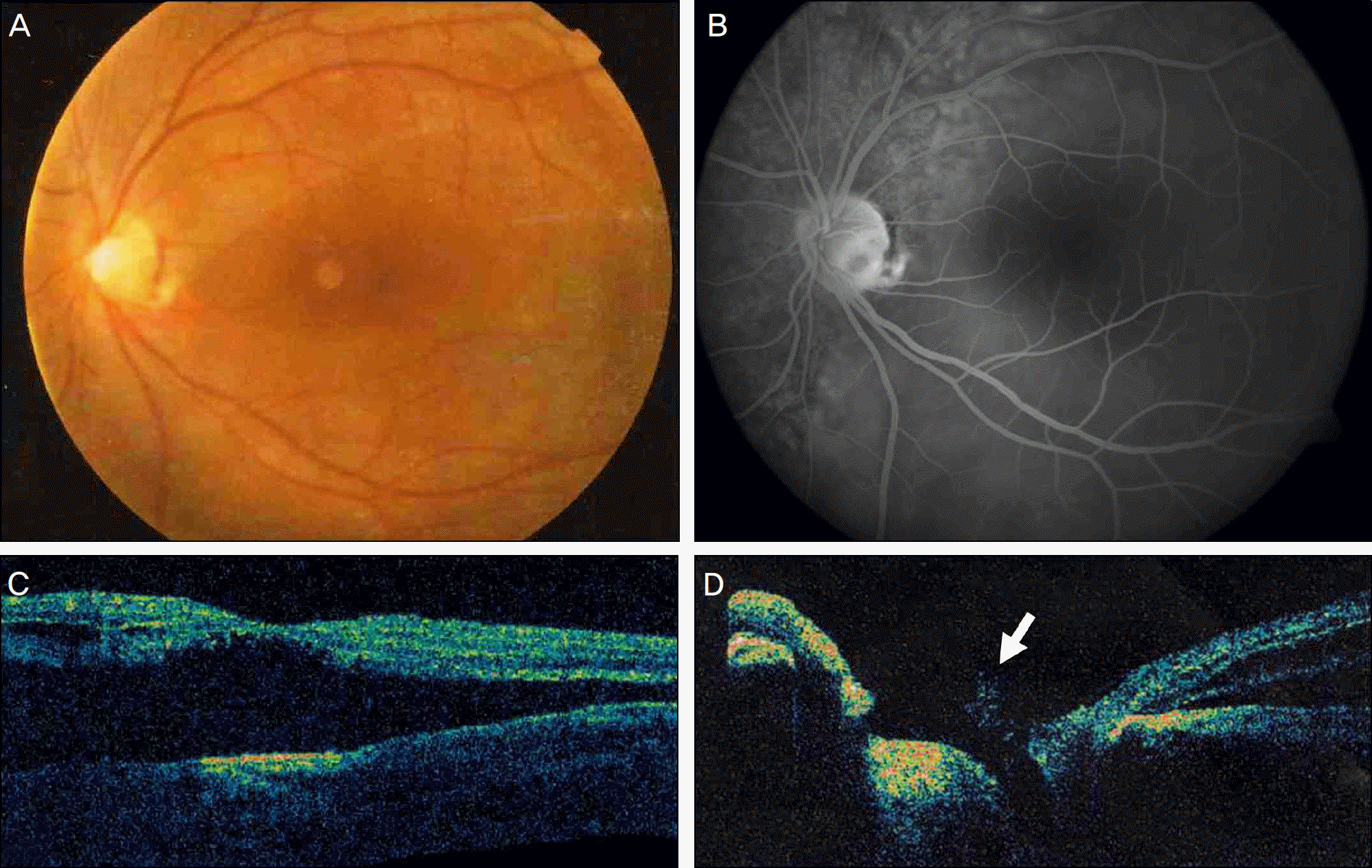 | Figure 1.(A) Preoperative fundus photograph of the left eye showing inferotemporal optic disc pit associated serous macular detachment. (B) Late phase of the fluorescein angiography showing hypofluorescence of the optic disc pit and dye pooling into sub-retinal fluid. (C, D) Optical coherence tomography image showing serous macular detachment, schisis-like separation of the retinal layers and dense vitreous strands (white arrow) attached to the optic disc pit. |
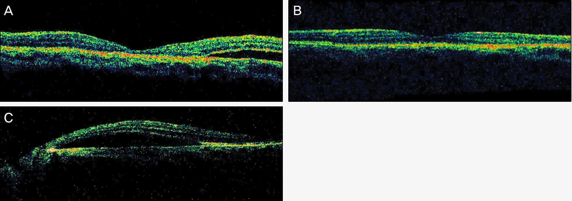 | Figure 2.(A) Optical coherence tomography (OCT) image showing macular attachment but residual subretinal fluid inferior to the macula. (B) OCT image showing complete resorption of subretinal fluid at 6 months after first operation and (C) re-accumulation of subretinal fluid in the papillomacular area at 15 months after first operation. |
 XML Download