

 PDF
PDF ePub
ePub Citation
 Print
Print


Abstract
Purpose
To report the presentation and management of an atypical and advanced case of nodular hidradenoma of the eyelid with ptosis.
Case summary
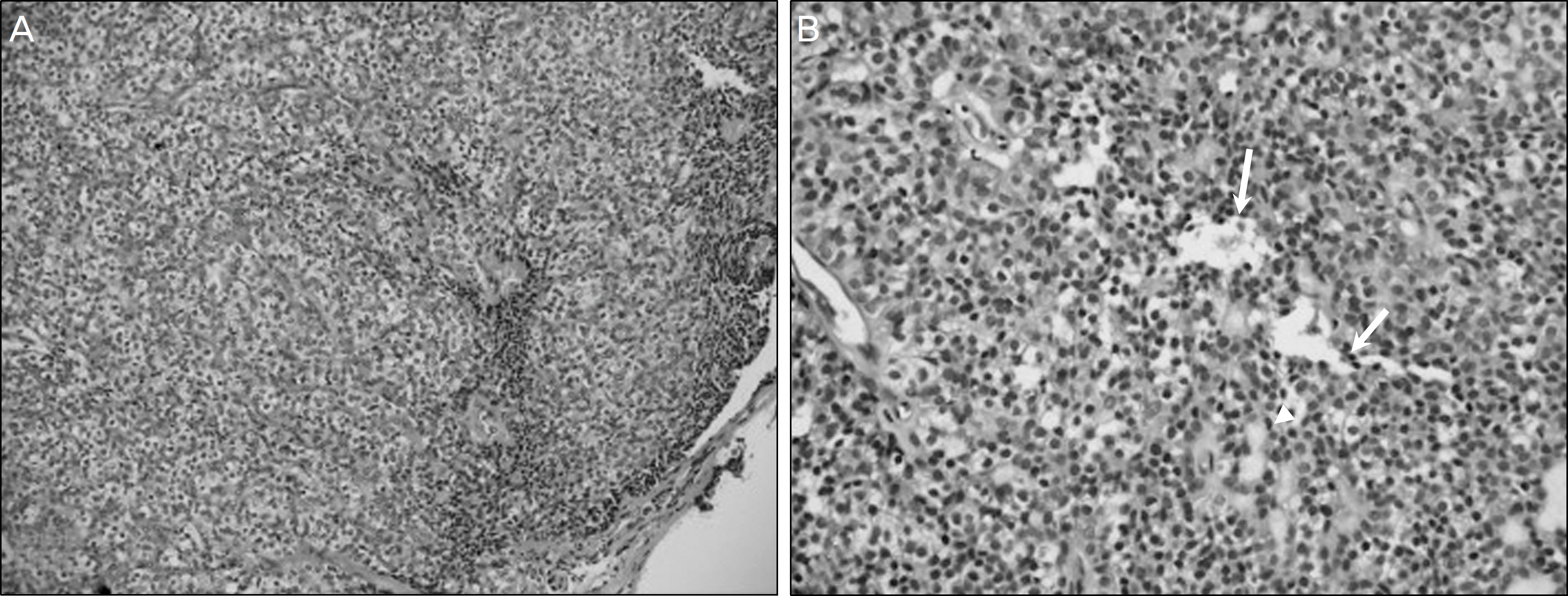
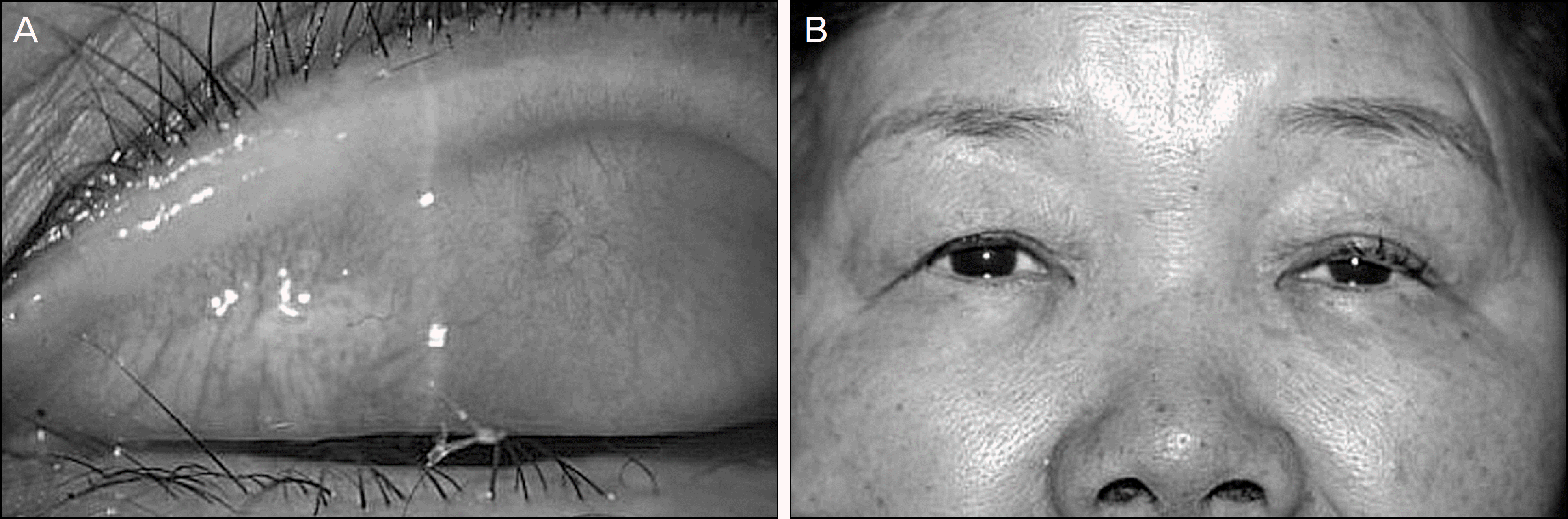
A 64-year-old woman who presented with a palpable growing nodular mass and ptosis was tested with marginal reflex distance 1 as right eye 1 mm, left eye −1.5 mm and levator function test as 12 mm and 10 mm, respectively during a hospital visit. The patient was tentatively diagnosed with eyelid adnexal tumor with mechanical ptosis and was managed by surgical excision of the lesion. Histology confirmed hidradenoma.
Conclusions
Hidradenomas are benign adnexal tumors originating from the eccrine gland and rarely detectable in the eyelid. However, rudimentary glandular structures can be a possible tumor source. Nodular hidradenoma should be considered in the differential diagnosis of adnexal masses and such lesions may cause significant functional and cosmetic morbidity despite their histologically benign nature.
References
1. The Text Compilation Committee of Korean Dermatological Association. Dermatology. 4th ed.Seoul: Yemongak;2001. p. 700–1.
2. Duairaj VD, Bartlett HM, Said S. Malignant hidradenoma of the medial canthus with orbital and intracranial extension. Ophthal Plast Reconstr Surg. 2006; 22:229–32.
3. Sheth HG, Raina J. Giant eccrine hidrocystoma presenting with unilateral ptosis and epiphora. Int Ophthalmol. 2008; 28:429–31.

4. Yasser H, Abdul Rahman, Mohammed O. Periocular hidradenoma papilliferum. Saudi Journal of Ophthalmology. 2009; 23:211–3.
5. Hernández-Pérez E, Cestoni-Parducci R. Nodular hidradenoma and hidradenocarcinoma. A 10-year review. J Am Acad Dermatol. 1985; 12:15–20.
6. Yanoff M, Fine BS. Ocular Pathology Skin and Lacrimal Drainage System. 5th ed.6. Missouri: Mosby Inc.;2002. p. 204–5.
7. Durairaj VD, Bartlett HM, Said S. Malignant hidradenoma of the medial canthus with orbital and intracranial extension. Ophthal Plast Reconstr Surg. 2006; 22:229–32.
8. Dailey RA, Saulny SM, Tower RN. Treatment of multiple apocrine hidrocystomas with trichloroacetic acid. Ophthal Plast Reconstr Surg. 2005; 21:148–50.
9. Yaghoobi R, Saboktakin M, Feily A, Mehri M. Bilateral multiple apocrine hidrocystoma of the eyelids. Acta Dermatovenerol Alp Panonica Adriat. 2009; 18:138–40.
10. Vignes JR, Franco-Vidal V, Eimer S, Liguoro D. Intraorbital apocrine hidrocystoma. Clin Neurol Neurosurg. 2007; 109:631–3.
11. Callahan M, Beard C. Beard's Ptosis. 4th ed.Alabama: Aesculapius Publishing;1990. p. 78–9.
12. Nerad JA. Oculoplastic Surgery; the Requisites in Ophthalmology. Philadelphia: Mosby;2001. p. 39–47.
Figure 1.
The photograph of the nodular mass protruding from the left upper tarsal conjunctiva that is measured as 10 × 8 mm in size.
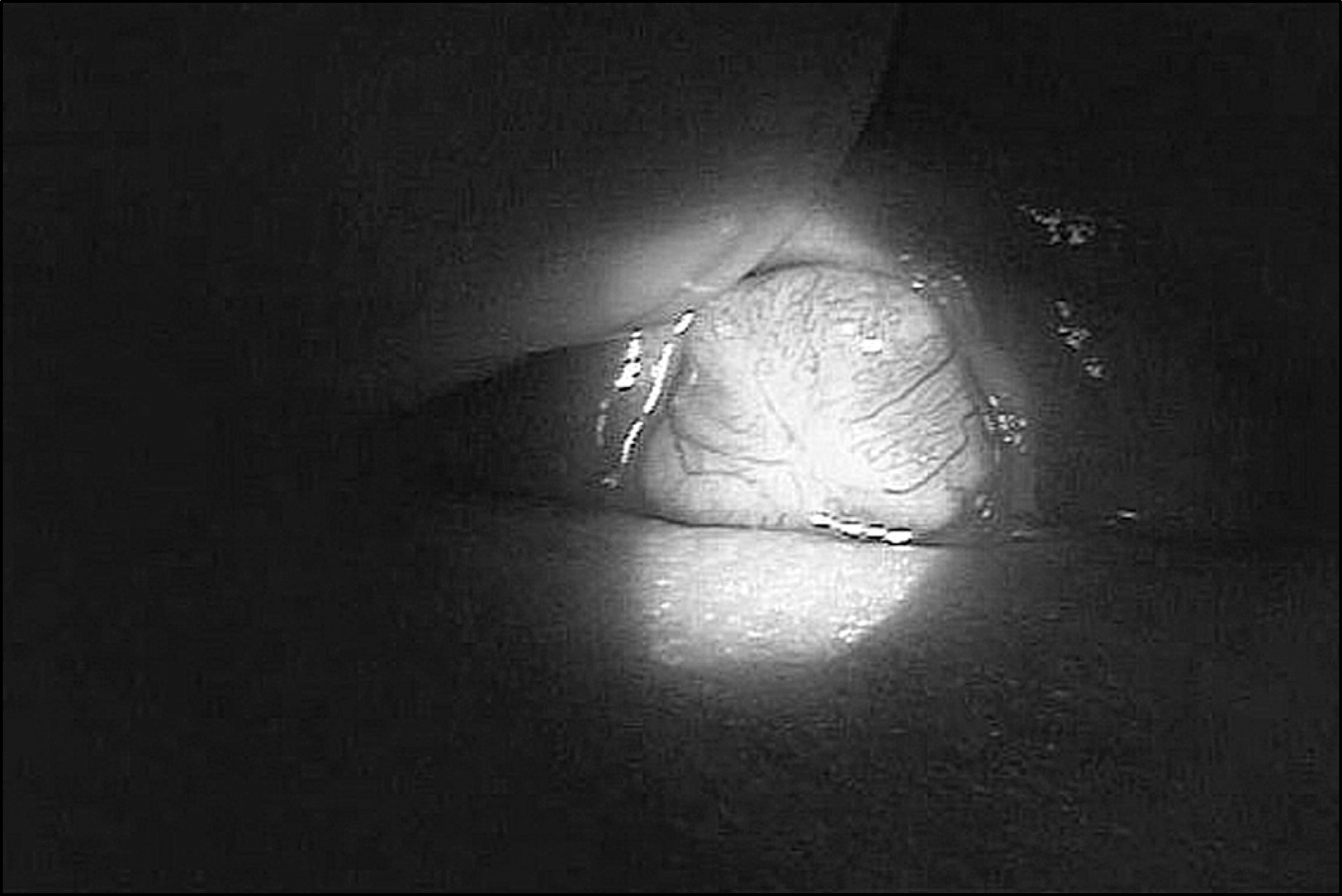
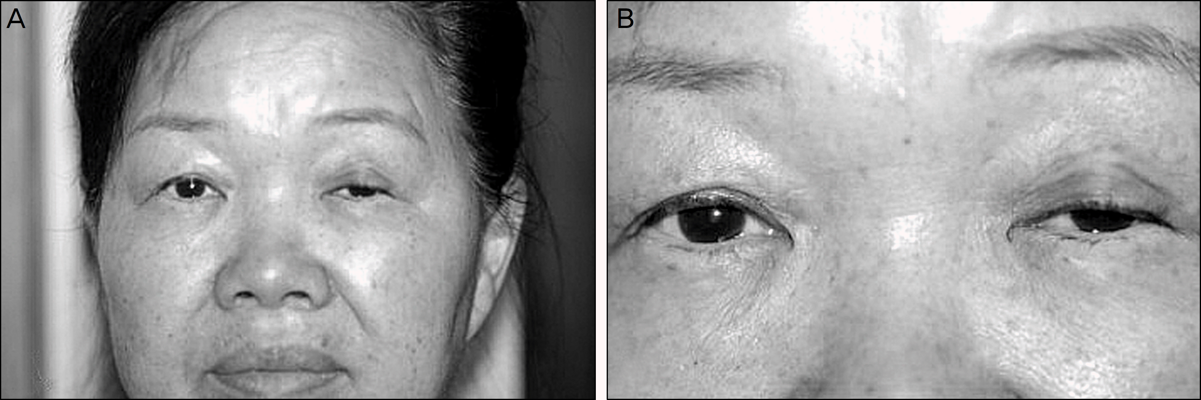
Figure 2.
On her first hospital visit. (A) She presented with left upper eyelid swelling causing mechanical ptosis. (B) Marginal reflex distance (MRD) 1 was measured as 1.0 mm in the right eye and −1.5 mm in the left.
 XML Download