

 PDF
PDF ePub
ePub Citation
 Print
Print


Abstract
Purpose
To evaluate the changes in the clinical signs and symptoms of dry eye syndrome before and after cataract surgery according to the main incision location and the type of artificial tears.
Methods
Twenty-four eyes of 17 patients who underwent phacoemulsification and posterior chamber lens insertion from May 2009 to July 2009 were enrolled in the present study prospectively. The main incision location (temporal or superior incision) was determined according to the axis of astigmatism and the postoperative artificial tears (sodium hyaluronate with or without preservatives) were determined randomly. The tear film breakup time (TF-BUT), Schirmer test, esthesiometer, corneal surface grading with Oxford system and ocular surface disease index (OSDI) questionnaire before and 1, 2 and 6 months after surgery were evaluated. The corneal nerve was also analyzed using corneal confocal microscopy (Confoscan 4, Nidek, Italy) before, and 1 and 6 months after surgery.
Results
The TF-BUT was significantly longer (p = 0.010) and the OSDI score was significantly lower (p = 0.007) in the patient group with preservative-free sodium hyaluronate than the group with sodium hyaluronate containing preservatives at 6 months after cataract surgery. There were no statistically significant differences according to the main incision location in the sodium hyaluronate without preservatives group.
References
1. Thylefors B, Negrel AD, Pararajasegaram R, Dadzie KY. Global data on blindness. Bull World Health Organ. 1995; 73:115–21.
2. Song KJ, Han MY, Kim SY, et al. Prevalence estimation of cataract based on a screening test. J Korean Ophthalmol Soc. 2007; 48:768–73.
3. Kohlhass M. Corneal sensation after cataract and refractive surgery. J Cataract Refract Surg. 1998; 24:1399–409.
4. Cho YK, Kim MS. Dry eye after cataract surgery and associated intraoperative risk factors. Korean J Ophthalmol. 2009; 23:65–73.

5. Ram J, Gupta A, Brar G, et al. Outcomes of phacoemulsification in patients with dry eye. J Cataract Refract Surg. 2002; 28:1386–9.
6. Li XM, Hu L, Hu J, Wang W. Investigation of dry eye disease and analysis of the pathogenic factors in patients after cataract surgery. Cornea. 2007; 26(9 Suppl 1):S16–20.
7. Roberts CW, Elie ER. Dry eye symptoms following cataract surgery. Insight. 2007; 32:14–21.
8. Sanchez MA, Arriola-Villalobos P, Torralbo-Jimenez P, et al. The effect of preservative-free HP-Guar on dry eye after phacoemulsification – a flow cytometric study. Eye. 2010; 24:1331–7.
9. Epstein SP, Chen D, Asbell PA. Evaluation of biomarkers of inflammation in response to benzalkonium chloride on corneal and conjunctival epithelial cells. J Ocul Pharmacol Ther. 2009; 25:415–24.
10. Epstein SP, Ahdoot M, Marcus E, Asbell PA. Comparative toxicity of preservatives on immortalized corneal and conjunctival epithelial cells. J Ocul Pharmacol Ther. 2009; 25:113–9.
11. Kim SJ, Kim EH, Lee JE, Lee JS. Effect of preservative-free artificial eye drop on human corneal epithelial cell in vitro. J Korean Ophthalmol Soc. 2010; 51:1113–20.
12. Maca SM, Amon M, Findl O, et al. Efficacy and tolerability of preservative-free and preserved diclofenac and preserved ketor-olac eyedrops after cataract surgery. Am J Ophthalmol. 2010; 149:777–84.
13. Muller LJ, Vrensen GF, Pels L, et al. Architecture of human corneal nerves. Invest Ophthlamol Vis Sci. 1997; 38:985–94.
14. Martin XD, Safran AB. Corneal hypoesthesia. Surv Ophthalmol. 1988; 33:28–40.
15. Jordan A, Baum J. Basic tear flow. Does it exist? Ophthalmology. 1998; 95:827–32.
16. Donnenfeld ED, Solomon K, Perry HD, et al. The effect of hinge position on corneal sensation and dry eye after LASIK. Ophthalmology. 2003; 110:1023–9.
17. Vroman DT, Sandoval HP, Fernandez de Castro LE, et al. Effect of hinge location on corneal sensation and dry eye after laser in situ keratomileusis for myopia. J Cataract Refract Surg. 2005; 31:1881–7.
18. Pflugfelder SC, Tseng SC, Sanabria O, et al. Evaluation of subjective assessments and objective diagnostic tests for diagnosing tear-film disorders known to cause ocular irritation. Cornea. 1998; 17:38–56.
19. Her J, Yu SI, Seo SG. Clinical effects of various antiinflammatory therapies in dry eye syndrome. J Korean Ophthalmol Soc. 2006; 47:1901–10.
20. Bron AJ. The Doyne Lecture. Reflections on the tears. Eye. 1997; 11:583–602.
21. Bron AJ, Evans VE, Smith JA. Grading of corneal and conjunctival staining in the context of other dry eye tests. Cornea. 2003; 22:640–50.
22. Khanal S, Tomlinson A, Esakowitz L, et al. Changes in corneal sensitivity and tear physiology after phacoemulsification. Ophthal Physiol Opt. 2008; 28:127–34.
23. Grene RB, Lankston P, Mordaunt J, et al. Unpreserved carboxymethylcellulose artificial tears evaluated in patients with keratoconjunctivitis sicca. Cornea. 1992; 11:294–301.
24. Muller LJ, Marfurt CF, Kruse F, Tervo TM. Corneal nerves:structure, contents, and function. Exp Eye Res. 2003; 76:521–42.
25. John T, Rao GN, Aquavella JV. Corneal sensitivity in aphakic and pseudophakic eyes. CLAO J. 1988; 14:101–4.
26. Kohlhaas M, Stahlhut O, Tholuck J, Richard G. Development of corneal sensitivity after phacoemulsification with scleral tunnel incision. Klin Monatsbl Augenheilkd. 1997; 211:32–6.
27. John T. Corneal sensation after small incision, sutureless, one-handed phacoemulsification. J Cataract Refrac Surg. 1995; 40:1511–9.
28. Zhang M, Chen J, Luo L, et al. Altered corneal nerves in aqueous tear deficiency viewed by in vivo confocal microscopy. Cornea. 2005; 24:818–24.
29. Ramirez M, Hernandez-Quintela E, Sanchez-Huerta V, Naranjo-Tackman R. Confocal microscopy of corneal flap microfolds after LASIK. J Refract Surg. 2006; 22:155–8.
30. Kim YM, Kim SW, Kim TI, et al. The change of corneal sensitivity and recovery of corneal nerve after cataract surgery. J Korean Ophthalmol Soc. 2007; 48:13–8.
31. Ghoreishi M, Aidenloo NS, Peyman A, et al. Does hinge position affect dry eye after laser in situ keratomileusis? Ophthalmologica. 2005; 219:276–80.
Figure 1.
The analysis of corneal subbasal nerve plexus using ImageJ software. A winding line of corneal nerve is visualized in the left original photograph and the nerve length was analyzed by “segmented line length analysis tool” in ImageJ software by tracing each nerve separately as seen in the right photograph. Arrow heads trace corneal subbasal nerve and the arrows indicates the sites of nerve branching.

Figure 2.
The presentative photographs of corneal subbasal nerve plexus. Each column represents subgroups: temporal incision with PSH (A), temporal incision with PFSH (B), and superior incision with PFSH (C) respectively. Each row represents the time of taken images: preoperative, postoperative 1 month, and postoperative 6 month respectively. Each column is the images in the same patient. They commonly showed a decrease of corneal nerve plexus at 1 month after cataract surgery and a restoration at 6 month after cataract surgery. PSH = preserved 0.1% sodium hyaluronate group; PFSH = preservative free 0.1% sodium hyaluronate group; preop = preoperative, POP = postoperative, mon = month.
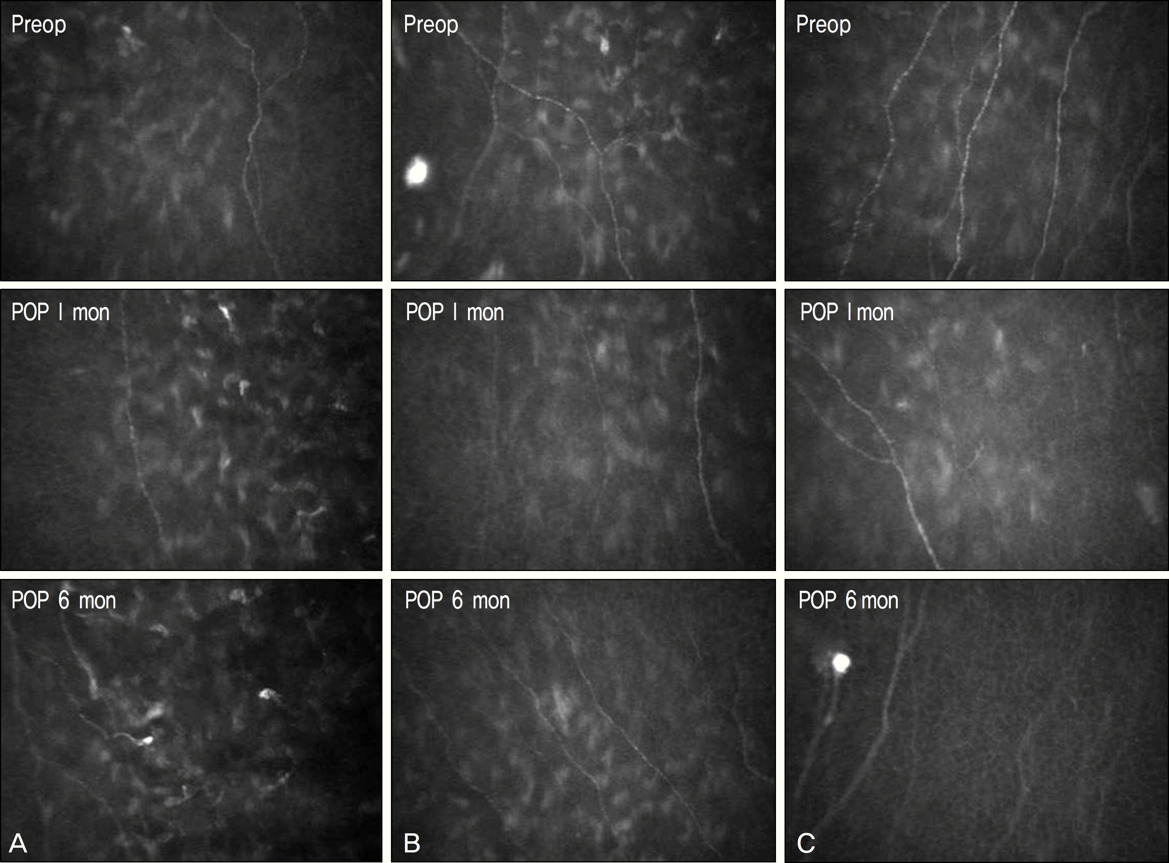
Table 1.
The changes in symptoms and signs of dry eye syndrome and corneal subbasal nerve plexus analysis before and after cataract surgery (mean ± SD)
| | Preop | Postop 1 mon | Postop 2 mon | Postop 6 mon |
|---|---|---|---|---|
| TF-BUT (sec) | 6.04 ± 2.80 | 4.75 ± 1.54 | 5.46 ± 1.28 | 6.25 ± 2.25 |
| Schirmer test (mm) | 14.10 ± 11.06 | 13.75 ± 8.86 | 14.83 ± 8.82 | 11.04 ± 4.77 |
| Esthesiometer (cm) | 5.81 ± 0.46 | 5.06 ± 1.19* | 5.00 ± 0.78* | 5.21 ± 0.94* |
| OSDI | 0.08 ± 0.12 | 0.11 ± 0.11* | 0.13 ± 0.12* | 0.14 ± 0.19* |
| Oxford grading of corneal surface (grade) | 0.38 ± 0.58 | 0.29 ± 0.46 | 0.13 ± 0.34 | 0.79 ± 0.66 |
| Nerve fiber density (number/mm2) | 18.77 ± 9.36 | 17.20 ± 7.72 | − | 17.85 ± 8.94 |
| Nerve fiber length (number/mm2) | 9.13 ± 3.83 | 7.37 ± 2.95* | − | 8.45 ± 3.06 |
| Nerve branch density (number/mm2) | 18.77 ± 16.1 | 15.10 ± 9.92 | − | 15.36 ± 9.02 |
Table 2.
The comparison of symptoms and signs of dry eye syndrome and corneal subbasal nerve plexus analysis between the groups with or without preservatives in the artificial tears (mean ± SD)
| | Group | Preop | Postop 1 mon | Postop 2 mon | Postop 6 mon |
|---|---|---|---|---|---|
| TF-BUT (sec) | PFSH | 5.71 ± 1.89 | 4.29 ± 0.95 | 4.86 ± 0.90 | 7.57 ± 1.90* |
| | PSH | 7.10 ± 3.67 | 5.50 ± 1.78 | 5.30 ± 1.34 | 5.60 ± 2.67* |
| Schirmer test (mm) | PFSH | 15.57 ± 13.89 | 16.71 ± 12.23 | 14.57 ± 10.77 | 11.29 ± 7.36 |
| | PSH | 13.80 ± 11.03 | 14.10 ± 8.53 | 18.60 ± 6.75 | 10.10 ± 1.73 |
| Esthesiometer (cm) | PFSH | 5.71 ± 0.76 | 4.79 ± 1.15 | 5.07 ± 0.67 | 5.43 ± 0.79 |
| | PSH | 5.90 ± 0.21 | 5.20 ± 0.63 | 4.75 ± 0.98 | 5.00 ± 1.25 |
| OSDI | PFSH | 0.09 ± 0.05 | 0.08 ± 0.03 | 0.11 ± 0.04 | 0.09 ± 0.05* |
| | PSH | 0.12 ± 0.18 | 0.17 ± 0.16 | 0.18 ± 0.17 | 0.24 ± 0.26* |
| Oxford grading of corneal surface (grade) | PFSH | 0.43 ± 0.53 | 0.29 ± 0.49 | 0.00 ± 0.00 | 1.14 ± 0.69 |
| | PSH | 0.40 ± 0.70 | 0.30 ± 0.48 | 0.10 ± 0.32 | 0.60 ± 0.70 |
| Nerve fiber density (number/mm2) | PFSH | 15.75 ± 6.30 | 16.65 ± 6.49 | | 20.25 ± 7.90 |
| | PSH | 18.90 ± 5.75 | 17.33 ± 8.69 | | 17.64 ± 8.69 |
| Nerve fiber length (number/mm2) | PFSH | 7.22 ± 2.31 | 7.65 ± 3.16 | | 8.33 ± 1.24 |
| | PSH | 9.89 ± 3.76 | 7.61 ± 3.11 | | 9.10 ± 3.94 |
| Nerve branch density (number/mm2) | PFSH | 17.10 ± 12.58 | 17.10 ± 13.72 | | 13.05 ± 7.14 |
| | PSH | 21.11 ± 21.37 | 15.44 ± 9.68 | | 15.75 ± 12.16 |
Table 3.
The comparison of symptoms and signs of dry eye syndrome and corneal subbasal nerve plexus analysis between the superior incision group and the temporal incision group (mean ± SD)
 XML Download