

 PDF
PDF ePub
ePub Citation
 Print
Print


Abstract
Purpose
To report a case of bilateral central retinal vein occlusion (CRVO) as one of the initial signs of disseminated intra-vascular coagulation (DIC).
Case summary
A 33-year-old woman with a history of pregnancy-induced thrombotic thrombocytopenic purpura presented to our hospital with bilateral visual loss. On her initial visit, visual acuity was counting fingers at 30 cm in both eyes. Based on the findings of a funduscopic examination, the patient was diagnosed with bilateral CRVO. Laboratory tests confirmed the diagnosis of DIC combined with thrombotic thrombocytopenic purpura (TTP). Plasma exchange and transfusion of cryoprecipitate with fresh frozen plasma was performed. The ocular fundus findings did not improve. Despite medical treatment, the patient's systemic condition deteriorated and she died of metabolic acidosis two weeks later.
Conclusions
Bilateral central retinal vein occlusion occurred as a sign of aggravation of preexisting TTP and progression to DIC in the presented case. In patients with severe bilateral retinal venous changes, there should be a very high level of suspicion for presence or progression of systemic disease, with the possibility of effective early systemic evaluation and therapy.
References
1. Levi M. Disseminated intravascular coagulation. Crit Care Med. 2007; 35:2191–5.

2. Lewis K, Herbert EN, Williamson TH. Severe ocular involvement in disseminated intravascular coagulation complicating meningococcaemia. Graefes Arch Clin Exp Ophthalmol. 2005; 243:1096–70.
3. Samples JR, Buettner H. Ocular involvement in disseminated intravascular coagulation (DIC). Ophthalmology. 1983; 90:914–6.
4. Lertsumitkul S, Whitcup SM, Chan CC. Ocular manifestations of disseminated intravascular coagulation in a patient with the acquired immunodeficiency syndrome. Arch Ophthalmol. 1997; 115:676–7.
5. Lahey JM, Tunç M, Kearney J, et al. Laboratory evaluation of hy-percoagulable states in patients with central retinal vein occlusion who are less than 56 years of age. Ophthalmology. 2002; 109:126–31.
6. Prisco D, Marcucci R. Retinal vein thrombosis: risk factors, pathogenesis and therapeutic approach. Pathophysiol Haemost Thromb. 2002; 32:308–11.
7. Schneider M. Thrombotic microangiopathy (TTP and HUS): advances in differentiation and diagnosis. Clin Lab Sci. 2007; 20:216–20.
8. Shappell KK, Gehrs KM, Keech RV, et al. Meningococcemia with vitreous opacities: endophthalmitis or vitreous hemorrhage? Arch Ophthalmol. 1999; 117:268–9.
9. Delaey C, Van De Voorde J. Regulatory mechanisms in the retinal and choroidal circulation. Ophthalmic Res. 2000; 32:249–56.
10. Casares PZ, Gillet DS, Verity DH, Rowson NR. Bilateral simultaneous central retinal vein occlusion (CRVO) caused by walden-strom's macroglobulinaemia with acquired von willebrand's disease. Br J Haematol. 2002; 118:344–7.
11. Kim SH, Kim J-Y. Bilateral central retinal vein occlusion in primary antiphospholipid syndrome. J Korean Ophthalmol Soc. 2007; 48:1433–7.
Figure 1.
Fundus photographs show dilated tortuous veins and diffuse retinal hemorrhages in the right eye (A) and left eye (B).
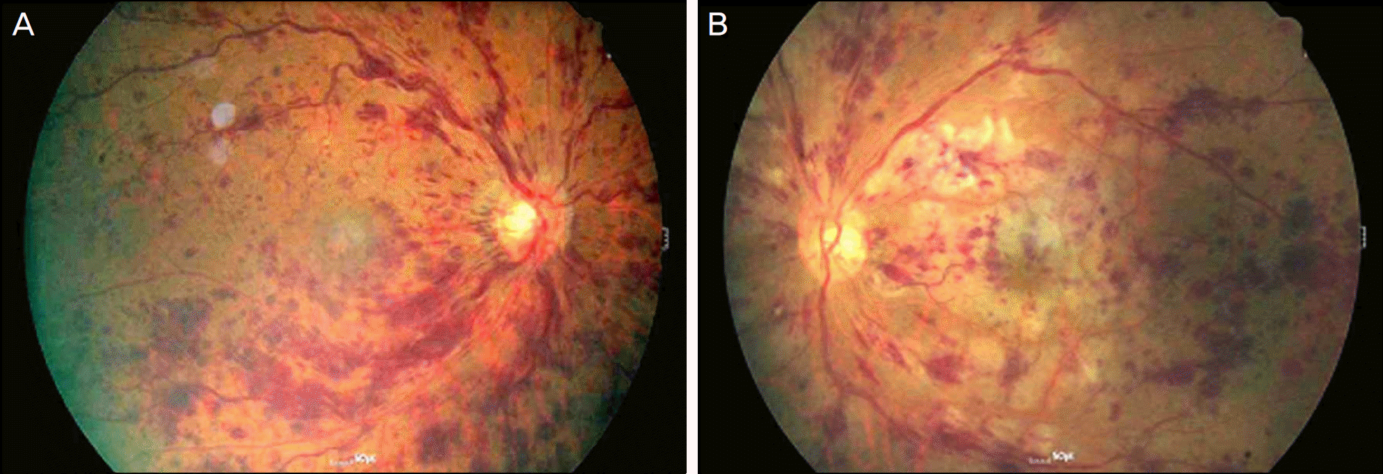
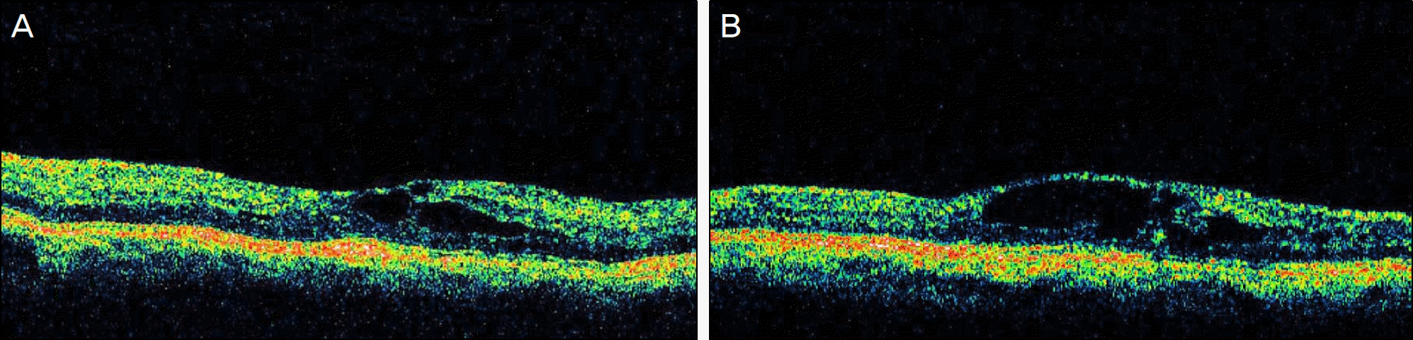
Table 1.
Algorithm for the diagnosis of disseminated intravascular coagulation (DIC)1
| Score global coagulation test results |
| 1. Platelet count (>100 × 103/μ l = 0, <100 × 103/μ l = 1, <50 × 103/μ l = 2) |
| 2. Elevated fibrin-related marker (e.g., fibrin degradation products or D-dimer*) (no increase, 0; moderate increase, 2; strong increase, 3) |
| 3. Prolonged prothrombin time (<3 secs = 0, >3 but <6 secs = 1, >6 secs = 2) |
| 4. Fibrinogen level (>1.0 g/l = 0, <1.0 g/l = 1) |
| Calculate score |
| If ≥ 5: compatible with overt DIC |
| If < 5: no overt DIC; repeat next 1–2 days |
 XML Download