Abstract
Purpose
The authors report a case of orbital mucocele lined with two types of histological epithelial cells developed after repair of the orbital wall fractures.
Case summary
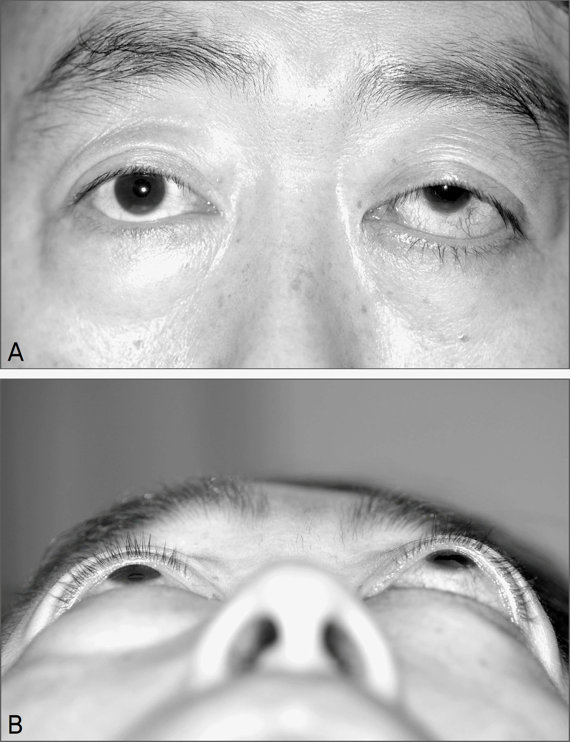
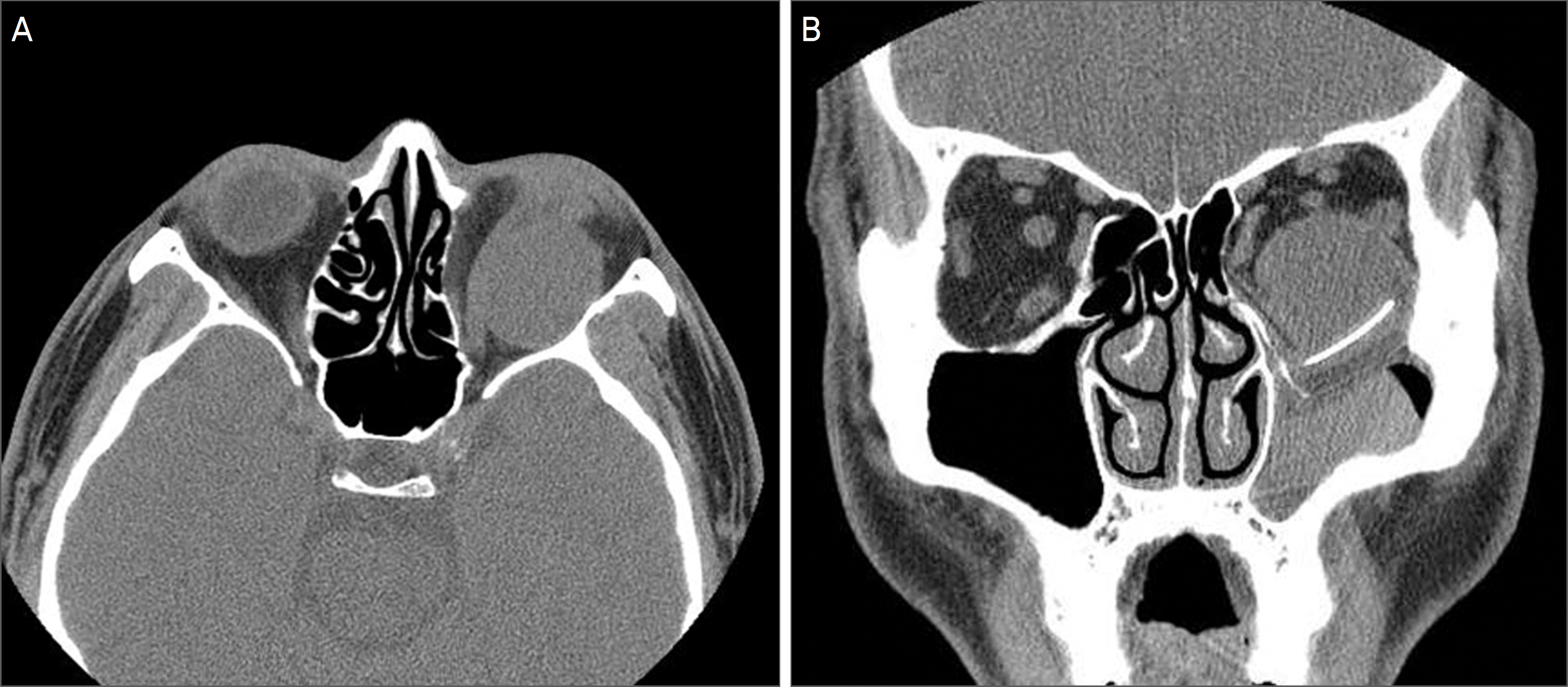
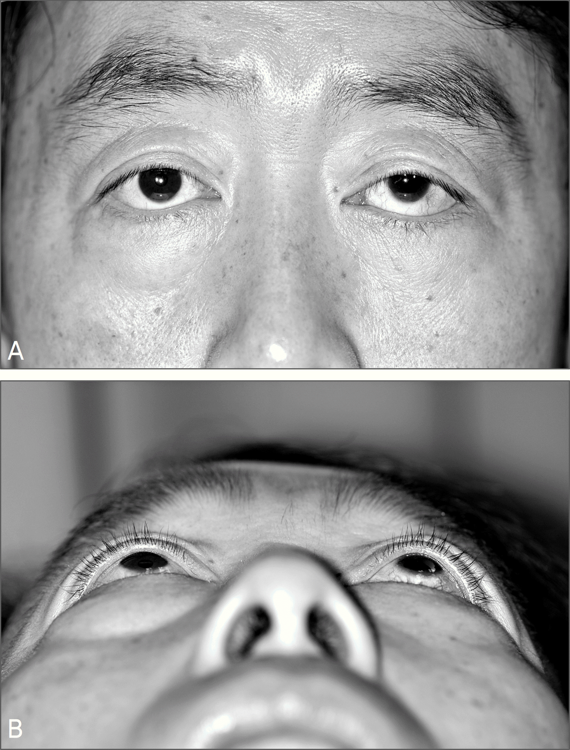
A 53-year-old man presented with proptosis of the left eye for two years. The patient had a history of left orbital inferomedial wall fracture repair ten years earlier at a different hospital. Examination revealed 4 mm proptosis and superior globe displacement of the left eye. Restriction of left ocular movements on elevation, depression and adduction were observed. A computed tomography scan demonstrated a large, non-enhancing, cystic tumor in the left inferior orbit with the inferior and medial wall displaced toward the paranasal sinus. An orbital cystic tumor was excised with the removal of previously-inserted orbital implant via a transconjunctival and transcaruncular approach. The inferior, and medial orbital walls were reconstructed using a MEDPOR® TITAN TM implant. The initial pathologic diagnosis was epidermal cyst. Histopathologic re-review revealed an orbital cyst lined with both stratified squamous epithelium and pseudostratified ciliated columnar epithelium, thus diagnosis was changed to orbital mucocele. Proptosis and restriction in ocular motility improved postoperatively.
Go to : 
References
1. Mauriello JA Jr, Fiore PM, Kotch M. Dacryocystitis. Late complication of orbital floor fracture repair with implant. Ophthalmology. 1987; 94:248–50.
2. Brown AE, Banks P. Late extrusion of alloplastic orbital floor implants. Br J Oral Maxillofac Surg. 1993; 31:154–7.

3. Rubin PA, Bilyk JR, Shore JW. Orbital reconstruction using porous polyethylene sheets. Ophthalmology. 1994; 101:1697–708.

4. Neves RB, Yeatts RP, Martin TJ. Pneumo-orbital cyst after orbital fracture repair. Am J Ophthalmol. 1998; 125:879–80.

5. Tahhan M, Alkhardaji F, Durrani OM, Price NJ. Intraorbital epithelial cyst formation: a rare complication of silastic implantation. Arch Ophthalmol. 2002; 120:1768–9.
6. Glavas I, Lissauer B, Hornblass A. Chronic subperiosteal hematic cyst formation twelve years after orbital fracture repair with alloplastic orbital floor implant. Orbit. 2005; 24:47–9.
7. Tan CS, Ang LP, Choo CT, et al. Orbital cysts lined with both stratified squamous and columnar epithelia: a late complication of silicone implants. Ophthal Plast Reconstr Surg. 2006; 22:398–400.

8. Kwun YK, Kim YD. Two cases of mucocele after orbital fracture repair. J Korean Ophthalmol Soc. 2009; 50:612–7.

9. Kang SJ, Kwak IH. Hematic cyst formation after repair of blow-out fracture. Korean J Ophthalmol. 1996; 10:60–2.

10. Lee SB, Park KS, Kim YD. Orbital cyst after repair of blow-out fracture. J Korean Ophthalmol Soc. 1999; 40:273–7.
11. James CR, Lyness R, Wright JE. Respiratory epithelium lined cysts presenting in the orbit without associated mucocele formation. Br J Ophthalmol. 1986; 70:387–90.

Go to : 
 | Figure 1.Preoperative appearance of the patient showing superior displacement (A) and exophthalmos (B) of the left eye. |
 | Figure 2.Preoperative Orbital CT. (A) Axial view demonstrates large, non-enhancing soft tissue mass with relative low density occupying inferolateral extraconal portion of the left orbit. (B) Coronal view showed haziness of the maxillary sinus and a large, non-enhancing, cystic tumor in the left inferior orbit with the inferior and medial wall displaced toward the paranasal sinus. Previously implanted silastic sheet (arrowhead) and polyethylene sheets (arrow) are observed. |
 | Figure 3.Histopathology of the cyst. (A) Low magnification view shows an unilocular cyst which had even wall thickness of 1 mm. (B) There are multiple needle-shaped spaces consistent with cholesterol clefts and inflammatory cells in the adjacent soft tissue of the cyst. (C) Stratified squamous epithelium lining the cystic cavity. (D) Pseudostratified ciliated columnar epithelium in the other area of the cyst wall (H&E staining, magnification ×40 (A, B), ×400 (C, D)). |


 PDF
PDF ePub
ePub Citation
 Print
Print


 XML Download