Abstract
Purpose
To evaluate the efficacy and safety of the combination of cataract surgery and intravitreal bevacizumab injection in patients with cataract and diabetic macular edema.
Methods
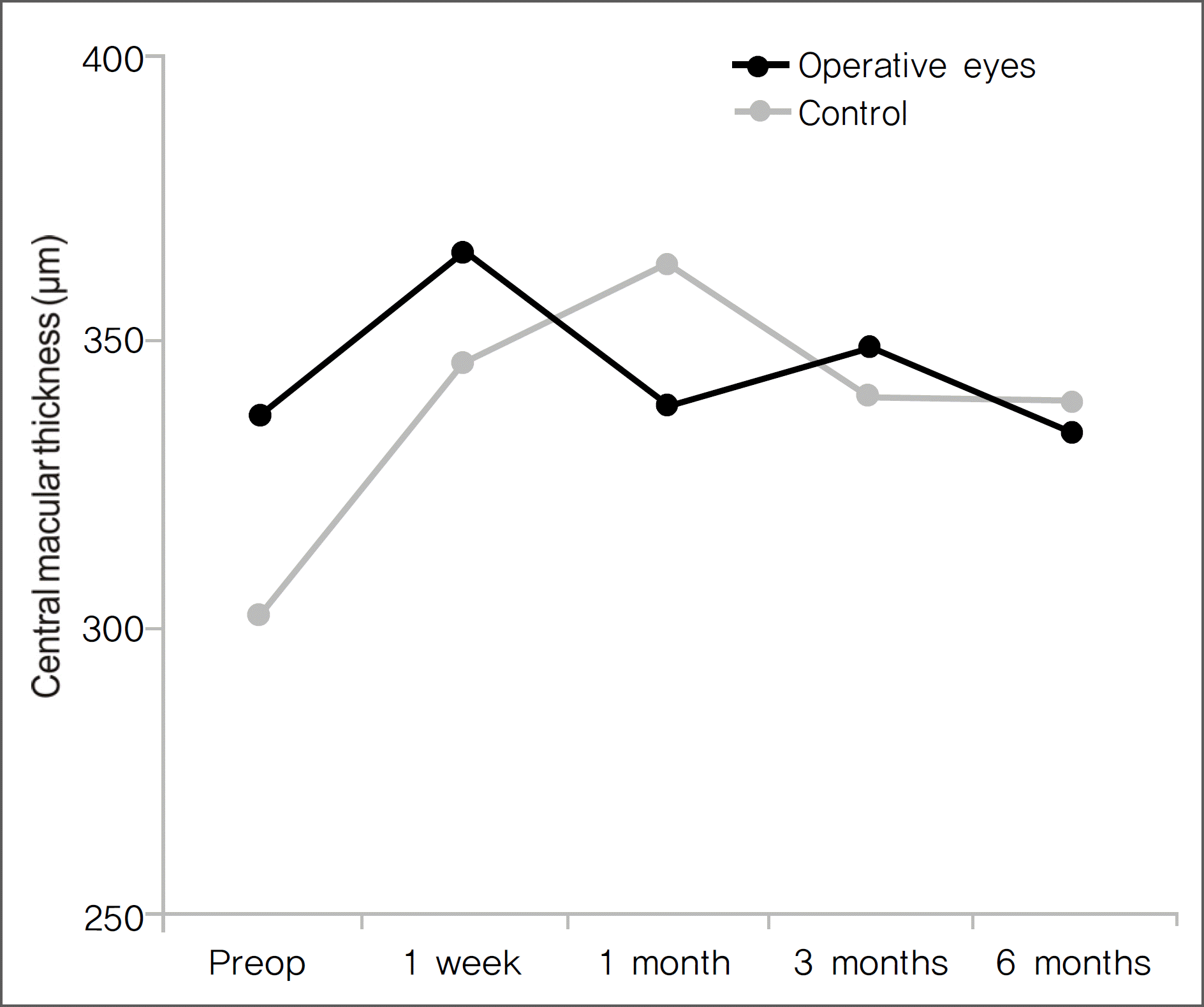
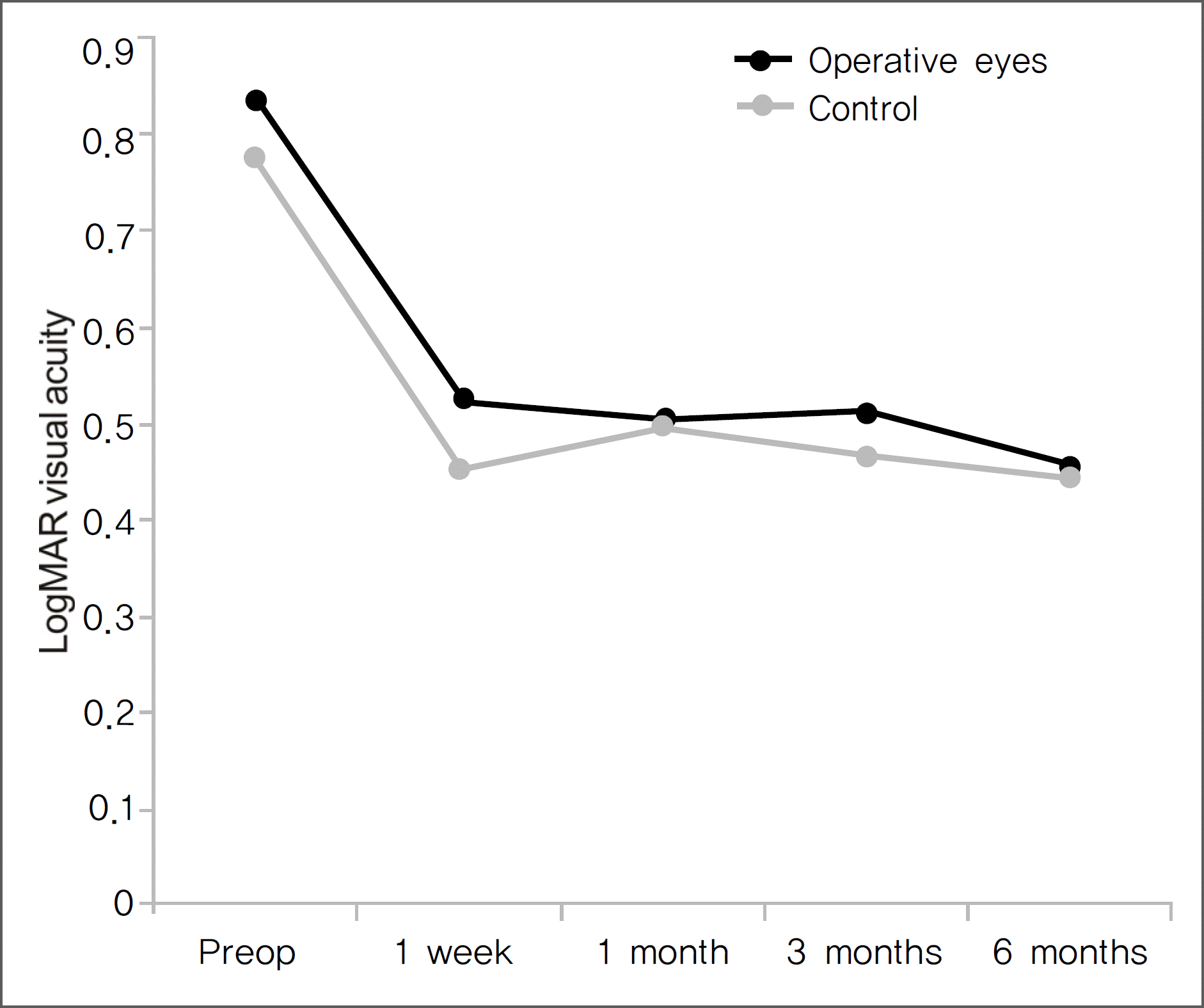
Patients received an intravitreal injection of bevacizumab(1.25 mg) combined with phacoemulsification and implantation of a posterior chamber intraocular lens. Best corrected visual acuity (BCVA, LogMAR) and, central macular thickness (CMT) were measured using OCT at baseline and at one week, one, three, and six months after surgery, and adverse events were recorded. Results: The mean baseline LogMAR BCVA was 0.84±0.50 and mean CMT was 337.1±57.50 μ m. At one week, one, three, and six months after surgery, the mean BCVAs were 0.52±0.40, 0.51±0.42, 0.52±0.34, and 0.46±0.37, and the mean CMTs were 356.4±86.44 μ m, 338.8±138.4 μ m, 349.0±122.9 μ m, and 334.2±100.4 μ m, respectively. No adverse events associated with cataract surgery or intravitreal bevacizumab injection were observed.
Conclusions
The short-term results from the present study suggest the combination of cataract surgery and intravitreal bevacizumab injection are safe and effective for the prevention of macular edema aggravation for one month, but has little effect on prevention of macular edema aggravation three months after surgery for diabetic macular edema patients.
Go to : 
References
1. Ederer F, Hiller R, Taylor HR. Senile lens changes and diabetes in two population studies. Am J Ophthalmol. 1981; 91:381–95.

2. Hamilton AM, Ulbig MVV, Polkinghorne P, et al. Epidemiology of diabetic retinopathy. In management of diabetic retinopathy. London: BMJ Publishing Group;1996. p. 1–15.
3. Hykin PG, Gregson RM, Hamilton AM. Extracapsular cataract extraction in diabetics rubeosis iridis. Eye. 1992; 6:296–9.
4. Kato S, Oshika T, Numaga J, et al. Anterior capsular contraction after cataract surgery in eyes of diabetic patients. Br J Ophthalmol. 2001; 85:21–3.

5. Dowler JG, Hykin PG, Lightman SL, Hamilton AM. Visual acuity following extracapsular cataract extraction in diabetes: a meta-analysis. Eye. 1995; 9:313–7.

6. Chung J, Kim MY, Kim HS, et al. Effect of cataract surgery on the progression of diabetic retinopathy. J Cataract Refract Surg. 2002; 28:626–30.

7. Rossetti L, Autelitano A. Cystoid macular edema following cataract surgery. Curr Opin Ophthalmol. 2000; 11:65–72.

8. Dowler JG, Sehmi KS, Hykin PG, Hamilton AM. The natural history of macular edema after cataract surgery in diabetes. Ophthalmology. 1999; 106:663–8.

9. Zaczek A, Olivestedt G, Zetterstrom C. Visual outcome after phacoe-mulsification and IOL implantation in diabetic patients. Br J Ophthalmol. 1999; 83:1036–41.

10. Krepler K, Biowski R, Schrey S, et al. Cataract surgery in patients with diabetic retinopathy: visual outcome, progression of diabetic retinopathy, and incidence of diabetic macular oedema. Graefes Arch Clin Exp Ophthalomol. 2002; 240:735–8.

11. Chew EY, Benson WE, Remaley NA, et al. Results after lens extraction in patients with diabetic retinopathy: early treatment diabetic retinopathy study report number 25. Arch Ophthalmol. 1999; 117:1600–6.
12. Flach AJ. The incidence, pathogenesis and treatment of cystoid macular edema following cataract surgery. Trans Am Ophthalmol Soc. 1998; 96:557–634.
13. Milch FA, Yannuzzi LA. Medical and surgical treatment of aphakic cystoid macular edema. Int Ophthalmol Clin. 1987; 27:205–17.

14. Early Treatment Diabetic Retinopahty Study Research Group. Photocoagulation for diabetic macular edema. Early Treatment Diabetic Retinopathy Study report number 1. Arch Ophthalmol. 1985; 103:1796–806.
15. Pendergast SD, Hassan TS, Williams GA, et al. Vitrectomy for diffuse diabetic macular edema associated with a taut premacular posterior hyaloids. Am J Ophthalmol. 2000; 130:178–86.
16. Martidis A, Duker JS, Greenberg PB, et al. Intravitreal triamcinolone for refractory diabetic macular edema. Ophthalmology. 2002; 109:920–7.

17. Ferrara N. Vascular endothelial growth factor: Basic science and clinical progress. Endocr Rev. 2004; 25:581–611.

18. Aiello LP, Avery RL, Arrigg PG, et al. Vascular endothelial growth factor in ocular fluid of patients with diabetic retinopathy and other retinal disorders. N Engl J Med. 1994; 331:1480–7.

19. Tolentino MJ, McLeod DS, Taomoto M, et al. Pathologic features of vascular endothelial growth factor-induced retinopathy in the nonhuman primate. Am J Ophthalmol. 2002; 133:373–85.

20. Patel JI, Hykin PG, Cree IA. Diabetic cataract removal: postoperative progression of maculopathy-growth factor and clinical analysis. Br J Ophthalmol. 2006; 90:697–701.

21. Barone A, Prascina F, Russo V, et al. Successful treatment of pseudophakic cystoid macular edema with intravitreal bevacizumab. J Cataract Refract Surg. 2008; 34:1210–2.

22. Arevalo JF, Garcia-Amaris RA, Roca JA, et al. Primary intravitreal bevacizumab for the management of pseudophakic cystoid macular ede-ma: pilot study of the Pan-American Collaborative Retina Study Group. J Cataract Refract Surg. 2007; 33:2098–105.
23. Kook D, Wolf A, Kreutzer T, et al. Long-term effect of intravitreal bevacizumab (Avastin) in patients with chronic diffuse diabetic macular edema. Retina. 2008; 28:1053–60.

24. Haritoglou C, Kook D, Neubauer A, et al. Intravitreal bevacizumab (Avastin) therapy for persistent diffuse diabetic macular edema. Retina. 2006; 26:999–1005.

25. Ah-Fat FG, Sharma MK, Majid MA, Yang YC. Vitreous loss during conversion from conventional extracapsular cataract extraction to phacoemulsification. J Cataract Refract Surg. 1998; 24:801–5.

26. Fogla R, Biswas J, Ganesh SK, Ravishnkar K. Evaluation of cataract surgery in intermediate uveitis. Ophthalmic Surg Lasers. 1999; 30:191–8.

27. Warwar RE, Bullock JD, Ballal D. Cystoid macular edema and anterior uveitis associated with latanoprost use. Experience and incidence in a retrospective review of 94 patients. Ophthalmology. 1998; 105:263–8.
28. Henry MM, Henry LM. A possible cause of chronic cystic maculopathy. Ann Ophthalmol. 1977; 9:455–7.
29. Ursell PG, Spalton DJ, Whitcup SM, Nussenblatt RB. Cystoid macular edema after phacoemulsification: relationship to blood-aqueous barrier damage and visual acuity. J Cataract Refract Surg. 1999; 25:1492–7.

30. Schalnus RW, Ohrloff C, Magone T. The aqueous humor-vitreous body barrier and the blood-aqueous humor barrier after YAG laser capsulotomy in capsular sac vs ciliary sulcus fixation of the intraocular lens. Ophthalmologe. 1995; 92:289–92.
31. Miyak K. Indomethacin in the treatment of postoperative cystoid macular edema. Surv Ophthalmol. 1984; 28:554–68.
32. Yannauzzi LA, Landau AN, Turtz AI. Incidence of aphakic cystoid macular edema with the use of topical indomethacin. Ophthalmology. 1981; 88:947–54.

33. von Jagow B, Ohrloff C, Kohnen T. Macular thickness after un-eventful cataract surgery determined by optical coherence tomography. Graefes Arch Clin Exp Ophthalmol. 2007; 245:1765–71.

34. Kim SJ, Equi R, Bressler NM. Analysis of macular edema after cataract surgery in patients with diabetes using optical coherence tomography. Ophthalmology. 2007; 114:881–9.

35. Wang SJ, Choi SH. The changes in macular thickness after phacoe-mulsification in patients with non-diabetes and nonproliferative diabetic retinopathy. J Korean Ophthalmol Soc. 2008; 49:57–64.

36. Rosetti L, Autelitano A. Cystoid macular edema following cataract surgery. Curr Opin Ophthalmol. 2000; 11:65–72.
37. Tano Y, Sugita G, Abrams G, Machemer R. Inhibition of intraocular proliferations with intravitreal corticosteroids. Am J Ophthalmol. 1980; 89:131–6.
38. Tano Y, Chandler D, Machemer R. Treatment of intraocular proliferation with intravitreal injection of triamcinolone acetonide. Am J Ophthalmol. 1980; 90:810–6.

39. Gillies MC, Simpson JM, Billson FA, et al. Safety of an intravitreal injection of Triamcinolone. Arch Ophthalmol. 2004; 122:336–40.

40. Jonas JB, Kreissig I, Degenring R. Intraocular pressure after intra-vitreal injection of triamcinolone acetonide. Br J Ophthalmol. 2003; 87:24–7.

41. Moshfeghi DM, Kaiser PK, Scott IU, et al. Acute endophthalmitis following intravitreal triamcinolone acetonide injection. Am J Ophthalmol. 2003; 136:791–6.

42. Islam MS, Vernon SA, Negi A. Intravitreal triamcinolone will cause posterior subcapsular cataract in most eyes with diabetic maculopathy within 2 years. Eye. 2007; 21:321–3.

43. Lam DS, Chan CK, Mohamed S, et al. Phacoemulsifi cation with intra-vitreal triamcinolone in patients with cataract and coexisting diabetic macular oedema: A 6-month prospective pilot study. Eye. 2005; 19:885–90.
44. Habib MS, Cannon PS, Steel DH. The combination of intravitreal triamcinolone and phacoemulsification surgery in patients with diabetic foveal oedema and cataract. BMC Ophthalmol. 2005; 5:15.

45. Jonas JB. Intravitreal triamcinolone acetonide for treatment of intra-ocular oedematous and neovascular diseases. Acta Ophthalmol Scand. 2005; 83:645–63.

46. Jonas JB, Degenring R, Kreissig I, Akkoyun I. Safety of intravitreal high-dose reinjections of triamcinolone acetonide. Am J Ophthalmol. 2004; 138:1054–5.

47. Chen CH, Liu YC, Wu PC. The combination of intravitreal bevacizumab and phacoemulsification surgery in patients with cataract and coexisting diabetic macular edema. J Ocul Pharmacol Ther. 2009; 25:83–9.

48. Seo JW, Park IW. Intravitreal bevacizumab for treatment of diabetic macular edema. Korean J Ophthalmol. 2009; 23:17–22.

Go to : 
 | Figure 1.Changes in central macular thickness with OCT after cataract surgery combined with intravitreal bevacizumab injection. CMT=central macular thickness; OCT=optical coherence tomography. |
 | Figure 2.Changes in BCVA (LogMAR) after cataract surgery combined with intravitreal bevacizumab injection. BCVA=best corrected visual acuity; LogMAR=logarithm of the minimum angle of resolution. |
Table 1.
Characteristics of patients
| Characteristics | Operated eyes | Control |
|---|---|---|
| Age (yrs) | ||
| Mean± SD* | 65.14±7.86 | 67.90±5.97 |
| Range | 52∼80 | 56∼74 |
| Gender (n) | ||
| Male | 8 | 4 |
| Female | 6 | 8 |
| Foollow-up time (days) | ||
| Mean± SD | 297.12±84.14 | 316±90.57 |
| Range | 191∼342 | 205∼498 |
| Preoperative BCVA† (LogMAR‡ | ||
| Mean± SD | 0.84±0.50 | 0.77±0.48 |
| Range | 0.3∼2.3 | 0.4∼1.0 |
| p-value | 0.3 | 37 |
| Preoperative CMT§ (μm) | ||
| Mean± SD | 337.1±57.50 | 302.1±56.65 |
| Range | 281∼434 | 210∼387 |
| p-value | 0.0 | 07 |
Table 2.
Changes in BCVA and CMT after cataract surgery combined with intravitreal bevacizumab injection
| Preop | 1 week | 1 month | 3 months | 6 months | |
|---|---|---|---|---|---|
| BCVA* (LogMAR†) | |||||
| Operated eyes | 0.84±0.50 | 0.52±0.40 | 0.51±0.42 | 0.52±0.34 | 0.46±0.37 |
| (P=0.003) | (P<0.001) | (P<0.001) | (P<0.001) | ||
| Control | 0.77±0.48 | 0.45±0.25 | 0.49±0.34 | 0.47±0.32 | 0.44±0.32 |
| (P=0.004) | (P=0.062) | (P=0.052) | (P=0.042) | ||
| CMT‡ (μm) | |||||
| Operated eyes | 337.1±57.50 | 356.4±86.44 | 338.8±138.4 | 349.0±122.9 | 334.2±100.4 |
| (P=0.093) | (P=0.477) | (P=0.334) | (P=0.495) | ||
| Control | 302.1±56.65 | 346.4±62.32 | 363.6±92.01 | 340.3±58.89 | 339.9±82.11 |
| (P=0.061) | (P=0.038) | (P=0.056) | (P=0.041) | ||
Table 3.
Difference in CMT after cataract operation from preoperative CMT
| Operated eyes | Control | |
|---|---|---|
| 1 week after operation | ||
| Mean± SD* (μm) | 28.21±75.76 | 44.25±72.98 |
| p-value | 0.211 | |
| 1 month after operation | ||
| Mean± SD (μm) | 1.64±88.54 | 61.5±104.35 |
| p-value | 0.032 | |
| 3 months after operation | ||
| Mean± SD (μm) | 11.85±76.81 | 38.17±77.07 |
| p-value | 0.194 | |
| 6 months after operation | ||
| Mean± SD (μm) | −3±69.87 | 37.58±59.31 |
| p-value | 0.061 | |


 PDF
PDF ePub
ePub Citation
 Print
Print


 XML Download